




Abstract
Hematopoietic cells derived from human embryonic stem cells (hESCs) have a number of potential utilities, including the modeling of hematological disorders in vitro, whereas the use for cell replacement therapies has proved to be a loftier goal. This is due to the failure of differentiated hematopoietic cells, derived from human pluripotent stem cells (hPSCs), to functionally recapitulate the in vivo properties of bona fide adult hematopoietic stem/progenitor cells (HSPCs). To better understand the limitations of differentiation programming at the molecular level, we have utilized differential gene expression analysis of highly purified cells that are enriched for hematopoietic repopulating activity across embryonic, fetal, and adult human samples, including in vivo explants of human HSPCs 8-weeks post-transplantation. We reveal that hESC-derived hematopoietic progenitor cells (eHPCs) fail to express critical transcription factors which are known to govern self-renewal and myeloid/lymphoid development and instead retain the expression of Polycomb Group (PcG) and Trithorax Group (TrxG) factors which are more prevalent in embryonic cell types that include EZH1 and ASH1L, respectively. These molecular profiles indicate that the differential expression of the core epigenetic machinery comprising PcGs/TrxGs in eHPCs may serve as previously unexplored molecular targets that direct hematopoietic differentiation of PSCs toward functional HSPCs in humans.
Introduction
Human HSC function is defined by hematopoietic repopulation and multilineage differentiation capacity. The gold standard method that assesses HSC function is the severe combined immunodeficiency (SCID)-mouse repopulating cell (SRC) assay [23 –25]. Putative HSPCs remain functionally heterogeneous, but SRCs can be enriched using CD34+CD38−Lin− phenotyping and are capable of long-term (>6 months) multi-lineage in vivo engraftment in immunodeficient mice [24,26]. As such, molecular comparisons to SRCs become of utmost importance to distil candidate molecular targets that contribute to in vivo engraftment. Here, we use a global hierarchical cluster analysis of the genes that are present in highly purified HSPC enriched for repopulating activity across embryonic, fetal, and adult samples. Unlike previous studies that compared purified populations of HSPCs with hPSC-derived blood cells, we have compared the expression profiles from human SRCs 8-weeks post-transplantation with isolated HSPCs and differentiated hESCs. We reveal that eHPCs and fetal HSPCs cluster with undifferentiated hESCs due to the maintenance of an embryonic set of genes. In particular, a set of epigenetic transcriptional regulators, such as core Polycomb Group (PcG) and Trithorax Group (TrxG) genes, were over-expressed in eHPCs compared with phenotypically isolated HSPCs and SRCs. Furthermore, select PcG/TrxG genes were maintained throughout in vitro hematopoietic differentiation. Based on these molecular analyses, we suggest that aberrant expression of epigenetic regulators in putative blood cells derived from hPSCs may coordinate to suppress molecular signatures that are essential for in vivo hematopoietic reconstitution.
Materials and Methods
Culture of hESCs and formation of human embryoid bodies
H9 hESC lines were cultured on Matrigel (BD Biosciences)-coated plates and maintained in a mouse embryonic fibroblast conditioned medium supplemented with 8 ng/mL of human recombinant basic fibroblast growth factor (Invitrogen) as previously described [9,27]. Formation of human embryoid bodies (hEBs) and hematopoietic differentiation were performed as previously reported [9].
Colony-forming unit assay
EBs were dissociated, and 15,000 cells or single cells were seeded in single wells of a 24-well plate containing methylcellulose H4230 supplemented with stem cell factor (SCF), Flt-3L, interleukin (IL)-3, IL-6, granulocyte colony-stimulating factor (G-CSF), and BMP4. Emergent hematopoietic cell clusters displaying more than 50 cells were counted as colonies [colony forming unit (CFU)] after 14 days.
Microarray expression analysis
Affymetrix CEL files were imported into dChIP software for data normalization, extraction of signal intensities, differential gene expression, and hierarchical cluster analysis. Gene set enrichment analysis was performed using GSEA software and the Molecular Signatures Database [28,29]. Biological networks were generated using Cytoscape [30]. Results were presented as mean±standard error of the mean, and statistical significance was determined using paired or unpaired Student t-test where applicable; P value cutoff at or below 0.05.
Quantitative reverse transcription-polymerase chain reaction
Total RNA was extracted from cells using the Total RNA purification Kit (Norgen) according to the manufacturer's guidelines. First-strand cDNA was synthesized using the First-Strand cDNA Synthesis Kit (Amersham Biosciences). Polymerase chain reaction (PCR) in the absence of templates was used as a negative control. The following PCR conditions were used: 50°C for 2 min, 95°C for 2 min, and 40 cycles of 95°C for 15 s, 60°C for 30 s. Quantitative PCR was conducted using Platinum® SYBR® Green qPCR SuperMix-UDG (Invitrogen) on a Chromo4 Four-Color Real-Time PCR Detection System® (Bio-Rad) according to the manufacturer's guidelines. All values were normalized to GAPDH or GUSB genes.
Results
Epigenetic transcriptional regulators are differentially expressed in eHPCs versus HSPCs
An established methodology for the stage-wise generation of hematopoietic cells from hEBs uses a cocktail of hematopoietic cytokines, resulting in the emergence of small round precursor cells 7–10 days after initial hEB formation [9
–11] (Fig. 1a, b). These precursor cells could be isolated from culture based on cell surface expression of platelet/endothelial cell adhesion molecule 1 (PECAM1), fetal liver kinase 1 (FLK1/KDR), VE-Cadherin, and absence of the pan-hematopoietic marker CD45 (termed CD45-PFV cells) (Fig. 1c). Furthermore, CD45-PFV cells have the ability to give rise to either blood or endothelial cells and are, thus, bipotent in nature [10]. CD45− PFV cells give rise to primitive eHPCs, which co-express CD34 and CD45 in a similar manner to adult HPCs based on their phenotype and in vitro colony-forming (CFU) capacity (Fig. 1d) [9
–11]. However, functional analysis from several groups reveals that these cells are devoid of in vivo repopulating capacity similar to somatic HSPCs with SRC capacity [8,11
–13,15
–17,19,22]. Thus, an in-depth analysis of the molecular basis for in vitro blood development is required to better understand the failure to generate cells with HSC behavior in vivo. As such, we analyzed global expression data generated from cell populations taken at different stages during hESC hematopoietic differentiation from 2 independent studies [10,11]. We compared hESC-derived cells to transcriptome profiles from multiple sources of CD34+CD38− HSPCs and in vivo explants of SRCs to identify candidate genes required for hematopoietic progenitors versus stem cells (all arrays and corresponding cell sources are provided in Supplementary Table S1; Supplementary Data are available online at
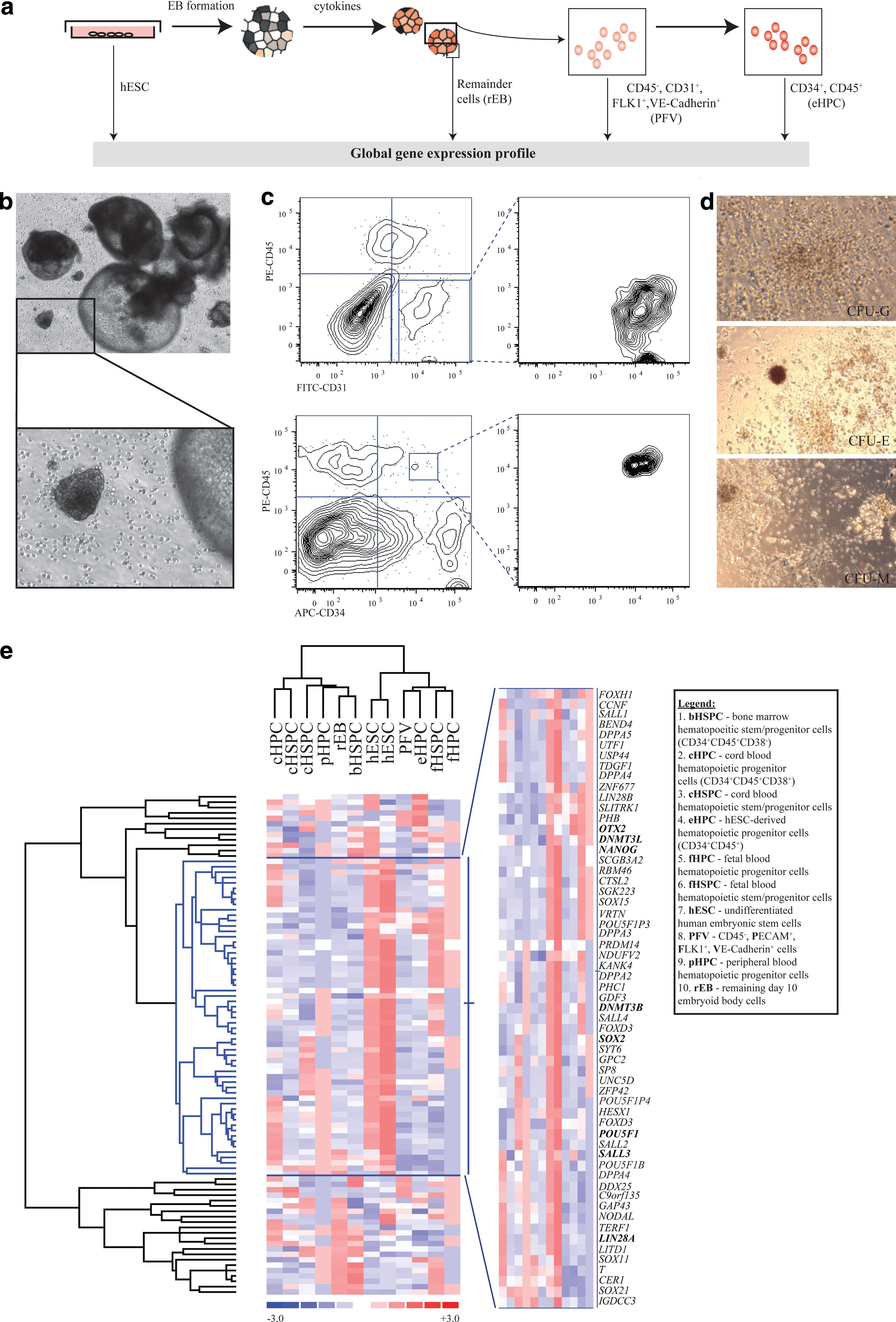
Hematopoietic differentiation from hESCs progresses from a hemogenic/endothelial precursor stage to a primitive hematopoietic stage.
During EB formation and hematopoietic differentiation, core pluripotency factors such as POU5F1, SOX2, NANOG, and LIN28A are expectedly down-regulated as shown by hierarchical clustering of hESC-enriched genes [31] (Fig. 1e). hESCs and their hematopoietic derivatives clustered together among fetal sources of HSPCs (CD34+CD38−) and HPCs (CD34+CD38+), based on the preservation of a small subset of genes that define a fetal/embryonic signature (Fig. 1e). In particular, critical regulators of embryonic and neural development such as DNMT3B/DNMT3L, SALL3 and OTX2 were maintained in eHPCs. The resultant cluster demonstrates that although eHPCs express hematopoietic initiation factors (ie, RUNX1 and TAL1), they still express a subset of PSC genes at a level that maintains their close relationship to embryonic cells (Fig. 1e).
Next, we looked at the stage-specific expression of all genes as hESCs progressed from the undifferentiated state to hematopoietic progenitor-like cells. Genes that were present at all stages of the differentiation process and significantly down-regulated at least 1.5-fold (P value≤0.05) in the EB-derived cells, which are devoid of hematopoietic potential, were filtered to identify the factors enriched during in vitro blood development. This provided a negative control for hematopoietic differentiation signatures not previously exploited to better distill significant gene profiles. From this rigorous criterion, 833 genes in total were present and up-regulated across all hESC and hESC-derivatives with hematopoietic potential, of which 747 were categorized using the Gene Ontology database (GO;
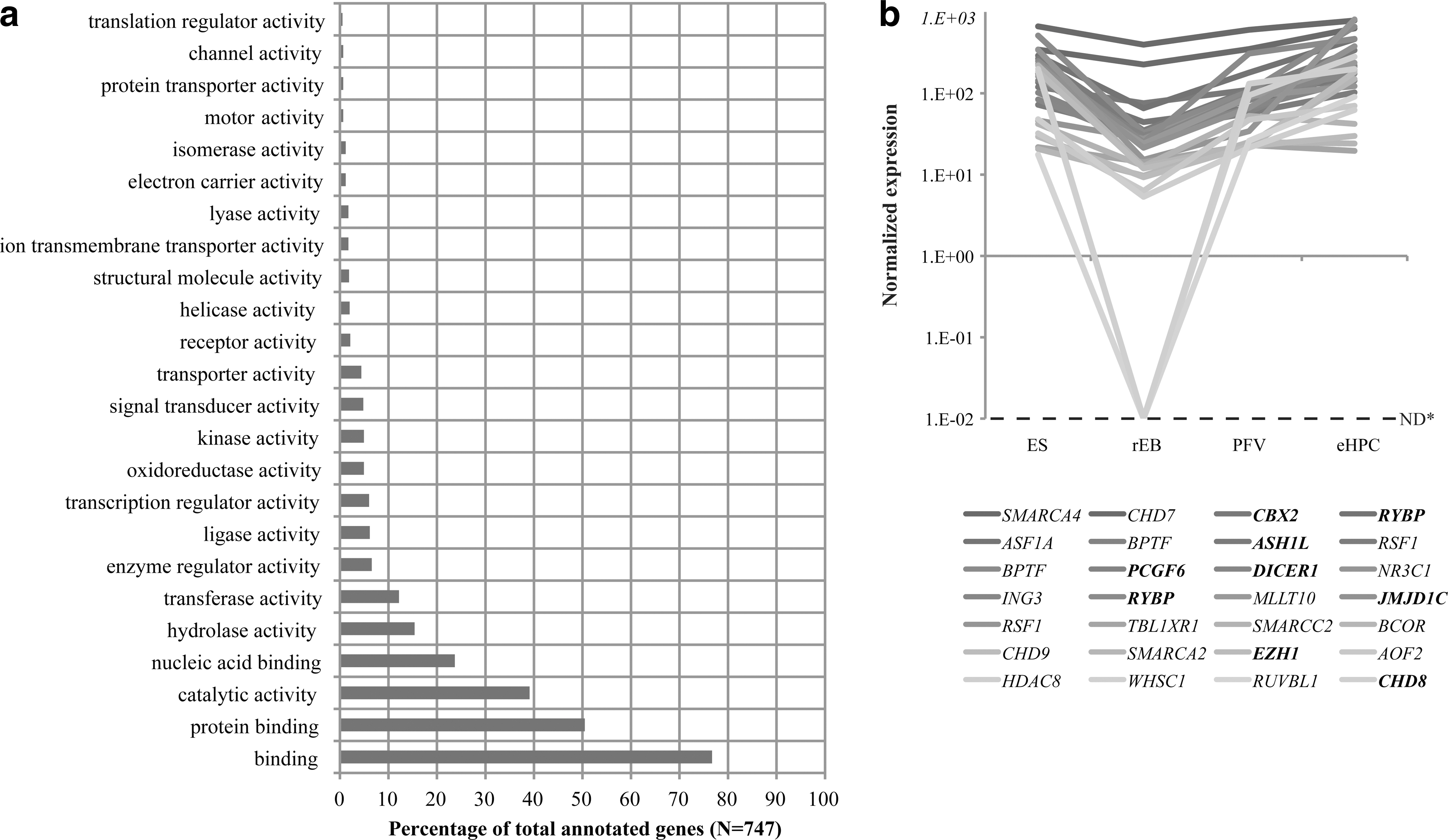
Genes expressed/up-regulated exclusively during hESC hematopoietic differentiation.
ES, embryonic stem; EB, embryoid body; eHPC, hESC-derived hematopoietic progenitor cells; nd, not done.
Among these 104, genes associated with PSC maintenance such as DICER1 [33], JMJD1C [34], and KLF2 [35] were identified. KLF2 has been shown to be critical for the maintenance of primitive erythropoiesis and embryonic globin expression [36]; thus, its expression is consistent with the absence of adult globin expression, contributing to the primitive nature of eHPCs [11] (Fig. 1e). A major proportion of genes involved in transcriptional regulation (∼24%) were epigenetic and chromatin-related genes such as Polycomb (CBX2, EZH1, PCGF6, and RYBP) and Trithorax genes (ASH1L and CHD8), which maintained expression throughout hESC differentiation (Fig. 2b).
hESC hematopoietic derivatives have limited expression of HSC self-renewal and differentiation genes
We compared the expression profiles generated from hESC derivatives, with emphasis on eHPCs, with heterogeneous HSPCs (CD34+CD38− cells) and SRCs (similarly enriched for CD34+CD38− expression post-transplantation) (see Supplementary Fig. S1 for the level of human engraftment) to identify the differentially expressed genes that underlie the inability to derive functional HSCs on hESC differentiation (Fig. 3). Linear regression analysis of genes expressed during hematopoiesis (defined by GO annotation) between eHPCs and isolated CD34+CD38− HSPCs yielded correlation coefficients ranging from 0.600 to 0.678 (Fig. 4a), while pair-wise comparisons between different sources of HSPCs (ie, from bone marrow, cord blood, or fetal blood) were much more closely related (R 2=0.75) (Supplementary Fig. S2). In fact, the expression of hematopoietic gene sets was not highly correlated between SRCs and either eHPCs or isolated HSPCs (R 2=0.449 and 0.473 respectively) (Fig. 4a and Supplementary Fig. S2), which is consistent with the functional divergence between hESC derivatives and bona fide HSCs.
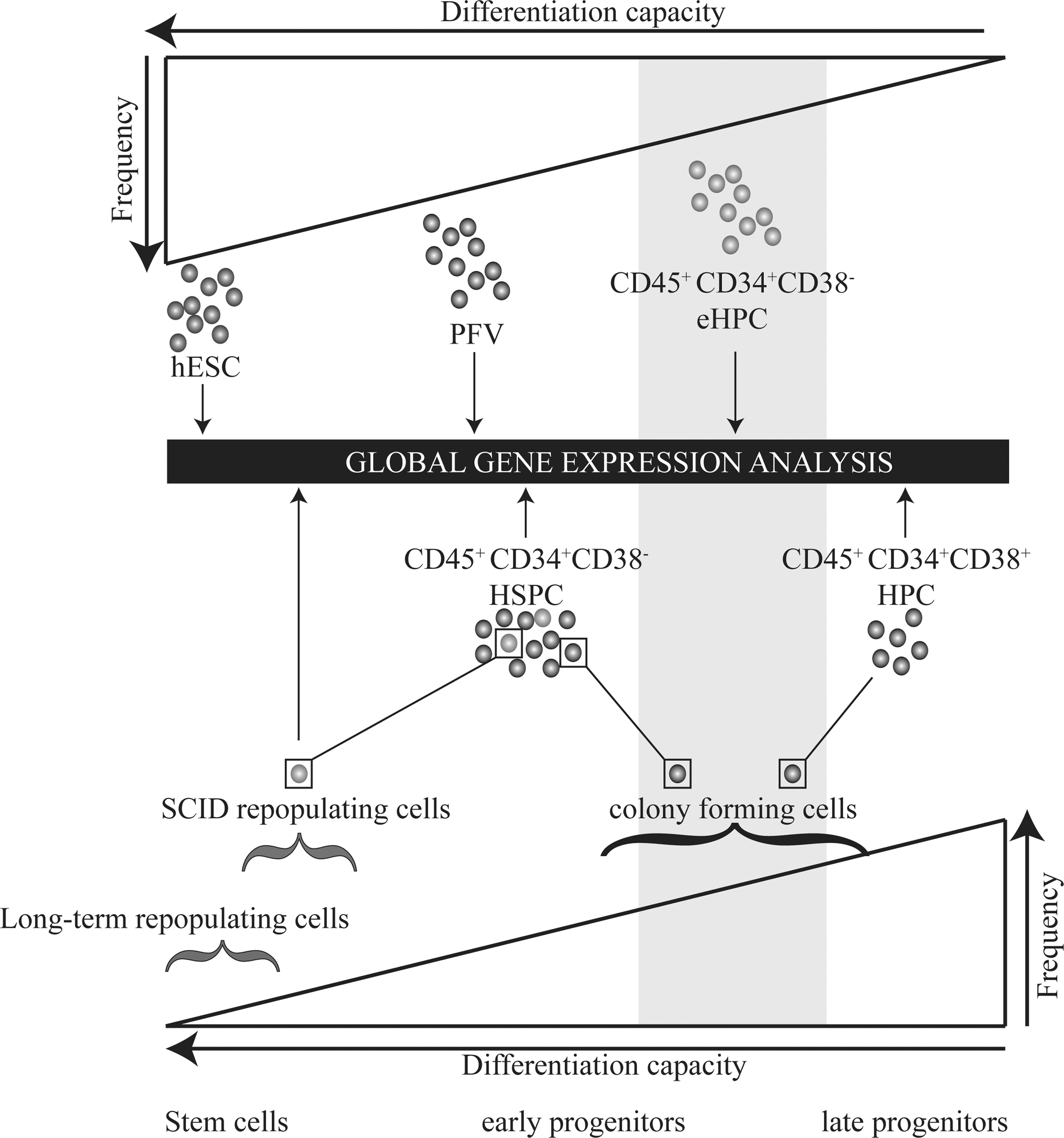
Schematic representation of expression profiling during hESC hematopoietic differentiation compared with phenotypically and functionally defined HSCs (heterogeneous stem/progenitor cells and SRCs respectively). HSPC, hematopoietic stem/progenitor cells; HPC, hematopoietic progenitor cells; SRC, NOD/SCID mouse repopulating cells; SCID, severe combined immunodeficiency.
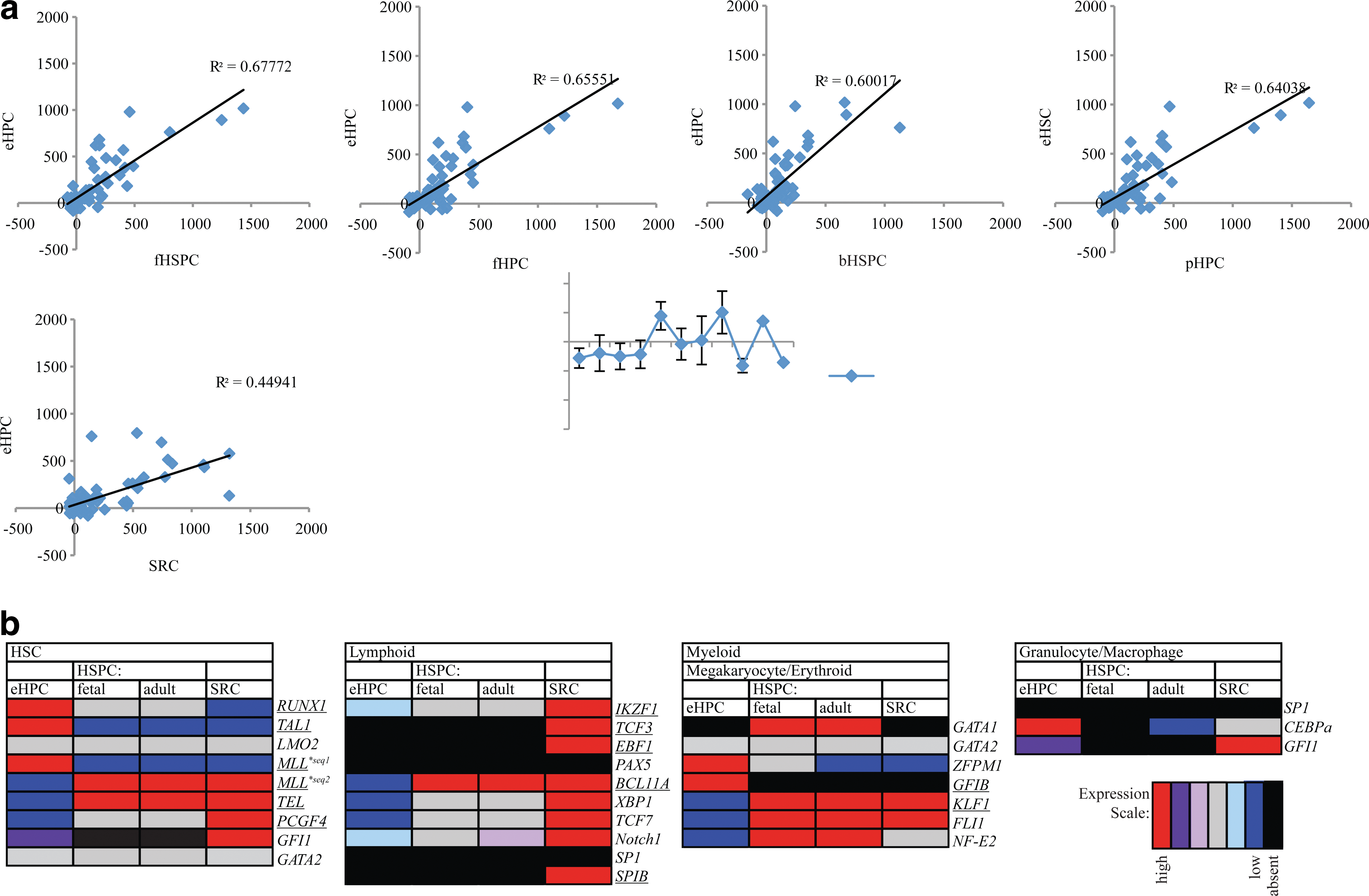
Hematopoietic gene signature expression in eHPCs compared with isolated HSPCs.
We next dissected the expression of critical transcription factors expressed at different stages of hematopoietic development (Fig. 4b). The expression of HSC self-renewal genes (ie, MLL and TEL) and lineage-specific genes (ie, KLF1, FLI1, and BCL11A) were common among SRCs and HSPCs (Fig. 4b). Genes important for hematopoietic establishment such as RUNX1, TAL1, and MLL [37] were significantly more abundant in eHPCs compared with SRCs (P<0.05). Notably, differential expression of MLL isoforms was observed between hESC-derived cells and SRCs. Full-length MLL (probeset 212078_s_at) is more abundant in eHPCs, but still expressed in SRCs, while an alternative partial MLL transcript (probeset 212079_s_at; GenBank D14540.1) is more highly expressed in SRCs. MLL functions in the maintenance of HOX expression throughout development and particularly in HSC specification. The functional consequence of the up-regulation of truncated MLL across CD34+CD38−cells and SRCs is unknown but may underlie the opposed HOXA and HOXB cluster expression observed between eHPCs and HSPCs [11]. Factors important for HSC self-renewal and progenitor differentiation, such as BMI1 [38,39], KLF1 [40], and IKZF1 [41,42], showed higher expression in HSPCs (P<0.05; Fig. 4b) than in eHPCs. Furthermore, several transcription factors that were considered critical for lymphopoiesis (ie, IKZF1, TCF3, and EBF1) were more highly expressed in SRCs compared with either eHPCs or HSPCs, which provides a distinguishing gene signature that may define hematopoietic repopulation capacity. Taken together, the expression of critical transcription factors in different branches of the hematopoietic hierarchy suggest that eHPCs undergo hematopoietic specification but fail to appropriately express determinants of HSC self-renewal and differentiation, particularly lymphoid transcription factors.
De novo and in vivo human HSCs are molecularly distinct from eHPCs
Multiple sources of HSCs are used for clinical transplantation (bone marrow, cord blood, and mobilized peripheral blood), each with different propensities for hematopoietic reconstitution, with cord blood having the most robust engraftment [43]. The commonality between HSPCs from different blood sources is the ability to engraft, regardless of level, that eludes eHPCs. Previous comparisons between eHPCs and cord blood HSPCs present the 2 extremes of hematopoietic engraftment potential. Thus, we sought to identify a common set of candidate genes required for any level of engraftment by utilizing multiple sources of HSPCs and SRCs. Pair-wise comparisons of eHPCs versus putative HSCs (CD34+CD38− cells sorted from bone marrow, cord blood, or fetal blood samples) yielded 1,365 transcripts that were differentially expressed among all HSPC samples (Supplementary Fig. S3).
The 25 most significantly enriched genes in eHPCs compared with HSPCs had broad functions in metabolism, cell-cycle regulation, and signal transduction (Fig. 5a). Notably, LIN28A, a factor used in stem cell reprogramming from somatic cells (iPS generation) and a known post-transcriptional regulator of POU5F1 (Oct-4) [31,44], was highly up-regulated in eHPCs versus adult HSPCs. The expression of LIN28A is consistent with our observation of the maintenance of a subset of genes enriched in ESCs during hESC hematopoietic differentiation (Fig. 1e).

Differentially expressed genes between eHPCs and phenotypically and functionally defined HSCs (HSPCs and SRCs respectively) The top 25 most significant differentially expressed genes that were up-regulated in
Since the sole determinant of a functional HSC is hematopoietic engraftment, we compared eHPCs with CD34+CD38− expressing SRCs isolated 8 weeks post-transplantation. In total, 3,332 unique probe sets corresponded to 2,643 genes that were at least 2-fold differentially expressed (P≤0.05) and denoted as present across 50% or more replicate arrays between eHPCs and SRCs. Two genes involved in metabolism and signal transduction were highly abundant in eHPCs compared with both HSPCs and SRCs (CYP1B1 and PKIB respectively) (Fig. 5a, b). Furthermore, an alternate isoform of LIN28, namely LIN28B, was up-regulated in eHPCs than in SRCs. It should be noted that this is in contrast to eHPCs versus HSPCs, of which LIN28A was up-regulated. We also found that EZH1, a component of a non-canonical Polycomb repressive complex (PRC)-2 [45], was more than 21-fold highly expressed in eHPCs, placing it among the most significantly differentially expressed genes between eHPCs and SRCs (Fig. 5b). EZH1 displays functional redundancy in ESCs with the core PRC2 gene EZH2 and can mediate the methylation of histone H3 lysine 27, which is critical to pluripotency and developmental plasticity [45]. Persistent expression of EZH1 in eHPCs may be linked to the repression of critical hematopoietic genes and the inability to fully reproduce a bona fide HSC. Conversely, several major histocompatibility complex genes were among the 25 most highly up-regulated genes in HSPCs and SRCs (Fig. 5c, d). Furthermore, HOXA9 was consistently up-regulated across multiple sources of HSPCs and SRCs versus eHPCs. The homeobox transcription factor, HOXA9, is a known regulator of normal and malignant hematopoiesis [46 –48]. hESC-derived HPCs and HSPCs/SRCs express different levels of MLL isoforms (Fig. 4b), where the potential interaction of a partial MLL transcript with the MLL protein complex may be correlated with differential HOXA9 expression.
eHPCs maintain expression of epigenetic transcriptional regulators
To elucidate whether specific categories of molecular function or biological pathways were preferentially enriched among differentially expressed genes between hESC-derived hematopoietic cells and HSPCs or SRCs, we performed gene set enrichment analysis (GSEA) (
The classifications include Gene Ontology (GO) Molecular functions and Kyoto Encyclopedia of Genes and Genomes (KEGG) pathways that are enriched in eHPCs.
eHPCs, hESC-derived hematopoietic progenitor cells; SRCs, severe combined immunodeficiency (SCID)-mouse repopulating cells; HSPCs, hematopoietic stem/progenitor cells.
However, the cell adhesion gene ITGA6 (CD49f), which has recently been used to enhance enrichment of HSCs [49], was more abundant in SRCs compared with HSPCs, thus distinguishing SRC activity from functionally heterogeneous CD34+CD38− hematopoietic cells (Fig. 5g). In contrast, eHPCs were enriched for cell cycle-associated genes compared with both HSPCs and SRCs (P value 0.019 and 0.023 respectively). In particular, the preferential expression of genes that promote cell-cycle progression, such as various cyclin-dependant kinases and E2F transcription factors (E2F1 and E2F2) (Fig. 5e, f), may indicate the maintenance of hESC-like proliferative capabilities and/or an effect of cytokine stimulation due to differentiation conditions.
A further examination of the GSEA results revealed that transcription factor binding genes were enriched in eHPCs in comparison to SRCs (P value 0.023). Furthermore, gene categories involving transcriptional regulation were more abundant in eHPCs compared with both HSPCs and SRCs (Supplementary Tables S2 and S3). Dissection of the genes classified as transcriptional regulators revealed several Polycomb-associated genes (such as EZH1, PCGF2, RING1, RYBP, PCGF6, and EED) that were preferentially expressed in eHPCs (Fig. 5i–l), with EZH1 being highly differentially expressed (Fig. 5b). In contrast, a critical regulator of HSC self-renewal and a core component of PRC-1, BMI1 [38,39], was more abundant in SRCs versus eHPCs (Fig. 5j). Moreover, the TrxG gene ASH1L was among the 32 genes that were up-regulated throughout hESC blood development and over-expressed in eHPCs versus all HSCs (putative and in vivo explants) (Fig. 5h–j, l). These data suggest that embryonic-derived hematopoietic cells appear to have an imbalanced expression of core PcG and TrxG compared with SRCs, which may prevent the expression of genes that are necessary for the derivation of functional HSC with an in vivo engraftment capacity.
A network of PcG and TrxG genes are inappropriately expressed in eHPCs
Several epigenetic transcriptional regulators were highly abundant in eHPCs and during the in vitro hematopoietic differentiation. This suggests that the inability to down-regulate PSC-expressed epigenetic factors in hESC derived hematopoietic cells prevents programming of the ideal repertoire of genes required for functional reconstitution behavior similar to somatic HSC.
To examine this idea, we performed differential gene expression analysis and identified a core set of PcG genes that significantly distinguished hESC hematopoiesis from HSPCs and in vivo explanted human SRCs. Among these, EZH1, ASH1L, and PCGF6 were up-regulated in hESC-derived cells compared with HSPCs or SRCs (Fig. 5h–k). Using Cytoscape [30], we constructed a network of differentially expressed PcG and TrxG between eHPCs and HSPCs/SRCs, of which EZH1, ASH1L, and PCGF6 maintain their expression from undifferentiated hESCs to hematopoietic derivatives (Fig. 6). The interactome network analysis illustrates that a non-canonical PcG repressive complex, centering on EZH1, may be responsible for the maintenance of an embryonic molecular phenotype and the failure to relieve repression at lineage-specific hematopoietic genes in hESC-derived cells.
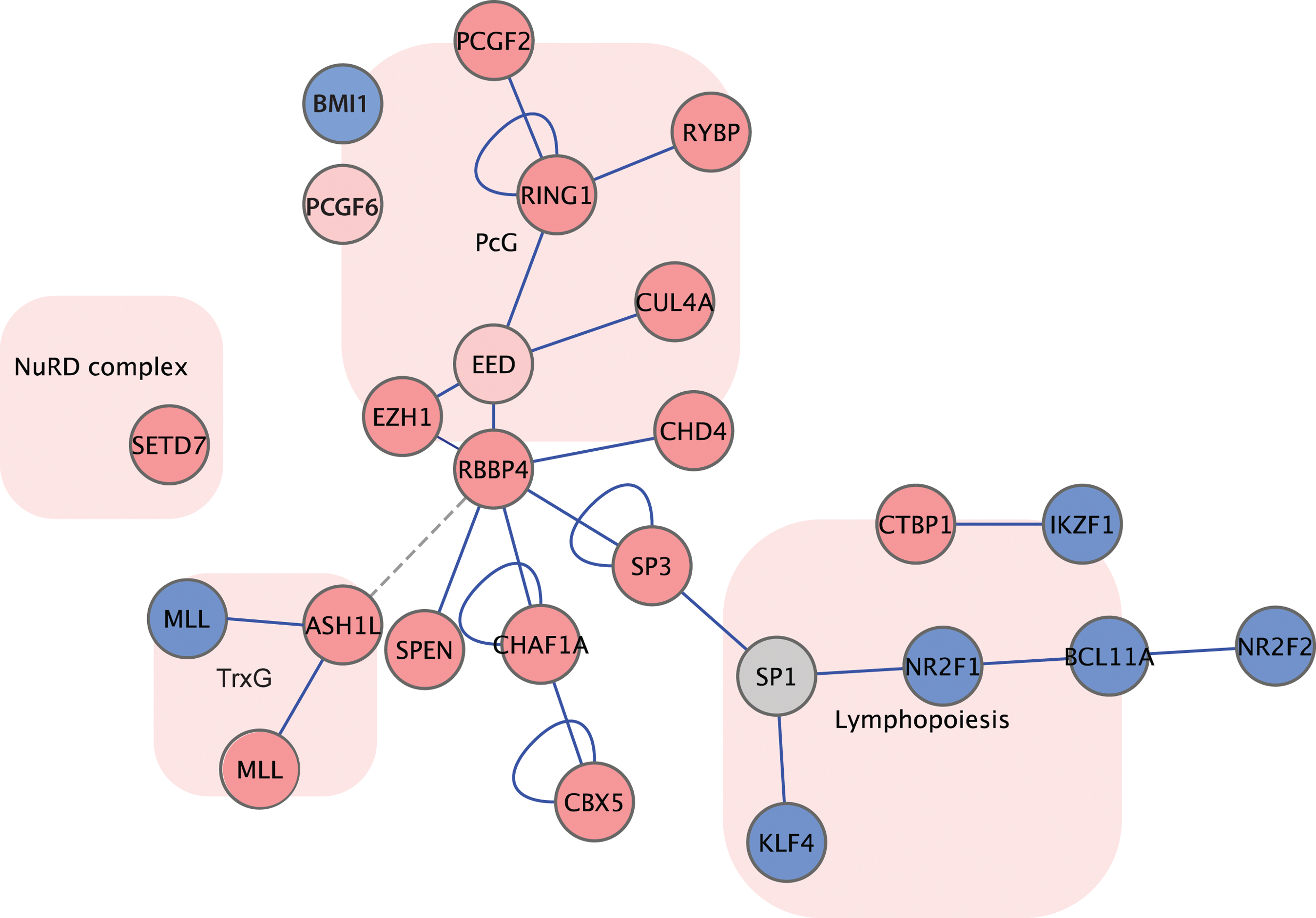
A biological network of differentially expressed genes in eHPCs versus HSPCs and SRCs. PRC network versus lymphopoiesis biological network. Dark pink corresponds to genes significantly more abundant in eHPCs versus SRCs; light pink corresponds to genes higher in eHPCs versus HSPCs; blue corresponds to genes higher in SRCs; and gray corresponds to no significant difference in expression. PRC, Polycomb repressive complex. Color images available online at
During hESC hematopoietic differentiation, it appears as if the persistent expression of a set of epigenetic regulators (EZH1, ASH1L, and PCGF6) and the lack of sufficient expression of BMI1 may predispose eHPCs to an intermediate ES/HSC cell fate. This semi-differentiated state sustains an embryonic expression pattern, resulting in a primitive hematopoietic phenotype that is incapable of in vivo reconstituting capacity, similar to yolk sac-derived hematopoietic progenitors in the mouse model [50]. Detailed functional analysis using gain-and-loss-function technologies to engineer and regulate these specific candidate epigenetic regulators in hESCs are emerging [51,52], and are being systemically applied to hematopoietic differentiating hESCs in our lab at present.
Clonal analysis reveals that hematopoietic development from hESCs may be paused at a primitive embryonic state in a cytokine-dependant manner
Based on our differential expression analyses, we focused on PcG and TrxG expression as a class of candidate genes that may play a causative role in the functional deficits observed in products of in vitro hematopoietic differentiation of PSCs. As in vitro methods that inadequately approximate an HSC, we examined how specific differentiation conditions affect the expression of PcG and TrxG genes, and ultimately how those may relate to HSC function. Thus, we investigated the aberrant expression of PcG/TrxG genes (ie, EZH1, PCGF6, BMI1, and ASH1L) during hESC hematopoietic differentiation with emphasis on genes with documented roles in normal and malignant hematopoietic cells (such as BMI1) [39,53,54]. The transcriptional repressors, BMI1 and PCGF6, in particular, are regulated in a cell cycle-dependant manner [55 –57]. Though the functional consequence of PCGF6 regulation is not clear, BMI1 is known to dissociate from target genes during G2/M phase as a consequence of its phosphorylation. Cell-cycle regulation is mediated by specific cytokines in hematopoietic cells, such as SCF, IL-3, IL6, and G-CSF, which promote HSCs to progress through the G2/M phase [58]. Interestingly, ex vivo expansion using hematopoietic cytokines leads to impaired hematopoietic reconstitution of mouse HSCs [59,60]. Similarly, human HSCs cultured for 9 days with SCF, IL3, IL6, and G-CSF do not result in SCID-repopulation on transplantation [24]. In studies of aged HSCs versus fetal HSCs, it is known that fetal HSCs provide better in vivo reconstitution [61]. In contrast, aged/adult HSCs lack functional competency and are shown to have a greater fraction of cells in cycle [61], which may relate to the over-expression of cell-cycle progression genes in eHPCs as another reason for their lack of hematopoietic reconstitution. Based on these observations, we sought to determine whether the addition of hematopoietic cytokines during in vitro differentiation is correlated with the inability of eHPCs to reconstitute the mouse hematopoietic system and whether this is associated with the differential expression of PcG/TrxG genes.
hESCs were differentiated to hematopoietic cells with or without the addition of cytokines during EB formation, and the emergence of CD45—PFV cells and eHPCs was measured. As previously shown, the addition of hematopoietic cytokines results in larger EBs from which a higher frequency of hESC hematopoietic derivatives can be obtained compared with spontaneous differentiation (Fig. 7a–d) [9]. Next, we examined the in vitro functional capacity of eHPCs derived with or without the addition of cytokines using the CFU assay. Equal numbers of cells from dissociated EBs treated with or without cytokines were seeded into methylcellulose to assess hematopoietic progenitor differentiation. The absolute number of hESC-CD34+CD45+ in the initial seeding density dramatically differed between differentiation conditions with a 5.5-fold higher frequency in populations treated with cytokines (Fig. 7e); this resulted in a larger number of hematopoietic colonies (Fig. 7f). However, though there were fewer colonies obtained from spontaneously differentiated hematopoietic cells, the average number of cells per CFU was greater without the addition of cytokines (Fig. 7f). To assess the quality of individual eHPCs generated spontaneously or with cytokines, we repeated the assay by seeding single CD34+CD45+ cells in methylcellulose in 96-well plates and monitored them for the emergence of colonies (Fig. 7g). Functional differences in the ability of eHPCs (CD34+CD45+) for single cell CFU production were observed. A marginal increase in the absolute number of wells that formed hematopoietic colonies was observed for eHPCs formed without the addition of cytokines (Fig. 7h, j). The most dramatic difference between derivation methods of hematopoietic cells was the increase in the total number of cells per CFU when cytokines were omitted (Fig. 7i, k). Next, using quantitative RT-PCR, we measured the expression of candidate genes that were differentially expressed between eHPCs and HSPCs/SRCs (BMI1, PCGF6, ASH1L, and EZH1) in hEBs treated with or without cytokines. Furthermore, we measured the expression of IKZF1, a transcription factor and a critical regulator of lymphopoiesis and HSC self-renewal, as it was more abundant in SRCs compared with HSPCs and eHPCs (Figs. 4b and 5j) [37]. During in vitro hematopoietic differentiation, BMI1 is down-regulated in day 15 and 19 hEBs treated with cytokines, while IKZF1, ASH1l, and EZH1 remain relatively unchanged between experimental conditions (Fig. 7l). PCGF6, which bears a sequence homology to BMI1 and is similarly regulated by cell cycle-dependent phosphorylation [55] (Fig. 7l), was also significantly lower in hEBs treated with cytokines compared with hEBs formed in the absence of cytokines. The expression pattern suggests that PCGF6 may be co-regulated with BMI1 during hESC differentiation. The role of BMI1 in regulating HSC self-renewal and function, particularly proliferation, is consistent with the increased number of CFUs and CFU cell number observed under hematopoietic differentiation without cytokines. Though PCGF6 and BMI1 expression is correlated during differential cytokine treatment in hEBs, PCGF6 is not differentially expressed between eHPCs and SRCs, suggesting that co-expression is only in the context of cytokine treatment and may or may not have functional significance. Moreover, PCGF6 is more highly expressed in eHPCs versus HSPCs, which may be attributed to the cellular and functional heterogeneity of putative HSCs. Although we cannot rule out the role that EZH1, ASH1L, and IKZF1 may play in the context of incomplete in vitro hematopoietic differentiation of hESCs, these results suggest that lower levels of BMI1 may be correlated with eHPC impaired hematopoietic function and potential stem cell exhaustion in a cytokine-dependant fashion.
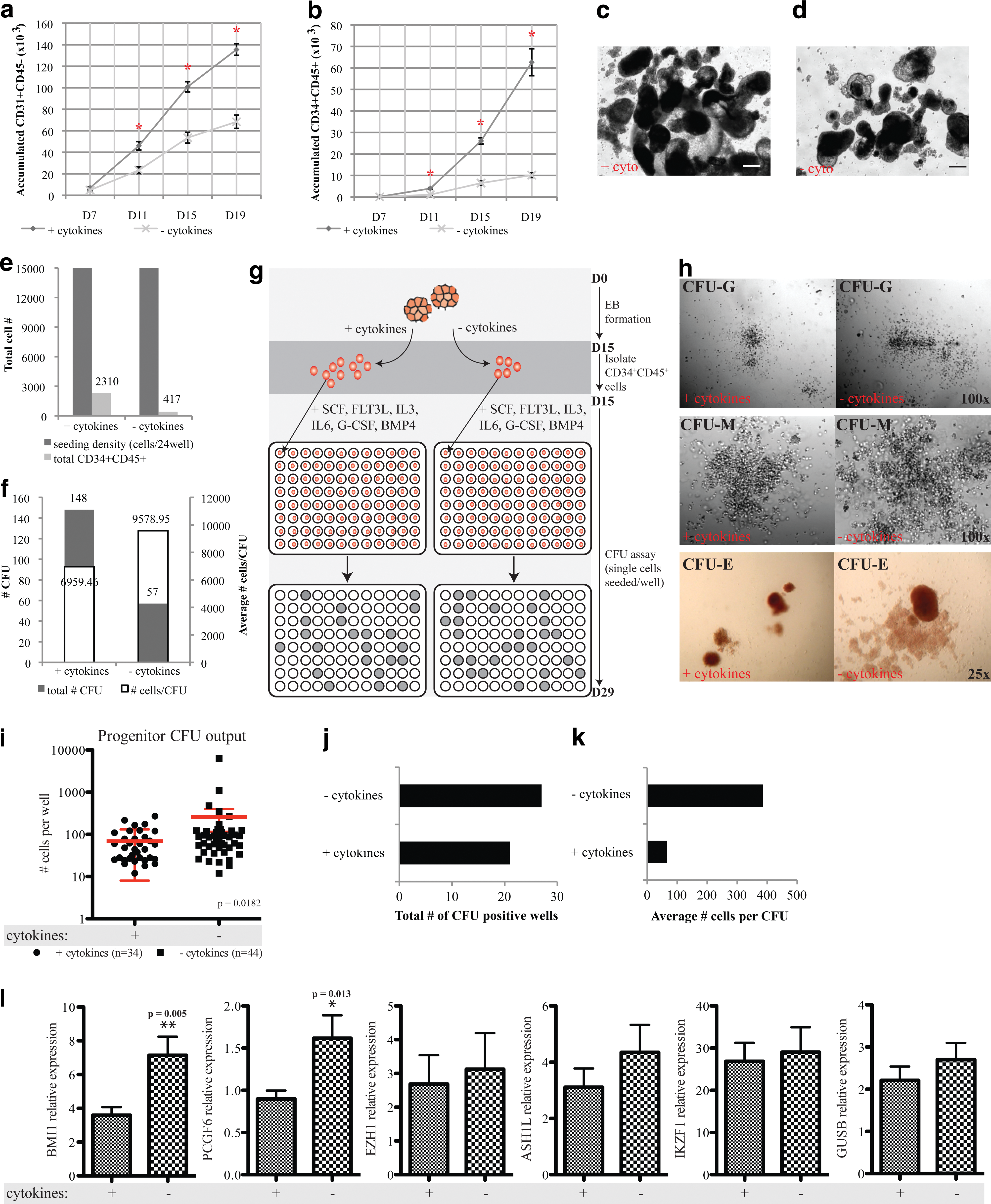
hESC hematopoietic differentiation with and without the addition of cytokines.
Discussion
Comparing the transcriptome of eHPCs with multiple sources of putative and engrafted HSCs is critical for the identification of conserved molecular determinants of hematopoietic function. Here, the inclusion of hematopoietic stem and progenitor cells throughout ontogeny also allowed for the classification of eHPCs according to the developmental timeline and hematopoietic hierarchy. In parallel, tracking the gene expression changes (or lack thereof) during stages of hESC hematopoiesis is a powerful tool that is used to critically evaluate our ability to mimic in vivo development. Thus far, attempts made at the directed differentiation of hESC toward functional HSC have failed. In this study, our interrogation of a unique compilation of transcriptome profiles reveals unexplored molecular targets that may hold eHPCs in a primitive hematopoietic state. The expression pattern of transcription factors defined at different branches of the hematopoietic hierarchy indicated that hESC differentiation can establish hematopoiesis, but in vivo hematopoietic reconstitution capacity is blocked. This blockade is most clearly evident by the deficient expression of a lymphopoiesis signature exemplified by nearly 3-fold lower expression of IKZF1 in eHPCs (Figs. 4b and 5j, l). Furthermore, during hESC differentiation, it appears as if the continued expression of epigenetic regulators, such as PCGF6 and ASH1L, may result in eHPCs that maintain an embryonic expression signature and are stalled in a primitive hematopoietic state, versus the desired definitive HSC state with in vivo function.
The members of the PcG repression complex that were highly abundant in eHPCs, compared with SRCs centers around the differential expression of EZH1, have been known to possess histone methyltransferase activity and were recently shown to coordinate with canonical PRC2 in maintaining mouse ESC pluripotency [45]. The importance of epigenetic marks, mediated by PcG and TrxG complexes, to directed differentiation and proper epiblast development is supported by studies conducted on both mESCs and mouse blastocysts, where the disruption of core PcG genes (Eed −/− and Suz12 −/−) results in loss of H3K27me3 and subsequent activation of developmental genes [62,63]. In addition, full-length MLL (the mammalian homolog of Drosophila trithorax) was up-regulated in eHPCs and has an important role in hematopoietic establishment [64] and hematologic malignancies [65 –67], while an alternative isoform of MLL is more highly expressed in SRCs. Significantly, the limited expression of core regulators of HSC function and progenitor differentiation in eHPCs may be related to the cell-cycle status and is highly influenced by hematopoietic cytokines during hESC hematopoietic differentiation. The up-regulation of BMI1 on the removal of cytokines during hESC-hematopoietic differentiation correlates with its cell-cycle dependent expression and supports the notion that the influence of cytokines on the cell cycle of hESC derivatives may functionally inhibit eHPCs. Though the addition of cytokines is important to increase the frequency of hESC-hematopoietic derivatives, the quality of these cells is questionable. The comparison of CFU formation from single eHPCs derived with or without cytokines resulted in a greater number of cells per CFU when cytokines are omitted. This suggests that the addition of growth factors works in effect as a proliferative steroid during hESC hematopoietic differentiation, but may have a crippling effect on hematopoietic function through the repression of HSC regulators. Thus, differential MLL isoform expression in conjunction with insufficient BMI1 levels may further predispose eHPCs to a primitive or incompletely programmed hematopoietic-like lineage.
Based on an adaptation of Waddington's epigenetic landscape model, a stem cell naturally travels down one of several valleys, illustrating the cells' declining developmental potential. Once a cell reaches each bifurcation point, its ability to choose different routes becomes increasingly restricted. We propose a model of in vitro hematopoietic development in which the inappropriate expression of epigenetic regulators (PCGF6, EZH1, EED, BMI1, and MLL) tethers eHPCs to a primitive hematopoietic state, through the maintenance of an ESC signature and failure to de-repress hematopoietic lineage genes such as IKZF1. Human eHPCs are, thus, prevented from moving down Waddington's valleys to a functional HSC (Fig. 8).
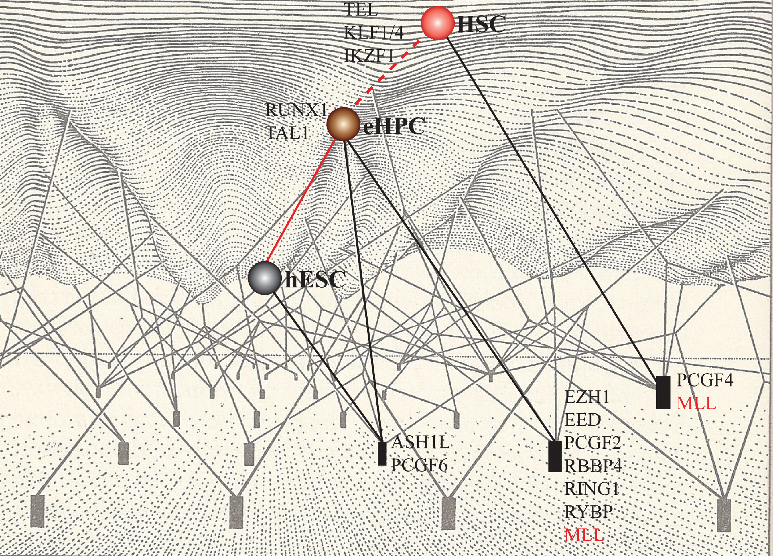
A model of epigenetic regulation of embryonic gene signatures in eHPCs and the blockade to bona fide HSC differentiation. Aspects of epigenetic regulation are active in hESCs and fail to be modulated in eHPCs, suggesting a predisposed condition. Gray=hESC, brown=eHPC; red=HSC. Color images available online at
We provide, through meta-analysis and an examination of biological networks, a plausible molecular mechanism for the inability to generate functional HSCs from human PSCs. We further propose that the expression of select PcG/TrxG genes may serve as novel targets for modulation during hESC hematopoietic differentiation and requires further functional validation that is on-going in our laboratory with the use of emerging technologies to engineer hESCs.
Footnotes
Acknowledgments
This work was supported by grants from the Canadian Institute of Health Research to M.B. M.B. is supported by the Canadian Chair Program holding the Canada Research Chair in human stem cell biology. The authors thank Dr. Eva Szabo and Dr. Brendan McIntyre from the Bhatia Lab for their constructive input toward this article and Marilyne Levadoux-Martin for technical assistance in flow cytometry.
Author Disclosure Statement
No competing financial interests exist.
References
Supplementary Material
Please find the following supplemental material available below.
For Open Access articles published under a Creative Commons License, all supplemental material carries the same license as the article it is associated with.
For non-Open Access articles published, all supplemental material carries a non-exclusive license, and permission requests for re-use of supplemental material or any part of supplemental material shall be sent directly to the copyright owner as specified in the copyright notice associated with the article.
