
Abstract
Neurogenesis in the adult brain is important for memory and learning, and the alterations in neural stem cells (NSCs) may be an important part of Alzheimer's disease pathogenesis. The phosphatidylinositol 3-kinase (PI3K) pathway has been suggested to play an important role in neuronal cell survival and is highly involved in adult neurogenesis. Recently, coenzyme Q10 (CoQ10) was found to affect the PI3K pathway. We investigated whether CoQ10 could restore amyloid β (Aβ)25-35 oligomer-inhibited proliferation of NSCs by focusing on the PI3K pathway. To evaluate the effects of CoQ10 on Aβ25-35 oligomer-inhibited proliferation of NSCs, NSCs were treated with several concentrations of CoQ10 and/or Aβ25-35 oligomers. BrdU labeling, Colony Formation Assays, and immunoreactivity of Ki-67, a marker of proliferative activity, showed that NSC proliferation decreased with Aβ25-35 oligomer treatment, but combined treatment with CoQ10 restored it. Western blotting showed that CoQ10 treatment increased the expression levels of p85α PI3K, phosphorylated Akt (Ser473), phosphorylated glycogen synthase kinase-3β (Ser9), and heat shock transcription factor, which are proteins related to the PI3K pathway in Aβ25-35 oligomers-treated NSCs. To confirm a direct role for the PI3K pathway in CoQ10-induced restoration of proliferation of NSCs inhibited by Aβ25-35 oligomers, NSCs were pretreated with a PI3K inhibitor, LY294002; the effects of CoQ10 on the proliferation of NSCs inhibited by Aβ25-35 oligomers were almost completely blocked. Together, these results suggest that CoQ10 restores Aβ25-35 oligomer-inhibited proliferation of NSCs by activating the PI3K pathway.
Introduction
N
Neurogenesis is reduced by a number of different factors including amyloid β (Aβ), and it has been reported that several cell signaling pathways associated with neurogenesis are decreased by Aβ, which plays very important roles in the pathogenesis of AD [9]. The phosphatidylinositol 3-kinase (PI3K) pathway is known to be important in neuronal survival and death [11 –15]. It is the main intracellular signal pathway responsible for the transmission of anti-apoptotic signals and the control of cell survival, and it is activated by several kinds of neuroprotective stimuli [16 –23]. This pathway also plays a pivotal role in control of tau phosphorylation in AD brains and is highly involved in adult neurogenesis [24].
Coenzyme Q10 (CoQ10), an endogenous proenzyme found in the inner mitochondrial membrane of cells [25], is a promising neuroprotectant. AD and other neurological disorders have demonstrated defects in the inner mitochondrial membrane and, as a result, in oxidative phosphorylation [26]. Many preclinical studies have suggested that CoQ10 inhibits oxidative stress and affects the expression of genes involved in cell signaling, metabolism, and transport [27]. In a previous study, we showed that CoQ10 has a protective effect on Aβ25–35-induced neuronal cell death by activating the PI3K signaling pathway [28]; thus, we hypothesize that CoQ10 might contribute to restoration of proliferative activity of NSCs injured by Aβ25–35.
In this study, we investigated whether and how CoQ10 restored Aβ25-35 oligomer-inhibited proliferation of NSCs, with a special focus on the PI3K pathway.
Materials and Methods
Materials
Dulbecco's modified Eagle's medium (DMEM, high glucose) was purchased from Gibco (Invitrogen Corporation). Amyloid-β25–35) (Aβ25–35), protein protease inhibitor cocktail, trypan blue solution, insulin, and DNase I were obtained from Sigma-Aldrich. CoQ10 (Ubidecarenone) was a generous gift from Daewoong Bio, Inc. Before use, CoQ10 was dissolved in methyl chloride to 500 mM, diluted with DMSO to 100 mM, and then diluted with culture medium to the desired concentrations.
Cultures of NSCs and treatment
All procedures using animals were consistent with Hanyang University's guidelines for the care and use of laboratory animals. We made every effort to minimize both the number of animals used and animal suffering. Each animal was utilized only once.
Embryonic brain tissue was dissected from the cortex, lateral ganglionic eminence (anlage of striatum), and ventral midbrain of embryonic day 12–14 (E12–E14) rats (Sprague-Dawley; Koateck) [29]. After mechanical trituration, 20,000 cells per cm2 were plated in culture dishes precoated with poly-L-ornithine (PO)/fibronectin (FN) in N2B medium [DMEM/F12, 4.4 μM insulin, 100 mg/L transferrin, 30 nM selenite, 0.6 μM putrescine, 20 nM progesterone, 0.2 mM ascorbic acid, 2 mM L-glutamine, 8.6 mM D(+) glucose, 20 mM NaHCO3, B27 (Invitrogen, Carlsbad, CA)] supplemented with basic fibroblast growth factor (bFGF, 20 ng/mL; R&D Systems) and epidermal growth factor (EGF, 20 ng/mL; R&D Systems) and were cultured for 4–6 days as a monolayer on the adherent surface. To obtain a uniform population of NSCs, clusters of cells formed by the proliferation of NSCs with mitogens (bFGF and EGF) were passaged by dissociating them into single cells and plating them onto freshly PO/FN-coated coverslips (12-mm diameter; Marienfeld GmbH & Co. KG) [30]. All data in this study were obtained from passaged cultures grown on the adherent surface. Cultures were maintained at 37°C in a 5% CO2 incubator. Media were changed every other day, and mitogens were added daily.
To measure the effects of Aβ25-35 oligomers on the proliferation of NSCs, NSCs were simultaneously treated with several concentrations of Aβ25-35 oligomers (0, 2.5, 5, 10, 20, or 40 μM) for 48 h. Soluble oligomeric forms of Aβ25-35 were prepared as reported by Dahlgren et al. [31]. Briefly, Aβ25-35 was dissolved at a concentration of 1 mM in hexafluoroisopropanol (Sigma) and separated into aliquots in sterile microcentrifuge tubes. Hexafluoroisopropanol was removed under vacuum in a Speed-Vac, and the peptide film was stored desiccated at −20°C. To prepare oligomers, the peptide was first suspended in dry dimethyl sulfoxide (Me2SO; Sigma) at a concentration of 5 mM, and Ham's F-12 (phenol red-free; BioSource) was added to bring the final concentration to 1 mM. This was followed by 24 h incubation at 4°C. Plates were carefully washed more than three times with phosphate-buffered saline (PBS), and cell viability was measured with MTT and trypan blue assays.
MTT assay and trypan blue staining to measure cell viability
MTT is absorbed into cells and transformed into formazan by mitochondrial succinate dehydrogenase. Accumulation of formazan directly reflects mitochondrial activity, which is an indirect measurement of cell viability. Cells were plated in 96-well plates at a density of 1×10⁴ cells/well in 200 μL of culture medium, and 50 μL of 2 mg/mL MTT (Sigma) was added to each well. An aliquot (220 μL) of the resulting solution was removed from each well, followed by the addition of 150 μL dimethyl sulfoxide. The precipitate from each well was resuspended on a microplate mixer for 10 min, and optical densities (OD) at 540 nm were measured using a plate reader. All results were normalized to OD values measured from an identically treated well without cultured cells. For trypan blue staining (TBS), 10 μL of trypan blue solution (BMS) was incubated for 2 min with 10 μL of dissociated cells from each sample. Unstained live cells were counted on a hemocytometer [15].
Immunostaining for Ki-67, nestin, and active caspase-3
The NSCs were seeded (1×10⁵ cells) on chamber well plates, and the cells were exposed to 10 μM Aβ25-35 oligomers and different concentrations of CoQ10 (0, 5, or 10 μM) for 24 h. The cells were fixed in 2% paraformaldehyde for 15 min and were permeabilized with 0.5% Triton X-100 in PBS for 5 min. The endogenous peroxidase activity was blocked using 3% H2O2 in PBS for 20 min. The cells were incubated with 5% normal serum in PBS for 60 min and were incubated overnight in 2% normal serum in PBS containing the following primary antibodies: mouse anti-nestin (1:100; Abcam), rabbit anti-Ki-67 (1:100; Abcam), and rabbit anti-cleaved caspase-3 (1:100; Cell Signaling). The next day, the cells were incubated in 2% normal serum in PBS containing the secondary antibodies goat-anti-rabbit Alexa Fluor 488 (Jackson Immunoresearch), anti-rabbit tetramethylrhodamine (Jackson Immunoresearch), and goat-anti-mouse Alexa Fluor 488 (Jackson Immunoresearch) for 60 min, washed several times with PBS, and mounted on glass slides using Moviol 488 solution. To stain the nuclei in the NSCs, the active caspase-3-stained cells were counterstained using 4′,6-diamidino-2-phenylindole (DAPI; Sigma). The cells were visualized under an Olympus fluorescence microscope at the appropriate excitation wavelengths.
BrdU cell proliferation assay
After 48 h of treatment, cells were incubated in BrdU-labeling medium (10 μM BrdU) for 1 h, and the cell proliferation assay was performed using the BrdU Labeling and Detection Kit (Roche Boehringer), according to the manufacturer's instructions. Results were calculated as the proportion of stained cells out of 1,000 cells counted under a light microscope at 400×magnification.
Colony formation assay
NSCs were used for colony forming assays. Approximately 1×10⁴ cells were seeded in a 100-mm culture dish and cultured for 14 days. Cells were fixed with 4% paraformaldehyde and stained with crystal violet (Sigma Aldrich). The number and size of the colonies (a colony was designated an aggregate of 50 or more cells) were measured and compared under a microscope (×100 magnification).
Western blot analyses
The levels of p85α PI3K, Akt, phosphorylated Akt (pAkt) (Ser473), GSK-3β, phosphorylated GSK-3β (pGSK-3β) (Ser9), heat shock transcription factor-1 (HSTF-1), cytosolic cytochrome C, and cleaved caspase-3 were analyzed by western blotting. Briefly, 5×10⁶ cells were washed twice in cold PBS and incubated in lysis buffer [50 mM Tris (pH 8.0), 150 mM NaCl, 0.02% sodium azide, 0.2% sodium dodecyl sulfate (SDS), 100 μg/mL phenylmethylsulfonylfluoride (PMSF), 50 μL/mL aprotinin, 1% Igepal 630, 100 mM NaF, 0.5% sodium deoxycholate, 0.5 mM EDTA, and 0.1 mM EGTA] for 10 min on ice. Cell lysates were centrifuged at 10,000 g and evaluated for levels of p85α PI3K, pAkt, pGSK-3β, and HSTF-1. Protein concentrations of cell lysates and postmitochondrial fractions were determined using a Bio-Rad protein assay kit (Bio-Rad, Hercules, CA, USA). Samples containing equal amounts (20 μg) of protein were resolved by 10% SDS-polyacrylamide gel electrophoresis (SDS-PAGE) and transferred to nitrocellulose membranes (Amersham Pharmacia Biotech). Membranes were blocked using 5% skim milk before incubation with specific primary antibodies against p85α PI3K (1:1,000; Sigma), Akt (1:1,000; Cell Signaling), pAkt (1:500; Cell Signaling), GSK-3β (1:1,000; Santa Cruz Biotech), pGSK-3β (Ser9) (1:1,000; Santa Cruz Biotech), HSTF-1 (1:1,000; Santa Cruz Biotech), and active caspase-3 (1:1,000; Santa Cruz Biotech). Membranes were washed with Tris-buffered saline containing 0.05% Tween-20 and then processed using a horseradish peroxidase-conjugated anti-rabbit or anti-mouse antibody (Amersham Pharmacia Biotech) followed by ECL detection (Amersham Pharmacia Biotech) [14]. The results from western blots were quantified using an image analyzer (Quantity One-4,2,0; Bio-Rad).
Statistical analysis
All data are presented as the mean±SD from five or more independent experiments. Statistical comparisons of viability among the different treatment groups were performed with Tukey's test after a one-way ANOVA. P values less than 0.05 were considered statistically significant.
Results
The viability and proliferation of NSCs injured by Aβ25-35 oligomers
To evaluate the effect of Aβ25-35 oligomers on the NSC viability, NSCs were treated with different concentrations of Aβ25-35 oligomers (0, 2.5, 5, 10, 20, or 40 μM) for 48 h. Cell viability was measured with the MTT assay, and surviving cells were counted with TBS. The viability of NSCs was not significantly decreased by treatment with 2.5 or 5 μM Aβ25-35 oligomer, but the viability of cells treated with concentrations greater than 10 μM was significantly decreased in a concentration-dependent manner (Fig. 1).
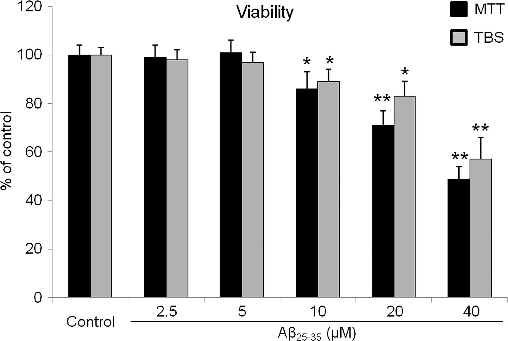
Viability of neural stem cells (NSCs) injured by the amyloid β (Aβ)25-35 oligomer. The data are mean (% of control)±SD from five independent experiments. Treatment groups were compared with the control group using Tukey's test after one-way ANOVA. MTT assay and trypan blue staining show that the viability of NSCs was decreased in a concentration-dependent manner when the cells were treated with more than 10 μM Aβ25-35 oligomer. *P<0.05 and **P<0.01 (vs. control group).
BrdU labeling showed that the proliferation of NSCs treated with Aβ25-35 oligomers at concentrations greater than 5 μM was significantly decreased in a concentration-dependent manner (Fig. 2). Based on these results, 10 μM Aβ25-35 oligomer was selected as the optimal concentration for subsequent experiments because, at this concentration, cell viability was only slightly decreased (to about 90%), but proliferation of NSCs was significantly decreased (to around 70%; P<0.01 when compared with the nontreated group).
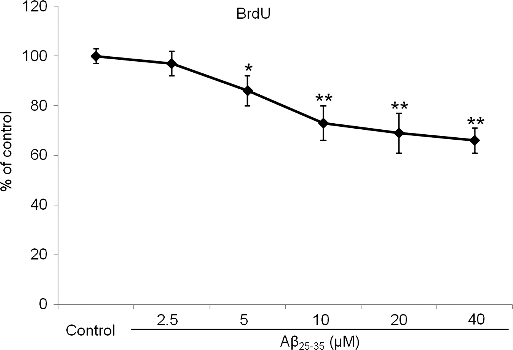
Proliferation of NSCs injured by the Aβ25-35 oligomer. The data are mean (% of control)±SD from five independent experiments. The treatment groups were compared with the control group using Tukey's test after one-way ANOVA. BrdU assay showed that the proliferation of NSCs was decreased in a concentration-dependent manner when the cells were treated with more than 5 μM Aβ25-35 oligomer. *P<0.05 and **P<0.01 (vs. control group).
The effects of CoQ10 on Aβ25-35 oligomer-inhibited proliferation of NSCs
To investigate the effects of CoQ10 on the Aβ25-35 oligomer-inhibited proliferation, the NSCs were treated with different concentrations of CoQ10 (0, 1, 2.5, 5, 10, 20, or 40 μM) and 10 μM of the Aβ25-35 oligomer for 48 h. The cell proliferation was assessed using BrdU labeling, the colony formation assay, and immunostaining for Ki-67 and nestin, which are markers of proliferative activity. Compared with the NSCs treated with 10 μM Aβ25-35 oligomer alone, the cells co-treated with CoQ10 plus the Aβ25-35 oligomer exhibited significantly higher cell proliferation rates (P<0.05). The proliferation rates were CoQ10-dependent up to a concentration of 10 μM (P<0.01) (Fig. 3). When a PI3K inhibitor (10 μM LY294002) was co-administered with CoQ10 and the oligomer for 48 h, the effect of CoQ10 on the proliferation of NSCs was blocked (Fig. 4).
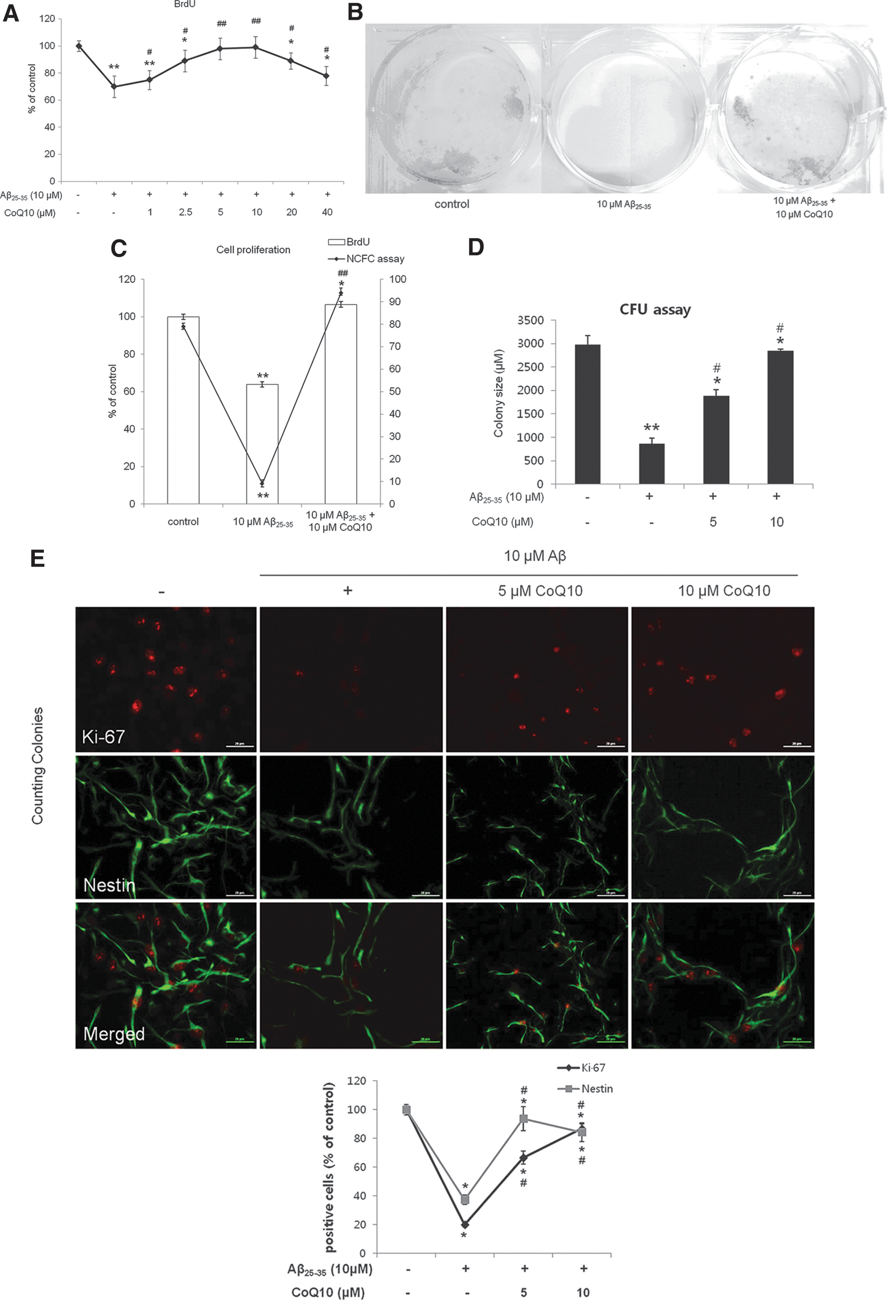
Effect of coenzyme Q10 (CoQ10) on proliferation of NSCs injured by Aβ25-35 oligomer. The data are mean (% of control)±SD from five independent experiments. Treatment groups were compared with the control group using Tukey's test after one-way ANOVA. BrdU assay showed that treatment with CoQ10 restored the proliferation of NSCs inhibited by 10 μM Aβ25-35 oligomers

Role of phosphatidylinositol 3-kinase activation in the effect of CoQ10 on proliferation of NSCs injured by Aβ25-35 oligomer. The data are mean (% of control)±SD from five independent experiments. The treatment groups were compared with the control group using Tukey's test after one-way ANOVA. BrdU assay showed that co-treatment with LY294002 almost completely blocked the effect of CoQ10 on the proliferation of NSCs inhibited by 10 μM Aβ25-35 oligomer. *P<0.05 and **P<0.01 (vs. control group), # P<0.05 and ## P<0.01 (vs. the group treated with only 10 μM Aβ25-35 oligomer), $ P<0.05 (vs. the group treated with 10 μM Aβ25-35 oligomer and 10 μM CoQ10).
The mechanisms of CoQ10 restoration of Aβ25-35 oligomer-inhibited proliferation of NSCs
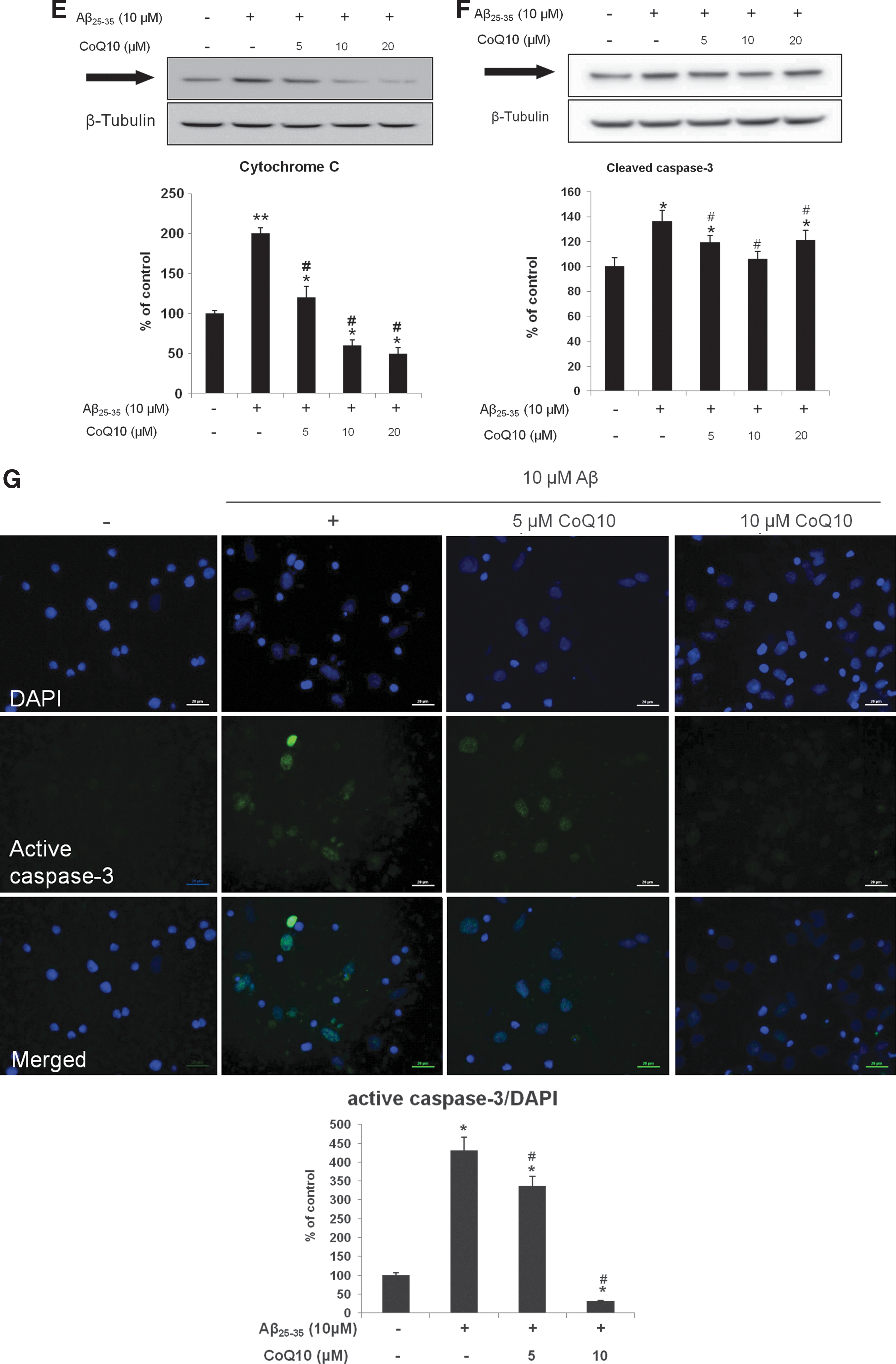
We determined the effects of CoQ10 on the intracellular signaling proteins, which are critical for the proliferation of NSCs, following treatment with the Aβ25-35 oligomers. The levels of p85α PI3K, Akt, pAkt (Ser473), GSK-3β, pGSK-3β (Ser9), HSTF-1, cytosolic cytochrome C, and cleaved caspase-3, which are part of the PI3K pathway that plays pivotal roles in the proliferation of NSCs, were investigated. Compared with the untreated cells (100%±10%, 100%±11%, 100%±9%, and 100%±8%, respectively), the immunoreactivities (IRs) of p85α PI3K, pAkt (Ser473), pGSK-3β (Ser9), and HSTF-1 were significantly decreased for the 10 μM Aβ25-35 oligomer-treated cells (73%±8%, 45%±9%, 67%±11%, and 75%±9%, respectively, P<0.01). However, this decrease was significantly attenuated by co-treatment with 10 μM CoQ10 (296%±14%, 179%±10%, 147%±12%, and 147%±10% at 10 μM CoQ10, respectively, P<0.01) (Fig. 5). In contrast, the IRs of the cytosolic cytochrome C and cleaved caspase-3 were significantly increased in the 10 μM Aβ25-35 oligomer-treated cells (196%±7% and 204%±13%, respectively, P<0.01), and this increase was significantly attenuated by co-treatment with 10 μM CoQ10 (55%±9% and 97%±5% at 10 μM CoQ10, P<0.01) (Fig. 5E–G).
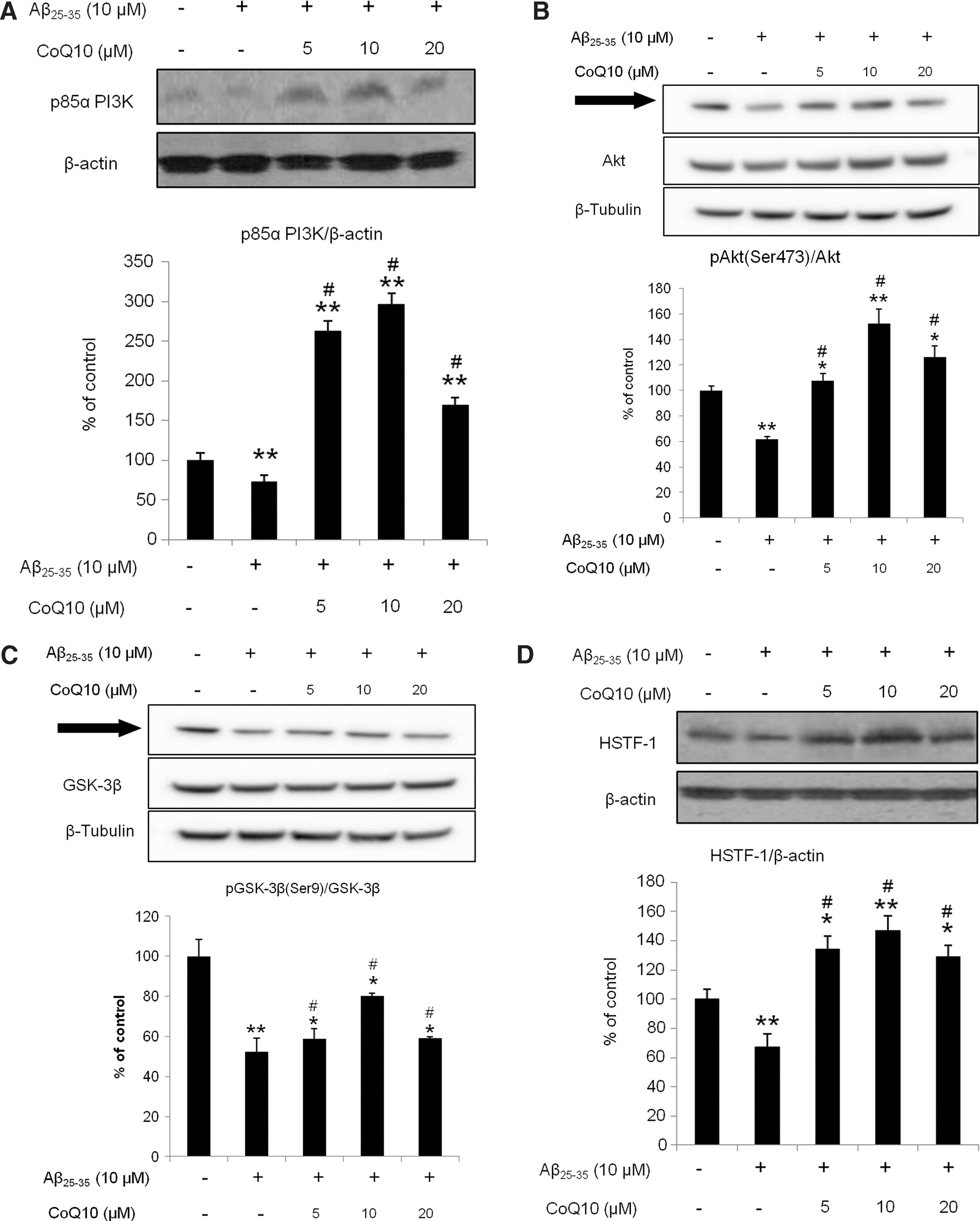
Effect of CoQ10 on proliferation- and death-related intracellular signaling proteins of NSCs injured by Aβ25-35 oligomers. The data represent the mean (% of control)±SD of five independent experiments. The treatment groups were compared with the control group using Tukey's test after one-way ANOVA. The western blotting demonstrated that the treatment with CoQ10 significantly enhanced the expression of the proliferation-related intracellular signaling proteins in the NSCs inhibited by 10 μM Aβ25-35 oligomer
Discussion
In the present study, NSC viability was significantly decreased by treatment with more than 20 μM oligomeric Aβ25-35 compared with untreated cells (Fig. 1). NSC proliferation was also decreased in a concentration-dependent manner, especially when NSCs were treated with more than 5 μM oligomeric Aβ25-35 (Fig. 2). Combined treatment with CoQ10 at concentrations ranging from 1 to 10 μM for 48 h increased the proliferation of NSCs injured by 10 μM Aβ25-35 oligomer in a concentration-dependent manner (Fig. 3), suggesting that CoQ10 treatment restores Aβ25-35 oligomer-inhibited proliferation of NSCs. A PI3K inhibitor (10 μM LY294002) blocked the effect of CoQ10 on the proliferation of NSCs (Fig. 4), indicating that the effect of CoQ10 on proliferation of NSCs is mediated by activation of the PI3K pathway. This finding and the fact that the PI3K pathway plays an important role in proliferation of NSCs led us to hypothesize that CoQ10 activation of the PI3K pathway restores Aβ25-35 oligomer-inhibited proliferation of NSCs, which we were able to confirm.
In the first step of the PI3K pathway, activated PI3K phosphorylates its downstream target, Akt/protein kinase B [32]. Phosphorylated Akt directly affects GSK-3β, Bcl-2-associated death promoter (BAD)/Bcl-2, caspase 9, IkB kinase, and Forkhead-related transcription factor [14,30]. pAkt also inhibits GSK-3β by phosphorylating it at Ser9 [32]. While the active form of GSK-3β inhibits HSTF-1 and activates the mitochondrial death pathway [13,32,33], inactivation of the phosphorylated GSK-3β by pAkt is important for function in a variety of stem cell types [34]. It was reported that the PI3K pathway is essential for the self-renewal of embryonic stem cells (ESCs) in mouse [35], and activation of this pathway is important for maintaining pluripotency in mouse and primate ESCs [36]. Several studies showed that the phosphatase and tensin homolog deleted on chromosome 10 (PTEN), an antagonist of PI3K, negatively regulates NSCs proliferation, survival, and self-renewal both in vivo and in vitro [37 –41]. Contrary to PTEN, components of the PI3K pathway were reported to be involved in the self-renewal of NSCs [42].
We demonstrated that CoQ10 affected the PI3K pathway in NSCs; treatment with 10 μM Aβ25-35 oligomer decreased the IRs of survival proteins such as p85α PI3K, Akt, pAkt (Ser473), GSK-3β, pGSK-3β (Ser9), and HSTF-1 and increased the activity of cytosolic cytochrome C and active caspase-3, cell death-related proteins (Fig. 5). However, the CoQ10 co-treatment significantly increased the expression of these survival proteins and inhibited the expression of cytosolic cytochrome C and active caspase-3 (Fig. 5). These findings provide strong support for our hypothesis that the effects of CoQ10 on the proliferative activity of NSCs and survival are mediated by the PI3K pathway.
There are some limitations to this study. First, these studies were initially performed in embryonic NSCs; however, because amyloid beta toxicity causes neurodegeneration in elderly individuals, it may be more appropriate to perform the studies in adult NSCs. Therefore, we attempted to investigate the effect of the Aβ25-35 oligomers and CoQ10 in adult hippocampal NSCs harvested from the adult brain tissue of 10-week-old rats [43]. Compared with the embryonic NSCs, the proliferation rate of the adult hippocampal NSCs was slow, which limited the use of the adult cells in these experiments. Therefore, we compared the effects of the Aβ25-35 oligomers and CoQ10 on the proliferative activity in embryonic and adult NSCs and demonstrated that in many cases, the effects were similar (See Supplementary Data; available online at
Taken together, the results of this study indicate that CoQ10 restores Aβ25-35 oligomer-inhibited proliferation of NSCs by activating the PI3K pathway and could be applied in potential NSC-based therapies for AD.
Footnotes
Acknowledgments
This work was supported by the NanoBio R&D Program of the Korea Science and Engineering Foundation, funded by the Ministry of Education, Science and Technology (2007-04717) and by the National Research Foundation of Korea (NRF) (2010-0009588).
Author Disclosure Statement
No competing financial interests exist.
References
Supplementary Material
Please find the following supplemental material available below.
For Open Access articles published under a Creative Commons License, all supplemental material carries the same license as the article it is associated with.
For non-Open Access articles published, all supplemental material carries a non-exclusive license, and permission requests for re-use of supplemental material or any part of supplemental material shall be sent directly to the copyright owner as specified in the copyright notice associated with the article.