
Abstract
Mechanical cues exert considerable influence on the fates of stem cells and terminally differentiated chondrocytes. The elucidation of the interactions between cell fate and mechanical cues in nuclear mechanotransduction will provide new clues to modulate tissue homeostasis and regeneration. In this study, we used an integrated microfluidic perfusion device to simultaneously generate multiple-parameter fluid shear stresses to investigate the role of fluid flow stimuli in the regulation of Yes-associated protein (YAP) expression and the fates of mesenchymal stem cells (MSCs) and primary chondrocytes. YAP expression was regulated by the level of fluid flow stimulus in both MSCs and chondrocytes. An increase in the magnitude of stimulation enhanced the expression of YAP, ultimately resulting in an increase in osteogenesis and a decrease in adipogenesis for MSCs, and initiating dedifferentiation for chondrocytes. Cytochalasin D not only repressed nuclear YAP accumulation in the flow state, but also abrogated flow-induced effects on MSC differentiation and the chondrocyte phenotype, resulting in MSC adipogenesis and the maintenance of the chondrocyte phenotype. Our findings reveal the connection between YAP and MSC/chondrocyte fates in a fluid flow-induced mechanical microenvironment and provide new insights into the mechanisms by which mechanical cues regulate the fates of MSCs and chondrocytes.
Introduction
T
The cells perceive and transmit mechanical cues via mechanotransduction mechanisms. It has been well established that the actin cytoskeleton plays a significant role in mechanosensing and mechanotransduction [8,9]. The lineage commitment of MSCs can be predicted based on cell geometry and cytoskeletal organization [10,11]. For example, flattened morphologies, stiffer substrates, and tensile stress result in high cytoskeletal tension, which promotes osteogenesis. In contrast, rounded morphologies, soft substrates, and a lack of extrinsic mechanical force contribute to adipogenesis [2,12]. Similarly, chondrocytes with a rounded shape or exposed to soft substrates can maintain their phenotype; otherwise, these cells are likely to dedifferentiate [13,14].
The Yes-associated protein (YAP) plays crucial roles in cell proliferation, survival, differentiation, tissue regeneration, and organ size control, and these effects are mediated through the Hippo pathway [15 –17]. Recently, it has been confirmed that YAP is a regulator of the nuclear transduction of mechanical cues [18]. The activity of YAP is regulated by intrinsically local cues, such as cell shape [18,19], cell–cell contact [17,20], extracellular matrix stiffness [18], and cytoskeleton tension [18,19,21], which have a distinct effect on the fate decision that occurs during stem cell differentiation [16,22 –24]. However, the responses of YAP to extrinsic mechanical force stimuli such as pressure stress, tensile stress, and fluid shear stress have not been well established. In particular, the relationships between YAP and the lineage commitment of MSCs and the chondrocyte phenotype in response to passive mechanical force remain incompletely understood.
Currently, microfluidic-based microdevices are attracting much attention in the field of cellular biomechanics, and their major advantage is the reduced time and resources they require due to their increased experimental throughput and the minimized size of the experimental platform compared with the conventional way [25,26]. In this study, we employed an integrated microfluidic device that can simultaneously generate multiple shear stresses to investigate the role of fluid flow stimuli in the regulation of YAP expression and the lineage commitment of MSCs and chondrocytes.
Materials and Methods
Microfluidic device design and fabrication
The design and operation of the microfluidic device are presented in Fig. 1A. The device had one inlet, one outlet, and four cell culture chambers connected to resistance channels of different dimensions. Each chamber was 100 μm in height, 1.2 mm in width, and 6 mm in length.
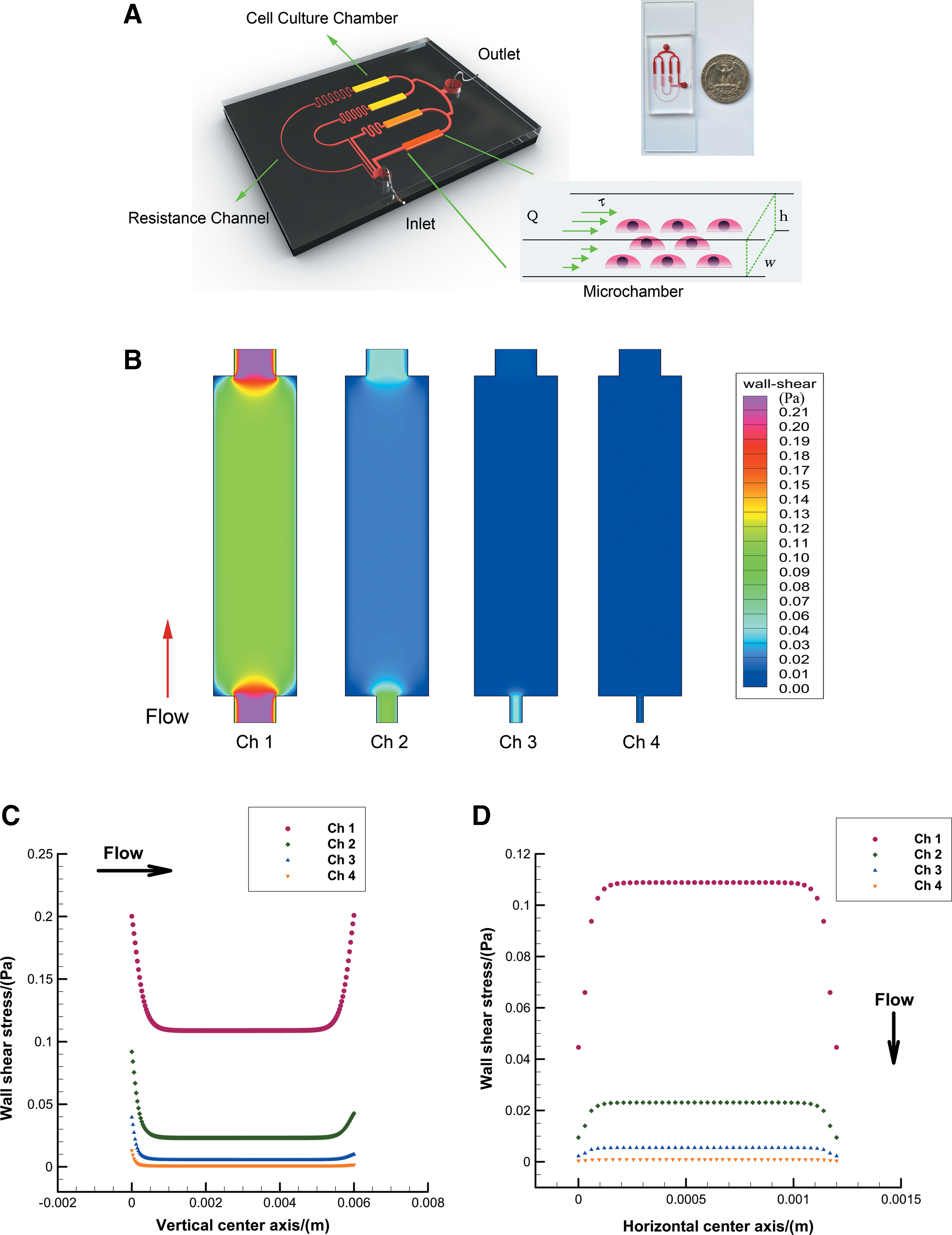
All devices were fabricated using conventional microfabrication techniques involving SU-8 (Microchem) photolithography and polydimethylsiloxane (PDMS) (Sylgard 184; Dow Corning) soft lithography [27]. Briefly, the mask was designed in AutoCAD 2007 (Autodesk) and printed on transparencies with a 4000-dpi resolution. The transparency mask was used in 1:1 contact photolithography with SU-8 photoresist to yield a negative master that was constructed from a photoresist relief on a silicon wafer. The PDMS was poured over the patterned wafer to completely cover the pattern, and the sample was placed in an oven at 80°C for 60 min. The PDMS replica and a clean glass substrate were irreversibly sealed using oxygen plasma (2 torr, 100 W) for 1 min.
Numerical modeling
To evaluate the local fluid shear stress distribution in the chamber, we simulated the 3D flow field in the chamber using a finite volume method (FVM)-based CFD code in FLUENT 6.3 (ANSYS, Inc.). We used a model in which the flow was assumed to be laminar, viscous, and incompressible, and we designed the microfluidic networks based on the electric circuit analogy [28]. Using this analogy, individual channel sections were treated as resistances within the flow circuit. A precise solution for the hydraulic resistance of the 3D rectangular channel was derived via Fourier series expansions. To avoid the computational rigor required to solve Fourier series expansions, we used an approximate version in algebraic form [29]:
where η is the fluid viscosity, w is the channel width, and h is the channel height, for h<w. For a square microchannel, the resistance can be calculated by
A constant pressure drop Δp results in a constant flow rate Q. This result can be summarized using the Hagen–Poiseuille equation, as follows [30]:
Using simple algebraic manipulation, we determined the pressure values at the inlets and outlets of the individual culture chambers. These pressure values were then used as the inlet and outlet pressure conditions to simulate the 3D flow field in each culture chamber using the CFD method. The incompressible Navier-Stokes equations were used to model the steady-state flow field in the culture chambers. It is worth noting that we did not consider the effect of the cells on the flows in the present simulations. The computational domain was discretized using approximately 52,000 hexahedral meshes and solved using FVM along with the aforementioned inlet/outlet pressure conditions and no-slip boundary conditions at the chamber walls. The density of the perfusion medium was 993.2 kg/m3, and its viscosity was 7.85×10⁻⁴ Pa s at 37°C.
The isolation and culture of MSCs and chondrocytes
All experimental procedures were approved by the Committee on Animal Use and Care of the Dalian Medical University. Articular cartilage chondrocytes were isolated from the humeral heads, femoral heads, and femoral condyles of male Sprague-Dawley rats weighing 80–120 g, as previously described [31]. Briefly, chondrocytes were isolated by digestion with 0.15% type II collagenase for 16 h and resuspended in the Dulbecco's modified Eagle's medium/F-12 (Hyclone) containing 10% fetal bovine serum (FBS; Hyclone), 50 mg/mL ascorbic acid-2-phosphate (Sigma), and 100 units/mL penicillin–streptomycin. The primary chondrocytes were used in the subsequent experiments. Primary rat MSCs were isolated from the bilateral femurs and tibias of the same rats. The distal ends of the bone were cut open, and the marrow cavities were lavaged with sterile phosphate-buffered saline (PBS). The cells were resuspended in the low-glucose Dulbecco's modified Eagle's medium (GIBCO Invitrogen) containing 10% FBS (Hyclone) and 100 units/mL penicillin–streptomycin. After 48 h of incubation at 37°C in 5% CO2, the medium was changed to remove the nonadherent cells. After two passages, the attached MSCs were devoid of any nonadhering cells and used in the following experiments.
The microdevice was sterilized in an autoclave, and then air-dried on a clean bench. The cell culture chambers were coated with 100 μg/mL fibronectin (Sigma) for 1 h at room temperature. Then, the chambers were washed with PBS. MSC suspension of 0.5×10⁵ cells/mL and a chondrocyte suspension of 1×10⁵ cells/mL were individually injected into chambers through the outlet, and the device was incubated at 37°C for 12 h to allow cell attachment. Then, the inlet of the device was connected to a peristaltic pump (Longer Pump BT100-2J, China), and the outlet was connected to a reservoir. MSCs were exposed to the adipogenic–osteogenic coinduction medium during perfusion culture. The mixed induction medium contained 1:1 adipogenic induction:osteogenic differentiation media (Cyagen Biosciences, Inc.), as previously described [12]. The cells were exposed to 1 μM cytochalasin D (CytoD) (Sigma) for 1 h to disrupt stress fibers before the application of the flow stimulus.
Immunofluorescence staining
Samples in the device were washed with PBS, fixed with 4% paraformaldehyde at room temperature for 15 min, and permeabilized with 0.1% Triton X-100 for 10 min. After washing with PBS 3 times, the samples were blocked with normal goat serum at room temperature for 30 min, incubated with primary antibodies against YAP (Santa Cruz), PPARγ (Santa Cruz), Runx2 (Santa Cruz), Sox9 (Santa Cruz), collagen II (Sigma), and collagen I (Sigma) at 4°C overnight; and then incubated with FITC-conjugated goat anti-rabbit IgG or TRITC-conjugated goat anti-rabbit secondary antibodies (Zhongshan) at room temperature for 1 h. The nucleus was stained with 4,6-diamino-2-phenyl indole (DAPI) (Invitrogen) for 10 min. After staining, the devices were washed with PBS 2–3 times and imaged using fluorescence microscopy (Olympus IX71). The fluorescence intensities of collagen I and collagen II were determined from the fluorescence photographs (N=10) using Image-Pro Plus 6.0 software to obtain relative fluorescence intensity (RFI) values.
Cell staining
After 5 days of perfusion, the cells were stained with Oil Red O and alkaline phosphatase (ALP) to determine adipogenic differentiation and osteogenic differentiation, respectively. Briefly, the cells were fixed with 4% paraformaldehyde, and then stained with Fast Blue RR/naphthol (Sigma) to visualize ALP. After washing with PBS, the cells were stained with 30 mg/mL of Oil Red O (Sigma) in 60% isopropanol to visualize the lipid droplets, and then rinsed in PBS.
Statistics and analysis
All experiments were performed at least in triplicate with different batches of devices. Differences among three groups were analyzed using one-way ANOVA. Comparisons of two groups were performed by using the two-sample t-test. P<0.05 was considered statistically significant.
Results
Computational simulation of the fluid dynamics in the microchambers
In this study, a microdevice was designed based on the principles of the Electric Circuit Analogy. This device was composed of one inlet, one outlet, resistance channels, and four cell culture chambers. The flow of fluid through the microdevice was typically driven by a peristaltic pump. Thus, the pressure-driven fluid flow through the chamber could be defined as steady-state flow and modeled as Poiseuille flow. The fluid shear stress on the cells could be assumed to be equal to the wall shear stress at the bottom of the chamber. When the perfusion flow rate of 30 μL/min was applied to the microdevice, different levels of fluid flow stimulus could be generated. The central area of these chambers exhibited a uniform distribution for the wall shear stress (Fig. 1B). The results demonstrated that the majority of the bottom of the chamber experienced uniform shear stress except the areas near the corners, the inlet, the outlet, and the side walls. To quantify the local distribution of shear stress in detail, we computed the wall shear stress along the vertical and horizontal center axes of each microchamber. This observation was further confirmed by the wall shear stress curves along the vertical center axis (Fig. 1C) and horizontal center axis (Fig. 1D). The average bottom wall shear stresses in Chamber 1 to Chamber 4 in these uniform regions were 1.089, 0.231, 0.055, and 0.009 dyne/cm2, respectively.
The proliferation rates of MSCs and chondrocytes exposed to different shear stresses
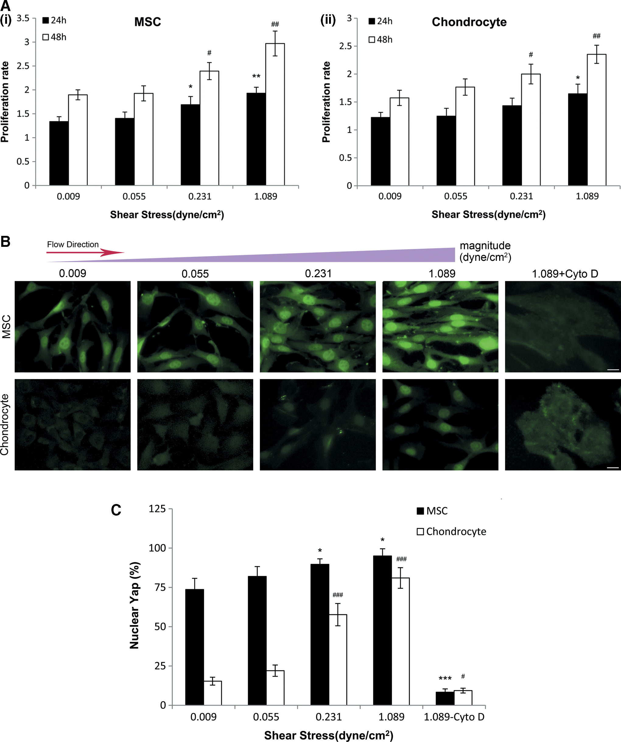
To examine the effects of different shear stresses on cell proliferation, the proliferation rates of MSCs and chondrocytes were analyzed by counting the number of cells present in the chambers under each condition. As shown in Fig. 2A, the proliferation rate of MSCs increased significantly in response to shear stress at 0.231 and 1.089 dyne/cm2 after 24 and 48 h of perfusion compared to the minimum stimulus (0.009 dyne/cm2). For chondrocytes, the proliferation rate increased significantly in response to shear stress at 1.089 dyne/cm2 after 24 h and shear stress at 0.231 and 1.089 dyne/cm2 after 48 h. These findings suggested that the proliferation rates of both MSCs and chondrocytes increased with an increasing magnitude of the fluid shear stress.
The changes in YAP expression in response to different shear stresses
To investigate the effects of different fluid shear stresses on YAP expression in MSCs and chondrocytes, the cells in the microdevices were stained for YAP after 5 days of perfusion for MSCs and 2 days of perfusion for chondrocytes. As shown in Fig. 2B, the distribution of YAP in MSCs was mainly in the nucleus with low levels in the cytoplasm. Although the increase in fluid shear stress did not alter YAP localization, the level of YAP in the nucleus tended to increase (Fig. 2B). In addition, we found that in primary chondrocytes, YAP was predominantly cytoplasmic at an extremely low level of shear stress. However, increased nuclear YAP accumulation was observed with an increasing magnitude of stimulation. At higher stimulus levels (0.231 and 1.089 dyne/cm2), the nuclear localization of YAP was markedly increased (Fig. 2B). Conversely, MSCs and chondrocytes treated with CytoD developed a more rounded morphology and exhibited YAP diffusion into the cytoplasm after exposure to the fluid flow. The quantification of nuclear YAP in MSCs and chondrocytes confirmed these findings (Fig. 2C). The percentage of nuclear YAP in these two types of cells was significantly higher under shear stresses of 0.231 and 1.089 dyne/cm2 than under the lowest level of shear stress (0.009 dyne/cm2). CytoD treatment was associated with a significant decrease in nuclear YAP relative to the lowest stimulus level.
The fate of MSCs changes under different shear stress conditions
To evaluate the effects of different fluid shear stresses on MSC differentiation, cells in the microdevices were stained for ALP and intracellular lipid droplets after 5 days of perfusion. As shown in Fig. 3A, cells exposed to an extremely low level of shear stress tended to undergo adipogenic differentiation, but with an increasing magnitude of shear stress, increased cells underwent osteogenic differentiation. Especially at the higher shear stress level of 1.089 dyne/cm2, cells tended to become committed to the osteogenic lineage. The ratio of osteogenic commitment to adipogenic commitment reflected a significant mechanical effect of different fluid shear stresses (Fig. 3B). However, CytoD-treated cells exposed to the fluid flow stimulus appeared to lose the ability to undergo osteogenic differentiation regardless of the level of flow. Interestingly, the pattern of adipogenic and osteogenic differentiation were consistent with the pattern of YAP distribution in the cytoplasm and nucleus.

MSC commitment varies with changes in the flow stimulus.
Next, we examined the expression of more lineage-specific regulators such as PPARγ, Runx2, and Sox9 in MSCs. As shown in Fig. 3C, PPARγ that promotes adipocyte differentiation in MSCs was predominantly localized in the nucleus, but a gradual decline in PPARγ expression was observed with an increasing magnitude of flow stimulus. In contrast, Runx2, a key regulator of osteogenic differentiation, displayed a gradually increasing expression in the cells, especially in the nucleus with an increasing magnitude of flow stimulus. However, CytoD-treated cells displayed high expression levels of PPARγ, but low expression levels of Runx2 regardless of the flow stimulus. For Sox9, an essential regulator of chondrogenesis, no obvious expression was detected under different fluid shear stress even when treated with CytoD. Taken together, we found that the changes in the expression of PPARγ and Runx2 were in accordance with the variation in lipid droplets and ALP.
The phenotypic variations of chondrocytes in response to different shear stresses
After 2 days of fluid flow stimulation, the phenotypic changes in primary chondrocytes were investigated by immunofluorescence staining for collagen I and collagen II. As shown in Fig. 4A, primary chondrocytes were nearly negative for collagen I and positive for collagen II when exposed to extremely low levels of fluid flow. However, with an increasing magnitude of shear stress, the cells began to synthesize collagen I, whereas the expression of collagen II diminished. When the cells were treated with CytoD, the hyaline cartilage phenotype of the cells was rescued, with low expression of collagen I. To further quantify the variation in the expression levels of collagen II and collagen I, the relative fluorescence intensities were plotted (Fig. 4B, C). We found that from 0.055 to 1.089 dyne/cm2, RFI values were significantly higher for collagen I and lower for collagen II than the values at the minimum level of stimulus. As shown in Figs. 2 and 4, we found that the chondrocyte dedifferentiation was concomitant with YAP accumulation in the nucleus, indicating that the mechanical cue of fluid flow leads to increased YAP expression in dedifferentiated chondrocytes.
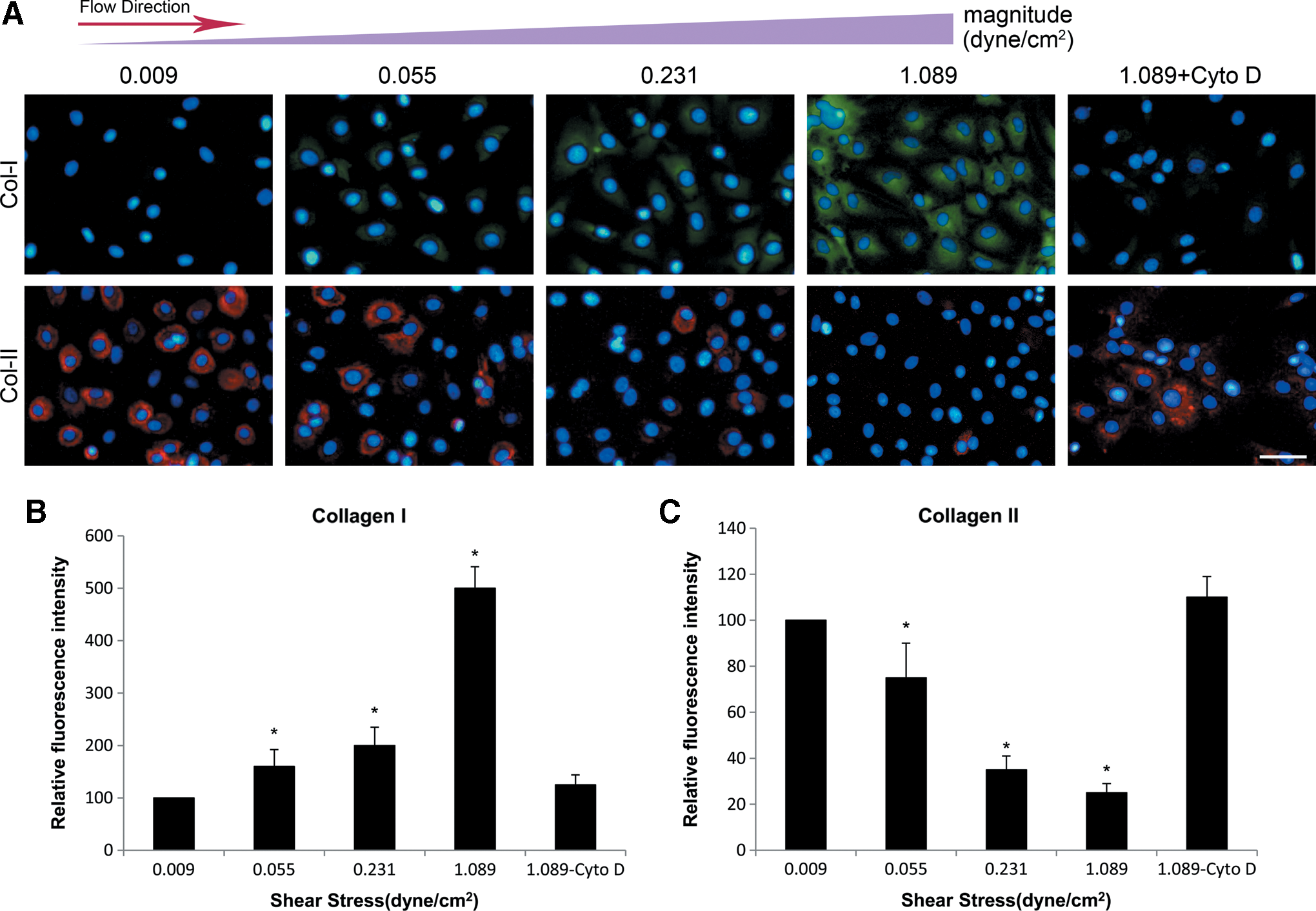
The phenotypic variations of chondrocytes in response to different shear stresses. Chondrocytes were immunostained for collagen I (green), collagen II (red), and DAPI (blue) after 2 days of perfusion.
Discussion
The YAP transcriptional coactivator has emerged as a key mediator of the regulation of cell behaviors such as proliferation, survival, apoptosis, and differentiation [16,17,21]. In this study, we evaluated the change in YAP expression and the consequent biological responses to flow-induced mechanical signals of different magnitudes. Our results suggest that YAP may mediate the effects of fluid flow-induced shear stress on the fates of MSCs and chondrocytes.
We characterized the changes in YAP expression along with the fates of MSCs and chondrocytes in response to different levels of the flow stimulus. Our data showed that YAP expression in MSCs was increased with an increasing magnitude of shear stress. Furthermore, we demonstrated that in chondrocytes, YAP was translocated into the nucleus in response to the fluid flow stimulus. The transduction of local mechanostimuli into biochemical signals occurs through several signaling pathways. Recently, the transcriptional regulators, YAP/TAZ, have been shown to act as not only sensors, but also mediators of physical signals, shuttling between the cytoplasm and the nucleus [22]. As the downstream effectors of mechanotransduction, YAP function depends on the tension of the actomyosin cytoskeleton and the Rho GTPase activity because proper cytoskeleton tension is necessary to maintain the YAP transcriptional activity. Moreover, increasing evidence has shown that shear stress can cause cytoskeleton reorganization in mechanically sensitive cells. Thus, the flow-dependent changes in YAP expression are likely due to an increase in cytoskeleton tension. When the flow shear force is transmitted to the intracellular space, the cells reorganize the cytoskeleton to gradually increase the strength of attachment with an increasing magnitude of stimulation. Meanwhile, cytoskeletal cues can mediate YAP nuclear localization to ultimately affect gene expression. It is well known that the actin cytoskeleton is disrupted when cells are treated with CytoD, thereby abrogating the ability of cells to sense external mechanical cues. In this study, the treatment of cells with CytoD resulted in YAP downregulation, and the flow-dependent response was abolished, further suggesting that shear flow plays a role in the regulation of YAP and the integrity of the actomyosin cytoskeleton is vital for shear stress-based regulation of YAP expression. Additionally, the effects of fluid shear stress on cells involve other pathways that may communicate with the YAP or Hippo pathway. The cell–cell contact or a high cell density inhibits the YAP activity by activating the Hippo pathway [20]. To avoid such an influence, a low seeding density was employed in our microdevice experiments. We assumed that this may limit the activation of the Hippo pathway and better investigate the effects of fluid shear stress on YAP. Consequently, we found that an increasing magnitude of fluid shear stress could promote cell proliferation and concomitantly enhance YAP expression in the nuclei.
Shear stress-induced proliferation of osteocytes, MSCs, as well as chondrocytes has been reported [32 –34], involving several mechanisms such as the ERK pathway, MAP kinase pathway, NO/cGMP/PKG, and calcium signaling [35]. In addition, mechanical signaling through the cytoskeleton linkage between focal adhesion and regulators of cellular contractility contribute to the regulation of cell proliferation [36]. YAP has been shown to act as a transcriptional coactivator of TEAD transcription factors to promote cell proliferation and survival in many tissues [37]. Here we propose that fluid flow upregulates YAP, which then promotes cell proliferation by binding to the TEAD family of transcription factors. Therefore, we conclude that nuclear YAP expression is a key regulator that has a correlation with mechanical signal-induced proliferation. Further investigations are required to determine whether these known pathways cooperate with the Hippo/YAP pathway to promote cell proliferation.
Control of the balance between adipogenesis and osteogenesis during MSC differentiation is necessary to maintain bone homeostasis. Stem cells are highly sensitive to mechanical cues and can convert mechanical stimuli into biochemical signals via mechanotransduction systems. In this study, we characterized the effects of distinct levels of flow stimulus on the regulation of MSC fate. We found that the expression level of YAP is correlated with the intensity of the stimulus experienced by the cells and the fates of the MSCs. Increased YAP expression is in accordance with the increase in the expression of Runx2 and ALP, indicating that increased YAP expression may contribute to osteogenesis in MSCs. Conversely, decreased YAP expression is concomitant with the increase in lipid droplets and the expression of PPARγ, which indicates adipogenesis in MSCs. It has been reported that YAP interacts with Runx2 and PPARγ to regulate the adipocyte/osteocyte gene expression [38]. Moreover, YAP modulated Runx2 and PPARγ activity by altering the activation of Wnt/β-catenin signaling [39]. It is therefore possible that fluid shear stress regulates the choice between adipogenesis and osteogenesis in MSCs by controlling the expression of YAP. Growing evidence has shown that the enhancement of mechanical or cytoskeletal cues would increase osteogenic differentiation and decrease adipogenic differentiation. In addition, cell density is an important factor that regulates the adipogenic and osteogenic differentiation of MSCs [12,40].
In a multilineage differentiation experiment, MSCs differentiated into adipocytes in response to the induction medium containing 10−6 M dexamethasone, but differentiated into osteocytes in response to the induction medium containing 10−7 M dexamethasone [41]. In this study, the mixed differentiation medium contained a relatively high concentration of dexamethasone because the two types of induction medium were mixed in equal proportions, thus causing an increase in the proportion of cells undergoing adipogenic differentiation. However, with increases in the shear stress magnitude, the proportions of cells undergoing adipogenic and osteogenic differentiation were reversed due to the upregulation of YAP expression, leading to an increase in osteogenesis and a decline in adipogenesis. Thus, by analyzing the YAP expression level, we can preliminarily estimate the lineage to which the MSCs have committed or will commit and determine which flow stimulus condition is appropriate for target differentiation.
Chondrocytes differentiate from mesenchymal cells during development and are apt to dedifferentiate in in vitro cultures [42]. An excess fluid flow stimulus has been shown to be detrimental to the maintenance of chondrocyte function [43 –45]. We first investigated the expression of YAP in response to distinct levels of fluid flow in primary chondrocytes, and then monitored the changes in the chondrocyte phenotype that are associated with the changes in YAP localization. We found that an increasing flow stimulus resulted in nuclear YAP accumulation and the loss of chondrocyte properties. When primary chondrocytes were exposed to an extremely low stimulus, no phenotypic variation was observed compared to the higher stimulus, suggesting that such a condition may be not enough to induce chondrocyte dedifferentiation in a short time. Instead, this condition apparently mimicked the flow of interstitial fluid in the cartilage and played an important role in nutrient transport. A previous study confirmed that YAP expression was suppressed by treatment with CytoD [19]. In our study, after the disruption of the cytoskeleton with CytoD, the cells undergoing YAP cytoplasmic translocation failed to respond to the flow stimulation, but better maintained their phenotype, suggesting that the process of regulating the chondrocyte phenotype is related to YAP expression. Taken together, our findings indicate that the maintenance of the phenotype of primary chondrocytes is associated with the exclusion of YAP from the nucleus, which may involve mechanical and cytoskeletal cues or the Hippo pathway. For instance, both pellet or monolayer culture with high cell density and soft substrate with low cytoskeleton tension can better maintain chondrocyte phenotypes in vitro. Such a high cell density induces the activation of Hippo signaling to suppress the expression of YAP [20], and low cytoskeleton tension also causes YAP inactivation [18]. The detailed biochemical mechanisms underlying these phenomena await further characterization. Notably, based on the localization of YAP in chondrocytes, we can estimate the stimulus intensity experienced by cells and evaluate whether chondrocytes would dedifferentiate when exposed to particular magnitudes of shear flow.
Microtechnology can be used to regulate biological stimuli at the cellular and subcellular scales, enabling us to decipher the roles of specific cues and mechanisms in the cells [46]. In our study, we combined the electric circuit theory with CFD analysis to design a lab-on-a-chip, and thus solve the problems related to complex microfluidic network-based devices. Using a physical analogy, the systematic networking of the microchannels from the inlet to outlet ports could readily be used to establish a relatively simple model. Thus, multiple shear stresses could be obtained through the different inlet channel resistances of the different chambers. Furthermore, this microdevice allowed the control of multiple shear stress values in a particular range by modifying the inlet resistance of each microchamber and the inlet flow rate. In addition, this microdevice provided a simple platform to rapidly screen cellular responses to different levels of flow stimulus and minimize the sample volume to the micron scale.
Conclusions
Using microfluidic techniques, we developed an integrated microdevice that simultaneously produces multiple fluid shear stresses comparable to or stronger compared with the interstitial flow. Using this device, the roles of the fluid flow stimulus in the regulation of YAP expression and the fates of MSCs and chondrocytes were investigated. Our results suggest that YAP expression in MSCs and chondrocytes is regulated by fluid shear stress and YAP mediates the fate determination of MSCs and chondrocytes in response to the fluid flow stimulus. Therefore, the modulation of YAP expression could be exploited to manipulate stem cell fate and increase the regenerative potential of terminally differentiated organs with poor intrinsic regenerative capacity.
Footnotes
Acknowledgment
This work was supported by the National Nature Science Foundation of China (no. 81171464, no. 81201212 and no. 81270052).
Author Disclosure Statement
No competing financial interests exist in this study.