
Abstract
Epigenetic asymmetry has been shown to be associated with the first lineage allocation event in preimplantation development, that is, the formation of the trophectoderm (TE) and inner cell mass (ICM) lineages in the blastocyst. Since parthenogenesis causes aberrant segregation between the TE and ICM lineages, we examined several development-associated histone modifications in parthenotes, including those involved in (i) transcriptional activation [acetylated histone H3 lysine 9 (H3K9Ac) and lysine 14 (H3K14Ac), trimethylated histone H3 lysine 4 (H3K4Me3), and dimethylated histone H3 arginine 26 (H3R26Me2)] and (ii) transcriptional repression [trimethylated histone H3 lysine 9 (H3K9Me3) and lysine 27 (H3K27Me3), and mono-ubiquitinated histone H2A lysine 119 (H2AK119u1)]. Here, we report that in parthenotes, H3R26Me2 expression decreased from the morula stage, while expression patterns and levels of H3K9Ac, H3K27Me3, and H2AK119u1 were unchanged until the blastocyst stage; whereas H3K14Ac, H3K4Me3, and H3K9Me3 showed normal patterns and levels of expressions. Relative to the decrease of H3K9Ac in the ICM and increase in the TE of parthenotes, we detected reduced expression of TAT-interactive protein 60 acetyltransferase and histone deacetylase 1 deacetylase in the ICM and TE of parthenotes, respectively. Relative to the decrease of H3R26Me2, we also observed decreased expression of coactivator-associated arginine methyltransferase 1 methyltransferase and increased expression of the Wnt effector transcription factor 7L2 and miR-181c microRNA in parthenotes. Furthermore, relative to the decrease in H3K27Me3 and H2AK119u1, we found increased phosphorylation of Akt1 and enhancer of zeste homolog 2 in parthenogenetic TE. Therefore, our findings that histone signatures are impaired in parthenotes provide a mechanistic explanation for aberrant lineage segregation and TE defects.
Introduction
P
It has been demonstrated that normal development of fetuses and placentae requires the proper expression of several imprinted genes [11,27 –29]. In addition, it was recently reported that the presence of paternal chromatin is essential for repressing nascent RNA synthesis during the two-cell to four-cell transition stage in the preimplantation mouse embryo, and this, in turn, is required for normal development [30]. Parthenogenetic embryos do not express paternal genes, but express maternal genes at twice the normal level [11]; such an imbalance of maternal–paternal gene dosage may be associated with the developmental defects observed in parthenotes [31 –33].
In contrast to the well-documented aberrations of gene expression in parthenogenetic embryos, histone remodeling and modifications during parthenogenetic development are less well understood. Epigenetic asymmetries have been demonstrated between paternal and maternal genomes at the pronuclear and two-cell stages, and between the ICM and TE in blastocysts during preimplantation development [34 –36]. Diploid parthenotes, which have two sets of maternal chromosomes and no paternal chromosomes, are known to lose the epigenetic asymmetry of DNA methylation and histone H3 lysine methylation between the two pronuclei, because both pronuclei inherit the same epigenetic profile from the mother [8,37]. It was previously reported that pronuclei of parthenotes at the one- and two-cell stages do not exhibit asymmetric distributions of heterochromatin protein 1β (Hp1β), trimethylated histone H3 lysine 9 (H3K9), or trimethylated histone H3 lysine 4 (H3K4) [37]. Interestingly, a separate study demonstrated that levels of Hp1 mRNA were elevated by almost four-fold in parthenotes as compared with fertilized embryos cultured in vitro to the zygote stage, but not to other developmental stages [22]. Parthenotes at the zygote stage exhibited perturbed expression of several histones and histone variants, including increased expression of histone H2B, H2afx, and H3.3b, and decreased expression of H2afz [22]. At later preimplantation stages (ie, the morula and blastocyst stages), only the mRNA levels of histone H2A variants H2afx and H2afz were affected in parthenotes, with both exhibiting an increase as compared with fertilized embryos [22]. Moreover, both DNA methylation and histone H4 acetylation were significantly decreased in the parthenote ICM as compared with the control ICM [38]. In addition to histone signatures, several histone-modifying proteins showed increased expression in parthenotes, including histone deacetylase 1 (HDAC1; at the two-cell and morula stages), DNA (cytosine-5)-methyltransferase 3B (at the two-cell stage), histone acetyltransferase 1 (at the blastocyst stage), and the histone-lysine N-methyltransferases SUV39H1 (at the morula and blastocyst stages) and G9A (at the blastocyst stage) [22,39].
Given that (i) the establishment of epigenetic asymmetry between the morula and blastocyst stages has been speculated to be associated with lineage allocation and commitment in mammalian embryos [34,35], and (ii) lineage segregation is impaired in mouse parthenogenetic embryos [19], we hypothesized that fertilized and parthenogenetic mouse embryos may exhibit different patterns of asymmetrical histone modifications at the morula and blastocyst stages. Since previous studies have reported differential expression of several histone variants, histone-modifying enzymes, or associated proteins between in vitro-fertilized and in vivo-fertilized mouse and bovine embryos [22,39], we chose to use in vivo- instead of in vitro-fertilized mouse embryos as controls in this study, to reflect on the authentic in vivo situation of epigenetic regulation. As compared with in vivo-fertilized control embryos, we observed (i) significantly decreased dimethylation of histone H3 arginine 26 (H3R26) in parthenogenetic morulae and ICM; (ii) altered levels of acetylation at histone H3 lysine 9 (H3K9) in the parthenote (decreased in the ICM and increased in the TE); (iii) loss of punctate signals of histone H3 lysine 27 (H3K27) trimethylation in the parthenote TE; and (iv) reduced mono-ubiquitination of histone H2A lysine 119 (H2AK119) in parthenogenetic blastocysts. We also found perturbed expression of several histone acetyltransferases, methyltransferases, and related signaling factors regulating the histone signatures mentioned earlier, including decreased expression of general control of nucleotide synthesis 5 (GCN5) and TAT-interactive protein 60 (Tip60) acetyltransferases, HDAC1 deacetylase and coactivator-associated arginine methyltransferase 1 (CARM1) methyltransferase, increased expression of CARM1 inhibitor miR-181c microRNA and its upstream Wnt effector transcription factor 7L2 (TCF7L2) [40 –42], and increased phosphorylation of enhancer of zeste homolog 2 (Ezh2) methyltransferase and its upstream Akt1 serine/threonine protein kinase [43].
Materials and Methods
Parthenogenetic activation and in vitro culture of preimplantation embryos
Animals used in this study were purchased from BioLASCO Taiwan, and approval was received from the Academia Sinica Institutional Animal Care and Utilization Committee. B6DBA female mice at 10–14 weeks old were superovulated by an intraperitoneal (i.p.) injection of 5 IU pregnant mare serum gonadotropin (Calbiochem®; Merck Millipore 367222), followed by an i.p. injection of 5 IU human chorionic gonadotropin (hCG) (Sigma-Aldrich C1063) at 48–50 h later; the mice were subsequently separated into two groups: One group was unmated, while mice of the other group were individually mated with B6DBA males of proven fertility. Hence, both unmated and mated female mice had been superovulated. At 14–15 h after the hCG injection mentioned earlier (counted as embryonic day 0.5 or E0.5), the oviducts of both unmated and mated females were collected into droplets of pre-equilibrated M2 media (EmbryoMax® M2 medium from Millipore; purchased from Level Biotechnology). Zygotes or unfertilized oocytes enclosed in cumulus masses were released from the ampullae, and cumulus cells were removed by pipetting using a mouth-controlled pipette (with an inner diameter of 200–300 μm) after 5 min of treatment with M2 media containing 50−100 U/mL hyaluronidase (EmbryoMax M2 medium with hyaluronidase; Millipore). Zygotes and oocytes were then washed and incubated in 35 μL droplets of potassium simplex optimized medium (KSOM) (EmbryoMax KSOM w/1/2 amino acids, glucose, and phenol red from Millipore), and covered with mineral oil at 37°C in a humidified atmosphere of 5% CO2 in air.
Parthenogenesis was activated after incubation of the unfertilized oocytes in KSOM for 1 h. Oocytes were then washed and incubated in droplets of CZBG medium containing 10 mM strontium chloride, which would stimulate the IP3-PIP2-medicated calcium oscillation mimicking fertilization [44,45], and 5 μg/mL cytochalasin B, which would inhibit the first cytokinesis and maintain the diploid chromosome set [46], at 37°C with 5% CO2 in air, as previously described [47]. After 5–6 h, successful parthenogenetic activation was confirmed by the presence of two pronuclei in the unfertilized oocytes, as previously described [48,49]. These parthenogenetic embryos at the pronuclei stage were then transferred to KSOM and incubated until they reached the developmental stage of interest (ie, the morula or blastocyst stage in this study).
Embryos usually cleave to two cells at E1.0–1.5, and form morulae at E2.5–3.0 (48–60 h in culture). Early blastocysts are formed at E3.5 (70–72 h in culture), blastocysts are formed at E4.0 (80–84 h in culture), and late expanded blastocysts are formed at E4.5 (92–96 h in culture). In this study, the embryos developed from fertilized oocytes that were extracted from mated superovulated female mice are defined as control embryos, while the embryos developed from unfertilized and strontium chloride-activated oocytes which were extracted from unmated superovulated female mice are defined as parthenogenetic embryos. The numbers of sacrificed female mice and embryos used as the control or parthenogenetic group for each analyzed marker were listed in Supplementary Table S1 (Supplementary Data are available online at
Immunofluorescence staining
For immunostaining, embryos were washed for 5–10 s in droplets of acidic Tyrode's solution (prepared by Sigma-Aldrich; purchased from Uni-onward) to remove the zona pellucida, and then fixed in 4% paraformaldehyde (Sigma-Aldrich) in 1× phosphate-buffered saline (PBS) for 15 min at room temperature. Embryos were then permeabilized with 0.25% Triton X-100 for 15 min, followed by washing and blocking for 1 h in blocking solution containing 0.05% Tween-20, 3% bovine serum albumin (BioXtra BSA suitable for mouse embryo cell culture; Sigma A3311), and 5% normal goat serum (Gibco®; Life Technologies PCN5000) in 1× PBS. After blocking, embryos were incubated at 4°C overnight with the following primary antibodies diluted in blocking solution: anti-acetylated histone H3 lysine 9 (rabbit polyclonal, 1:200 dilution; Abcam), anti-CARM1 (both mouse monoclonal and rabbit polyclonal, 1:200 dilution; Abcam), anti-Cdx2 (mouse monoclonal, 1:100 dilution; BioGenex), anti-dimethylated histone H3 arginine 26 (rabbit polyclonal, 1:200 dilution; Abcam), anti-Ezh2 (rabbit polyclonal, 1:200 dilution; Millipore), anti-mono-ubiquitinated histone H2A lysine 119 (mouse monoclonal, 1:200 dilution; Millipore), anti-phosphorylated Akt1 (phospho-S473) (rabbit polyclonal, 1:100 dilution; Abcam), anti-phosphorylated Ezh2 (phospho-S21) (rabbit polyclonal, 1:100 dilution; Abcam), anti-TCF7L2 (rabbit polyclonal, 1:100 dilution; Abcam), anti-trimethylated histone H3 lysine 4 (rabbit polyclonal, 1:200 dilution; Abcam), anti-trimethylated histone H3 lysine 9 (rabbit polyclonal, 1:200 dilution; Millipore), or anti-trimethylated histone H3 lysine 27 (rabbit polyclonal, 1:200 dilution; Millipore). On the second day, the embryos were washed and blocked for 1 h in blocking solution, followed by incubation at room temperature for 1 h with the following secondary antibodies conjugated to fluorophores: goat anti-mouse AlexaFluor 488 (green fluorescence) or goat anti-rabbit AlexaFluor 555 (red fluorescence) (Invitrogen Taiwan). After incubation, the embryos were washed for 10 min in washing solution containing 0.2% Triton X-100 in 1× PBS, and then counter-stained with 0.2 μg/mL 4′,6-diamidino-2-phenylindole (DAPI) in washing solution for 10 min, followed by mounting in VectaShield (Vector Laboratories) on glass slides.
In situ hybridization of miR-181c
An LNA (Locked Nucleic Acid)™-modified DNA oligonucleotide complementary to the mature microRNA miR-181c was used as a probe for in situ hybridization (Exiqon). The sequence of the LNA-miR181c probe was 5′-ACTCACCGACAGGTTGAATGTT-3′, while the sequence of the LNA-scrambled negative control probe was 5′-GCGTTATGACTGATGCTACTTACA-3′. Probes were labeled enzymatically using a 3′-end digoxigenin (DIG) labeling kit (Roche) according to the manufacturer's instructions, and purified using illustra® MicroSpin® G-25 Columns (GE Healthcare). The miRNA in situ hybridization procedure was performed using the DIG-AP Rembrandt® Universal RISH and Detection Kit (Life Technologies), in accordance with the manufacturer's protocol.
Confocal microscopy and statistical analyses
A series of confocal sections through three-dimensional (3D) preserved embryo nuclei were collected using a Leica TCS SP5 confocal microscope (Genomics Research Center, Academia Sinica) equipped with a Super Z galvanometer stage and Plan Apo 63×/1.4 NA oil immersion objectives. Fluorochromes were visualized using an argon laser with an excitation wavelength of 488 nm (for AlexaFluor 488), a diode-pumped solid-state laser with a laser line of 561 nm (for AlexaFluor 555), and a diode laser with a laser line of 405 nm (for DAPI). For each optical section, images were sequentially collected using the XYZ mode for two or three fluorochromes. The pinhole was set to 1–1.5 Airy units, with a 1.5× scan zoom. In order to compare the relative intensities of immunostaining between control and parthenogenetic embryos, identical scanning parameters (including the strength of laser emissions) were maintained for embryos stained with the same antibodies. Images of optical sections were then analyzed using Leica Application Suite, and 3D and maximum projections were constructed from serial stacks of sections for each embryo.
The image files of optical sections of each embryo were input into the Count Nuclei/Cell Sorting Application Module for MetaMorph (MetaMorph Offline vers. 7.0; Universal Imaging Corporation™) for quantitative analyses and a comparison between control and parthenogenetic embryos. Briefly, for cell counting, the numbers of blastomeres or nuclei stained with different colors of fluorescence were counted on the maximum projection pictures (which were stacked from all consecutive optical sections) from controls and parthenotes, respectively. For a comparison of fluorescent intensities, a specified unit of area (eg, a blastomere or a nucleus) in the control embryo was set up to quantify the fluorescent intensity within the area, which was recorded as a standard value, and the same unit of area was picked up from the parthenogenetic embryo, followed by quantification of fluorescent intensity within the area and comparison with the standard value. The relative intensity of an embryo was calculated as the summation of relative values obtained from all units of areas within the single embryo, which were quantified as a fold change of the standard value.
For statistical analyses, Student's t-tests were performed using Microsoft® Excel (Microsoft® Office software by Microsoft Corporation). Numbers of immunostained cells in parthenogenetic embryos were compared with the corresponding developmental stages of in vivo-fertilized control embryos.
Results
Perturbed H3K9 acetylation and normal H3K14 acetylationin parthenogenetic embryos
Immunostaining analyses of control blastocysts revealed a higher level of acetylated histone H3 lysine 9 (H3K9Ac) in the ICM as compared with the TE (Fig. 1B), as previously described [50]. On the other hand, asymmetrical H3K9 acetylation patterns were not observed among blastomeres or between Cdx2+ (green) and Cdx2− blastomeres in control morulae (Fig. 1A). At the morula stage, the pattern and level of H3K9 acetylation in parthenotes were similar to those in controls (Fig. 1A). However, the distribution of H3K9 acetylation in parthenogenetic blastocysts was the opposite of that in controls, with a higher level in the TE as compared with the ICM (indicated by arrows in Fig. 1B). Therefore, acetylation of H3K9 in parthenotes was increased in the TE but decreased in the ICM, as compared with controls. Abnormal distribution of H3K9 acetylation between the ICM and TE in mouse parthenogenetic blastocysts (Fig. 1B) is reminiscent of the loss of asymmetrical H3K9 acetylation in somatic cell nuclear transfer (SCNT)-cloned bovine blastocysts [50].
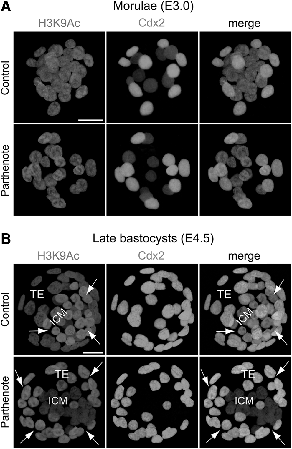
Reversed asymmetrical distribution of acetylated histone H3 lysine 9 (H3K9Ac) in parthenogenetic blastocysts. Double immunostaining of acetylated histone H3K9 and Cdx2 revealed relatively homogeneous staining among all nuclei of both control (normal fertilized) and parthenogenetic morulae (E3.0)
Since a previous study demonstrated that H3K9 and H3K14 acetylation co-occurred at several gene loci in mouse ES cells [51], we also examined H3K14 acetylation in parthenogenetic embryos. Unlike H3K9 acetylation, H3K14 acetylation was homogeneous among all blastomeres at both the morula and blastocyst stages, and its level and pattern were indistinguishable between controls and parthenotes (Fig. 2).
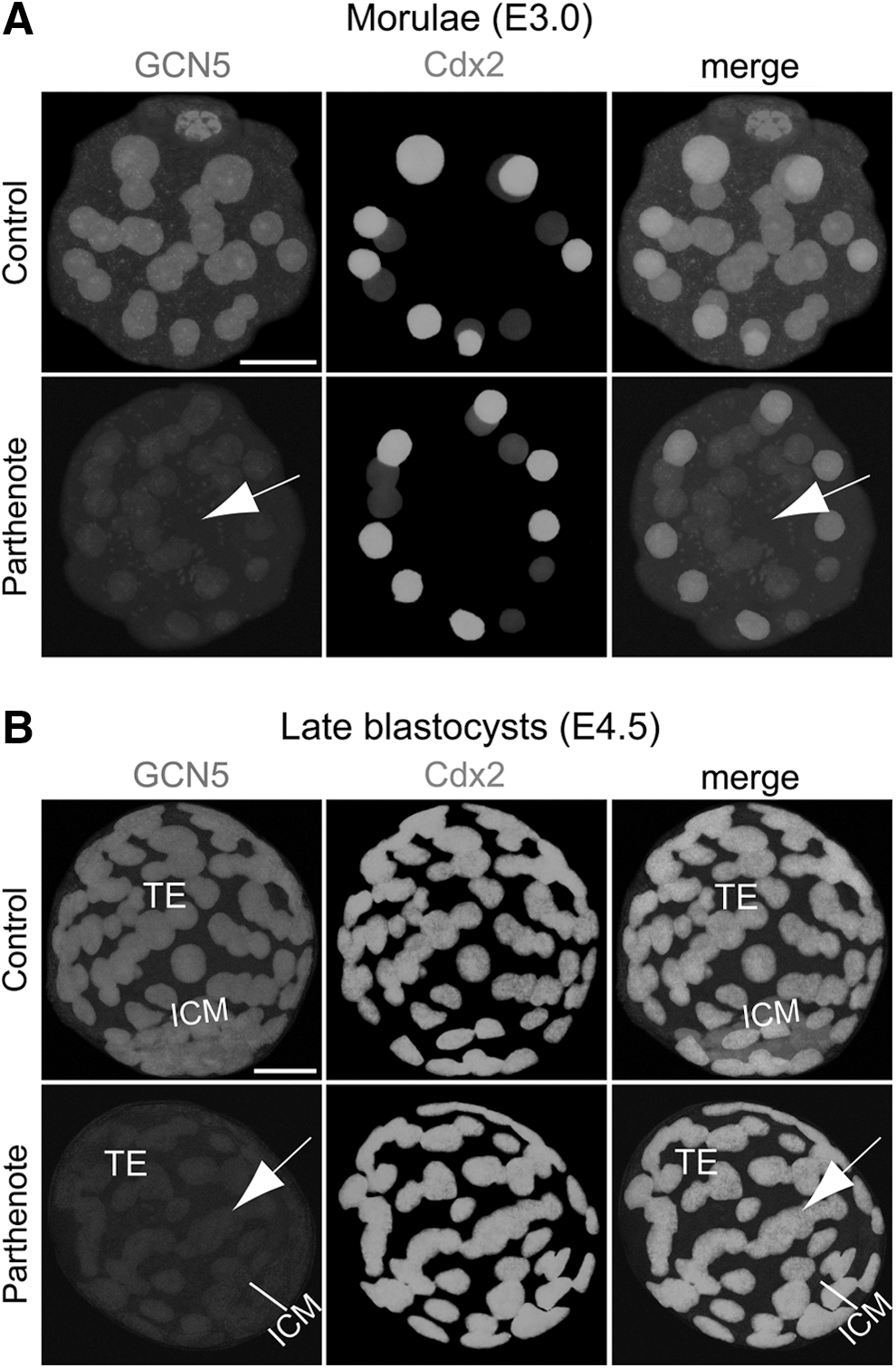
Decreased expression of GCN5 acetyltransferase in parthenogenetic morulae and blastocysts. Double immunostaining against GCN5 and Cdx2 revealed significantly decreased GCN5 expression (indicated by arrows) throughout the whole parthenogenetic embryos (including both the outer Cdx2+ and inner Cdx2- blastomeres) compared with control embryos at both the morula
Normal expression of p300 and decreased expression of GCN5, HDAC1, and Tip60 in parthenogenetic embryos
It has been reported that both H3K9 and H3K14 acetylation are regulated by the acetyltransferases GCN5/PCAF, while H3K9Ac is also regulated by Tip60 acetyltransferase, and H3K14 acetylation is also regulated by the acetyltransferases p300/CBP and/or Myst3 [51 –54]. Interestingly, a recent study demonstrated that the expression of GCN5 and HDAC1 was decreased in mouse parthenogenetic embryos at the four-cell stage, predominantly in or around the nuclei [55].
Consistent with the previous finding, we observed decreased GCN5 and HDAC1 expression in parthenotes compared with controls at both the morula and blastocyst stages (Figs. 2 and 3). While GCN5 expression showed a global reduction throughout the whole embryos (Fig. 2), HDAC1 expression was predominantly decreased in the Cdx2+ outer blastomeres and TE (indicated by arrows in Fig. 3), as HDAC1 was expressed at a much lower level in the Cdx2− inner blastomeres and ICM of both controls and parthenotes (Fig. 3). In contrast to the predominant reduction of HDAC1 expression in the outer blastomeres and TE of parthenotes, Tip60 showed a predominant decrease of expression in the inner blastomeres and ICM of parthenotes (indicated by arrows in Fig. 4). Similar to GCN5, Tip60 displayed a homogeneous expression pattern throughout the whole control embryos at both the morula and blastocyst stages (Fig. 4). It is noteworthy that the level of Tip60 expression in the Cdx2+ outer blastomeres and TE of parthenotes was comparable with the expression level in controls (Fig. 4). The reduction of HDAC1 and Tip60 expression in the outer and inner blastomeres of parthenotes, respectively, may impose an additive effect on H3K9 acetylation, leading to an increased and decreased level of H3K9Ac in the TE and ICM of parthenogenetic blastocysts (Fig. 1), respectively.
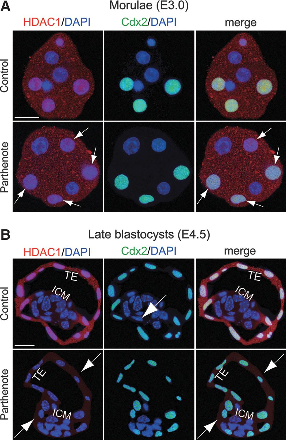
Decreased expression of histone deacetylase 1 (HDAC1) deacetylase in the outer blastomeres and TE of parthenogenetic embryos. Double immunostaining for HDAC1 (red) and Cdx2 (green) indicated that HDAC1 was expressed at a significantly higher level in the Cdx2+ blastomeres (ie, the outer blastomeres of morulae and TE of blastocysts)
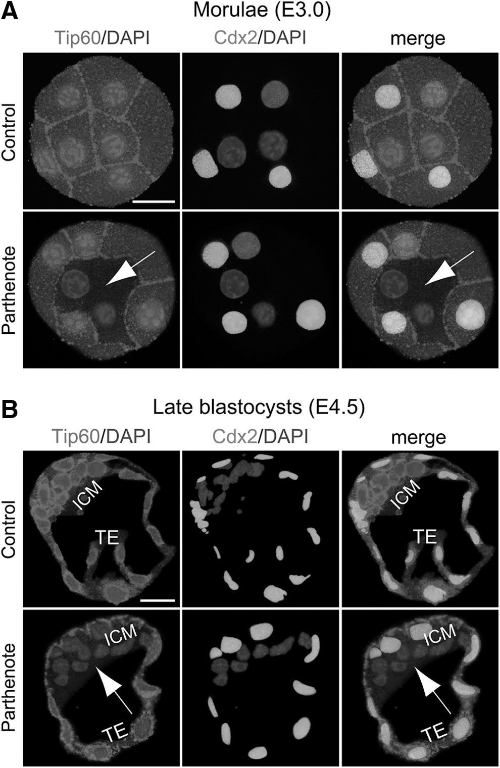
Decreased expression of TAT-interactive protein 60 (Tip60) acetyltransferase in the inner blastomeres and ICM of parthenogenetic embryos. Double immunostaining against Tip60 and Cdx2 indicated that, in parthenogenetic embryos, Tip60 expression was dramatically reduced in the Cdx2− blastomeres (indicated by arrows), including the inner blastomeres of morulae
H3K27 trimethylation is decreased in parthenogenetic TE
Since parthenogenetic embryos have been previously demonstrated to exhibit incomplete X inactivation and overexpression of X-linked genes [56 –60] and levels of trimethylation of histone H3 lysine 27 (H3K27Me3) have been reported to be associated with the inactivated paternal X chromosome [61 –64], we hypothesized that patterns of this epigenetic mark may be altered in parthenotes. H3K27 trimethylation is detectable in preimplantation embryos from the 16-cell morula stage onward [61 –66], and we observed multiple H3K27Me3 punctate signals in all nuclei of both control and parthenogenetic morulae (Fig. 5A). At the blastocyst stage (Fig. 5B), a single strong H3K27Me3 punctate signal was detected in all nuclei of the control TE, but no specific punctate signals above the background level were detected in nuclei of the parthenote TE, consistent with loss of X inactivation in the parthenote TE [56,57,60]. On the other hand, both control and parthenogenetic blastocysts displayed multiple, strong punctate signals of H3K27 trimethylation in the ICM (Fig. 5B). Therefore, trimethylation of H3K27 was normal in the parthenogenetic morula and ICM, but decreased in the parthenogenetic TE.
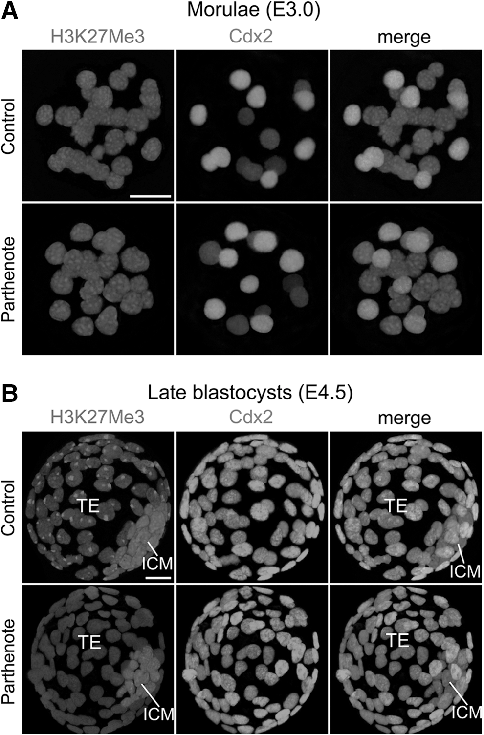
Absence of punctate staining against trimethylated histone H3 lysine 27 (H3K27Me3) in nuclei of the parthenogenetic TE. Immunostaining against dimethylated histone H3K27 revealed multiple punctate signals in all nuclei of both control (normally fertilized) and parthenogenetic morulae (E3.0)
Ezh2 phosphorylation is increased, while H2A mono-ubiquitination is decreased in parthenogenetic blastocysts
Previous studies have demonstrated that decreased H3K27 methylation results from increased phosphorylation of serine 21 of the Polycomb group methyltransferase Ezh2 [43,67]; thus, we examined the levels of total and phosphorylated Ezh2 proteins in parthenogenetic blastocysts. We found that the amounts of total Ezh2 in controls and parthenotes were comparable (Fig. 6A), whereas the level of Ser21-phosphorylated Ezh2 was considerably higher in the parthenote TE than in the control TE (Fig. 6B). On the other hand, the level of Ezh2 phosphorylation was very low in the ICM of both controls and parthenotes (Fig. 6B). Hence, the amount of phosphorylated Ezh2 was greatly increased in the parthenogenetic TE, while total Ezh2 was unaffected.
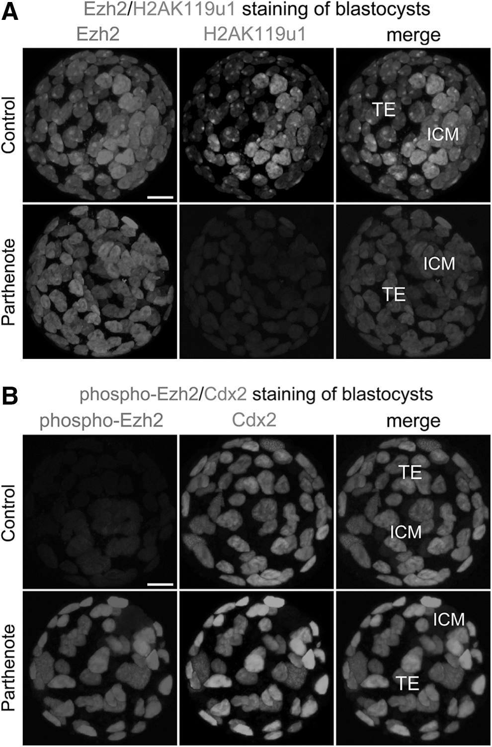
Dramatically decreased mono-ubiquitination of histone H2A lysine 119 (H2AK119u1) and phosphorylation of enhancer of zeste homolog 2 (Ezh2) methyltransferase in parthenogenetic blastocysts. Immunostaining was used to reveal that levels of total Ezh2 methyltransferase were similar between the nuclei of the TE of control and parthenogenetic blastocysts
H3K27 methylation recruits the Polycomb repressive complex 1 (PRC1) to histone H2A, resulting in mono-ubiquitination of histone H2A at lysine 119 (H2AK119u1) [68,69]; we, therefore, hypothesized that the level of H2AK119u1 would be reduced in parthenotes. As shown in Fig. 6A, we observed only background levels of H2AK119u1 immunostaining in both the TE and ICM of parthenogenetic blastocysts. In contrast, control blastocysts contained a specific H2AK119u1 punctate signal (green) in the nucleus of each TE cell, which co-localized with the punctate signal of Ezh2 (red); furthermore, multiple, strong punctate signals were observed in the nucleus of each ICM cell (Fig. 6A). Our results indicated that increased phosphorylation of Ezh2 in the parthenogenetic TE is concomitant with a significant decrease of both trimethylation of H3K27 and mono-ubiquitination of H2AK119.
Akt1 phosphorylation is increased in parthenogenetic embryos
The serine/threonine protein kinase Akt1 (also known as protein kinase B α) has been shown to suppress H3K27 trimethylation via phosphorylation of Ezh2 [43]. As such, we hypothesized that over-activation of Akt1 may accompany the increase in phosphorylated Ezh2 in parthenogenetic blastocysts. Previous studies have reported that Akt1 is maximally activated when its serine at position 473 is phosphorylated [70,71], and the level of Ser473-phosphorylated Akt1 is positively correlated with the level of Ser21-phosphorylated Ezh2, but inversely correlated with the level of trimethylated histone H3K27 [43]; we, therefore, investigated the level of Ser473-phosphorylated Akt1 in parthenotes. We found that Akt1 phosphorylation was greatly increased in parthenotes as compared with controls, at both the morula and blastocyst stages (Fig. 7). Ser473-phosphorylated Akt1 was increased primarily at the plasma membrane of parthenotes, but an increase was also observed in the cytoplasm at both the morula and blastocyst stages (indicated by arrows in Fig. 7), as well as in the nuclei of most outer blastomeres at the morula stage (Fig. 7A). Interestingly, by the blastocyst stage, phosphorylated Akt1 was no longer detected in the nuclei of parthenotes (Fig. 7B). Moreover, the level of Akt1 phosphorylation in controls was higher at the morula than at the blastocyst stage (compare Fig. 7A, B), and Ser473-phosphorylated Akt1 displayed an apical staining pattern on the TE at the blastocyst stage (Fig. 7B), consistent with the results of an earlier study [72].
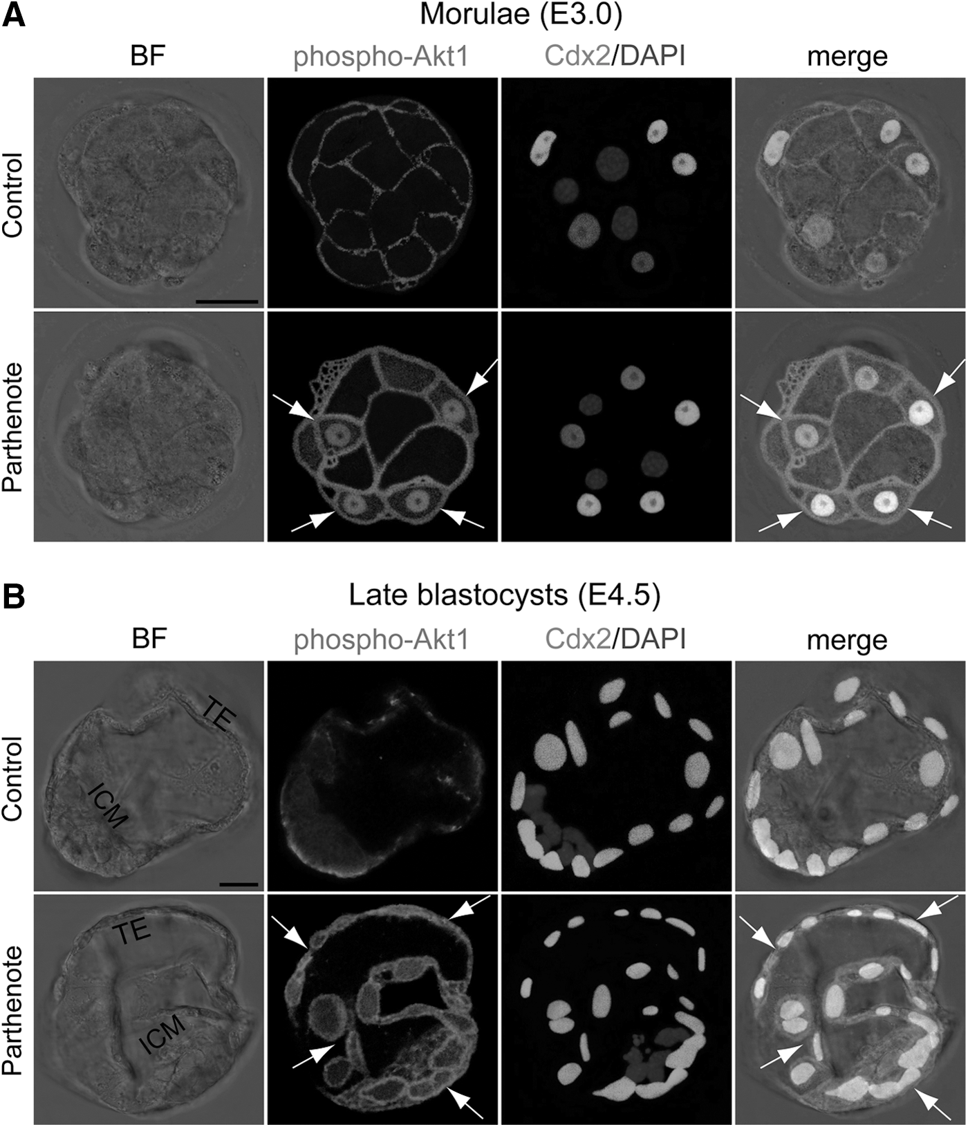
Increased phosphorylation of Akt1 in parthenogenetic morulae and blastocysts. Double immunostaining against serine 473-phosphorylated Akt1 and Cdx2 revealed that the phosphorylation level of Akt1 was greatly increased at the plasma membrane and in the cytoplasm of parthenogenetic Cdx2+ blastomeres, during both the morula (E3.0)
Trimethylation of H3K4 and H3K9 is unaffected in parthenogenetic embryos
In addition to the histone modifications mentioned earlier, trimethylation of lysine 4 and lysine 9 of histone H3 has also been demonstrated to be required for maintaining pluripotency and regulating ICM/TE lineage specification. Trimethylation of histone H3 lysine 4 (H3K4Me3) is observed at actively transcribed gene loci [73 –75], and is also associated with the promoters of poised developmental genes and active pluripotency genes, including Oct4, Nanog, and Sox2; of these genes, Nanog and Sox2 display aberrant expression in parthenogenetic morulae and blastocysts [19,76 –78]. Furthermore, parthenogenetic embryos at the one- and two-cell stages lose the asymmetrical staining of trimethylated H3K4 between the maternal and paternal genomes [37]. However, our immunofluorescence analyses revealed comparable levels of H3K4 trimethylation between controls and parthenotes at both the morula and blastocyst stages, and staining against trimethylated H3K4 was homogeneous throughout all nuclei of control and parthenogenetic morulae and blastocysts (Supplementary Fig. S1).
Trimethylation of histone H3 lysine 9 (H3K9Me3) is also a key epigenetic mark in developing embryos, and is associated with DNA methylation, inactivated paternal X chromosomes in blastomeres, and repression of TE-specific genes in the ICM [64,79 –82]. Immunostaining against H3K9Me3 was also homogeneous throughout the whole embryo at both the morula and blastocyst stages, with no difference between the ICM and TE [62,64,83,84]. Our results, thus, indicate that levels of H3K9 trimethylation are comparable between control and parthenogenetic embryos at both the morula and blastocyst stages (Supplementary Fig. S1).
H3R26 dimethylation and CARM1 expression are reduced in parthenogenetic embryos
In a previous study, we observed decreased Nanog and Sox2 expression in parthenotes at both the morula and blastocyst stages [19]; given that the expression of Nanog and Sox2 is upregulated by CARM1-mediated methylation of histone H3 arginine methylation [85,86], we hypothesized that histone H3 arginine methylation may be disrupted in parthenotes. Immunostaining was used to reveal that both CARM1 expression and dimethylation of histone H3 arginine 26 (H3R26Me2) were significantly decreased in parthenotes, with fewer positive nuclei and lower levels in each nucleus as compared with controls, at both the morula (Fig. 8B) and blastocyst (Fig. 8C, D) stages (indicated by arrows). We observed weak immunostaining signals against CARM1 at both the morula (Fig. 8A, B) and blastocyst (Fig. 8D) stages, in agreement with previous reports that CARM1 mRNA is downregulated by the two-cell stage [85] and protein is decreased by the morula stage [87]. At the morula stage, both CARM1 expression (Fig. 8A, B) and H3R26 dimethylation (Fig. 8B) were predominantly detected in the inner blastomeres, whereas a low level of H3R26Me2 was also observed in the outer blastomeres (especially in control morulae; Fig. 8B). The percentages of CARM1-positive blastomeres in control and parthenogenetic morulae were 36.5±2.8% and 10.4±2.9%, respectively, while the percentages of H3R26Me2-positive blastomeres in control and parthenogenetic morulae were 96.6±2.4% and 54.2±3.1%, respectively (n=12, p<0.01, Student's t test). At the blastocyst stage, H3R26 dimethylation colocalized with CARM1 expression (Fig. 8D); both signals were detected in both the ICM (higher level) and TE (lower level) of controls but detected only in the ICM of parthenotes (Fig. 8C, D). The percentages of CARM1-positive/H3R26-positive blastomeres in control and parthenogenetic blastocysts were 51.6±4.6% and 14.2±4.4%, respectively (n=12, p<0.01, Student's t test). Taken together, CARM1 expression and H3R26 dimethylation exhibited an ∼2–3.5-fold reduction in parthenotes as compared with controls during preimplantation development.
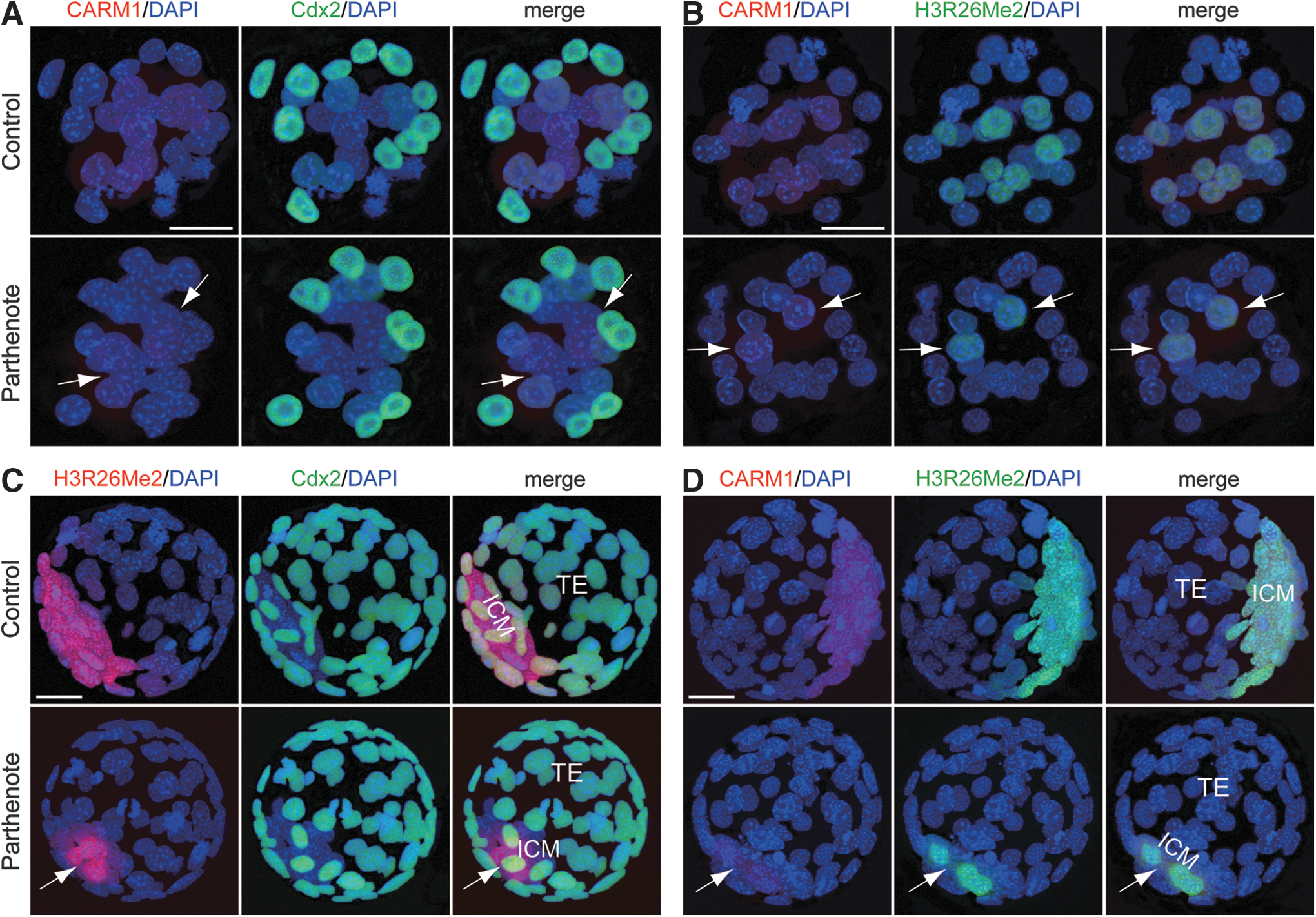
Decreased coactivator-associated arginine methyltransferase 1 (CARM1) expression and dimethylation of histone H3 arginine 26 (H3R26Me2) in parthenogenetic embryos.
TCF7L2 is decreased, while miR-181c is increased in parthenogenetic embryos
Recent studies have reported increased expression of Wnt signaling components, including the cAMP-dependent protein kinase PKA and transcription factor TCF7L2, in parthenogenetic blastocysts [88], as well as inhibition of CARM1 expression by miR-181c microRNA, a downstream effector of Wnt signaling [40,42]. We, thus, proceeded to investigate whether the observed decrease in CARM1 expression in parthenogenetic morulae and blastocysts correlated with expression of TCF7L2 and miR-181c. In control embryos, both TCF7L2 (Fig. 9) and miR-181c (Fig. 10) were predominantly expressed in the Cdx2-positive outer blastomeres of morulae and TE, with very weak expression in the inner blastomeres of morulae and ICM. In contrast, parthenotes exhibited strong expression of both TCF7L2 and miR-181c in the inner blastomeres of morulae and ICM (indicated by arrows in Figs. 9 and 10). Increased TCF7L2 expression in parthenotes may be associated with increased miR-181c expression, which would result in inhibition of CARM1 expression.
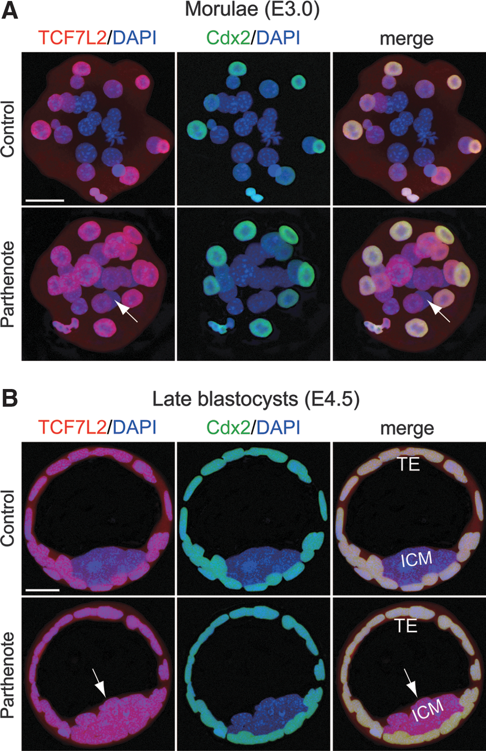
Increased expression of the Wnt effector transcription factor 7L2 (TCF7L2) in parthenogenetic morulae and blastocysts. Double immunostaining against TCF7L2 (red) and Cdx2 (green) revealed dramatically increased TCF7L2 expression (indicated by arrows) in the inner blastomeres of parthenogenetic morulae
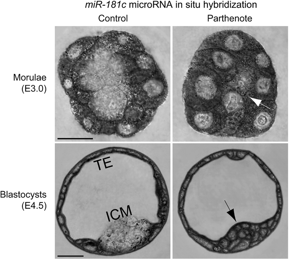
Increased expression of the CARM1 inhibitor miR-181c microRNA in parthenogenetic morulae and blastocysts. RNA in situ hybridization was performed using an LNA™-modified DNA oligonucleotide probe against miR-181c microRNA. Expression of miR-181c was dramatically increased in the inner blastomeres of parthenogenetic morulae and the ICM of parthenogenetic blastocysts (indicated by arrows), as compared with control embryos. On the other hand, miR-181c expression in the outer blastomeres of morulae and TE was comparable between control and parthenogenetic embryos. Scale bars: 20 μm.
Discussion
The presence of both paternal and maternal genomes is required to establish epigenetic asymmetry in the zygote, which is essential for correct nuclear reprogramming and restoration of cell totipotency during early preimplantation development [34,36]. The absence of one parental genome can contribute to aberrant expression of imprinted, development-related, and pluripotency genes, as alteration of epigenetic asymmetry has been suggested to change the developmental potency of cells [8,34,36,37,89]. However, aberrant nuclear reprogramming and loss of epigenetic asymmetry was also observed between zygote pronuclei of mammalian embryos cloned by SCNT, despite the nuclei carrying both paternal and maternal genomes [34,36,37,90]. This suggests that perturbed epigenetic asymmetry does not necessarily result from loss of a parental genome. It is likely that the in vitro activation and culture of oocytes per se is an important contributor to the aberrant nuclear reprogramming during early preimplantation development. In addition, superovulation per se has been demonstrated to perturb DNA methylation and expression of Line-1 retrotransposon and several imprinted genes, including PEG1/MEST and H19 [91 –94]. However, it remains to be unraveled whether superovulation affects histone modifications as well. In our study, the patterns and levels of H3K9Ac and H3K14Ac; methylation of H3 lysine 4, lysine 9, lysine 27, and arginine 26, as well as ubiquitylation of H2A lysine 119, are consistent with those shown in previous studies [50,62,64 –68,85,95,96]. Hence, the putative effect of superovulation on histone signatures may be negligible in this study.
Multiple lines of evidence have demonstrated that epigenetic asymmetry between blastomeres imposes heritable instructions for lineage-specific differentiation, and may, thus, direct lineage allocation between the morula and blastocyst stages [34,35,97]. Our findings of perturbed histone-modifying signaling proteins and aberrant histone signatures in parthenogenetic morulae and blastocysts are summarized in Fig. 11. Of the epigenetic modifications analyzed, asymmetrical DNA methylation between the ICM and TE lineages and differential methylation of histone H3 arginine among blastomeres since the four-cell stage were demonstrated to be the most important for lineage allocation and cell fate determination [85,97]. We observed a significant reduction in H3 arginine methylation and CARM1 expression in parthenogenetic embryos at both the morula and blastocyst stages; these findings, combined with our previous report that expression of Nanog and Sox2 is markedly decreased in such embryos [19], suggest a possible mechanism for impaired lineage allocation and cell fate determination in parthenotes. It was recently reported that CARM1 translation is downregulated by the miR-181 family of microRNAs [42], which are, in turn, upregulated by the Wnt/Tcf signaling pathway [40]. Interestingly, Wnt signaling was increased in parthenogenetic blastocysts, with increased expression of the TCF7L2 transcription factor and the cAMP-dependent protein kinase PKA [88]. We report here that the expression of both TCF7L2 and miR-181c is dramatically increased in the inner blastomeres of morulae and ICM in parthenotes, perhaps accounting for the decrease in CARM1 expression.
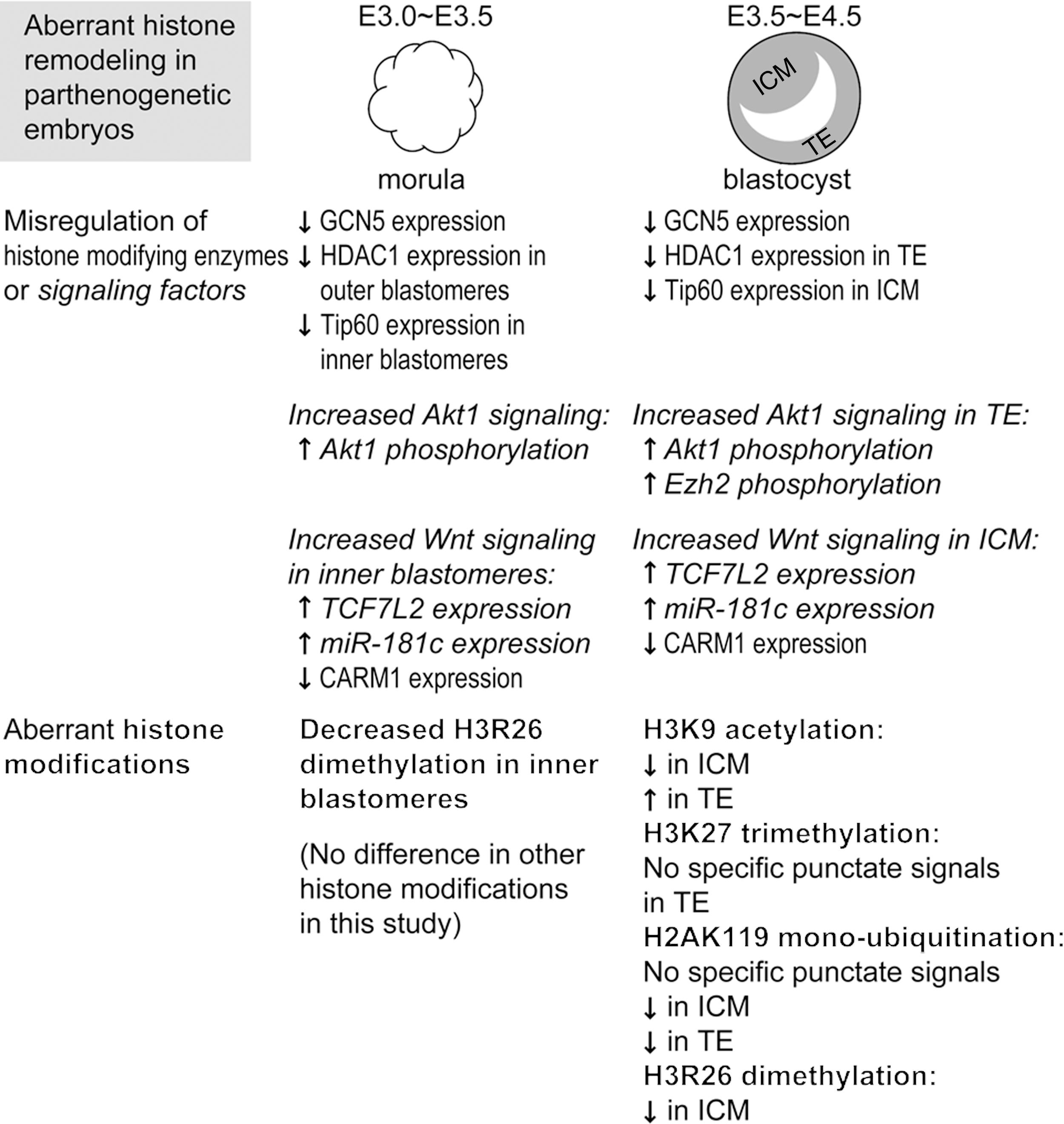
Summary of aberrant histone modifications and misregulation of histone-modifying enzymes and associated signaling factors in parthenogenetic morulae and blastocysts. Of the histone modifications analyzed in this study, only the dimethylation level of histone H3 arginine 26 (H3R26) was different between control and parthenogenetic embryos at the morula stage. At the blastocyst stage, however, levels of the following histone modifications were altered in parthenotes: H3R26 dimethylation, histone H3 lysine 9 (H3K9) acetylation, histone H3 lysine 27 (H3K27) trimethylation, and histone H2A lysine 119 (H2AK119) mono-ubiquitination. Decreased dimethylation of H3R26 in parthenotes may be due to decreased expression of CARM1 methyltransferase, which may result from the observed increase in TCF7L2 (a component of Wnt signaling) and expression of miR-181c microRNA at both the morula and blastocyst stages. Decreased trimethylation of H3K27 in parthenote TE, on the other hand, is associated with decreased mono-ubiquitination of H2AK119 and increased phosphorylation of Ezh2 methyltransferase, which may result from the observed increase in phosphorylation of Akt1 in parthenogenetic blastocysts. In addition, we found that H3K9 acetylation is decreased in parthenote ICM and increased in parthenote TE as compared with control blastocysts, whereas the underlying molecular mechanism remains to be elucidated.
Other asymmetric histone modifications in mammalian blastocysts include H3K9Ac and di-/tri-methylation of histone H3 lysine 27 (H3K27Me2/Me3) [34,35,50,62,64]. Previous studies reported that H3K9 acetylation was associated with active gene transcription [98 –101], and the expression of both Nanog and Sox2 was upregulated by H3K9 acetylation [73,102,103]. Our finding that levels of acetylated H3K9 are decreased in the parthenogenetic ICM (Fig. 1B) may be associated with reduced expression of Nanog and Sox2 [19]. The decrease of H3K9Ac in the parthenogenetic ICM may be associated with reduced expression of Tip60 acetyltransferase in the Cdx2− inner blastomeres of parthenotes (Fig. 4), whereas the increase of H3K9Ac in the parthenogenetic TE may be attributable to reduced expression of HDAC1 deacetylase in the Cdx2+ outer blastomeres of parthenotes (Fig. 3). Another histone modification, H3 lysine 14 acetylation (H3K14Ac), which has been shown to co-occur with H3K9Ac at multiple gene regulatory elements [51], did not show a significant change of pattern or level in parthenotes (Supplementary Fig. S2). Interestingly, H3K14Ac has been reported to be required for not only Nanog expression but also Oct4 expression in mouse preimplantation embryos or ES cells [95,104]. Unaffected acetylation of H3K14 in parthenotes, which may be attributable to unchanged expression of p300 [51,52] (Supplementary Fig. S3), may be associated with normal Oct4 expression in parthenotes as observed [19].
On the other hand, H3K27 methylation has been shown to be associated with the inactivation of the paternal X chromosome, as well as with promoters of pluripotency genes and poised developmental genes in TE cells of normal blastocysts [61,63,64,76 –78,105]. We found loss of specific punctate signals of H3K27 dimethylation in the TE, but not ICM, of parthenogenetic blastocysts (Fig. 5B), suggesting impaired X inactivation or aberrant expression of poised developmental genes in the parthenogenetic TE, for example, ectopic expression of Gata4 in the parthenote TE [19]. Interestingly, it was recently reported that H3K27Me3-positive signals were present in <10% of Gata4-positive hypoblast cells in in vitro-cultured human embryos [106]. In contrast to perturbed H3K9 acetylation and H3K27 trimethylation in parthenogenetic blastocysts, expression of both H3K9Ac and H3K27Me3 was normal in parthenogenetic morulae (Figs. 1A and 2A), despite the aberrant expression of lineage-specific genes Sox2, Nanog, and Gata4 [19] as well as acetylation-regulatory enzymes GCN5, HDAC1, and Tip60 in parthenotes at the morula stage. This suggests that restricted expression of lineage-specific genes may regulate the expression pattern of histone-modifying enzymes and, subsequently, contribute to the establishment of asymmetric patterns of H3K9Ac and H3K27Me3 between embryonic and extraembryonic lineages.
It has been reported that activated Akt1 phosphorylates Ezh2 methyltransferase and inhibits H3K27 trimethylation and H2AK119 ubiquitylation [43,67 –69]. We observed a dramatic increase of Ser21-phosphorylated Ezh2 and Ser473-phosphorylated Akt1 in parthenogenetic blastocysts, in association with decreased H3K27 trimethylation and H2AK119 mono-ubiquitination. It is noteworthy that increased phosphorylation of Akt1 was observed in parthenotes at the morula stage, preceding the occurrence of perturbedH3K27 trimethylation and H2AK119 mono-ubiquitination at the blastocyst stage (Fig. 5A and data not shown). In addition to Ezh2, both H3K27Me3 and H2AK119u1 are subject to regulation by the binding partner of Ezh2, the Polycomb protein embryonic ectoderm development (EED), which inhibits expression of TE lineage-specific genes, including Cdx2 and Gata3 [66]. Since Cdx2 expression is unchanged in parthenotes ([19] and this study), it is less likely that EED contributes to the perturbed pattern of H3K27Me3 in parthenogenetic TE. Another candidate of H3K27Me3-regulatory protein is KDM6B/JDJM3 (lysine-specific demethylase 6B, also known as Jumonji domain-containing protein 3) [66,107]. However, previous studies have reported that the histone demethylases JDJM3 and JDJM2C were expressed normally in parthenogenetic embryos [107,108], and depletion of these histone demethylases caused a developmental arrest before the blastocyst stage [107,108], unlike parthenotes, which could develop till the hatched blastocyst stage in vitro [19]. Therefore, KDM6B/JDJM3 demethylase may be ruled out as a misregulated candidate in parthenotes.
It is noteworthy that, although the levels of both H3K27 trimethylation and H2AK119 mono-ubiquitination significantly decreased in the parthenogenetic TE (Figs. 5B and 6A), H3K27Me3 was still present in the parthenogenetic ICM (Fig. 5B), where no specific signal of H2AK119u1 was detected (Fig. 6A). Since H3K27Me3 is directly recruited by the PRC2 and H2AK119u1 is directly recruited by PRC1, which is downstream of PRC2 [67 –69], it is plausible that perturbed expression or activity of the components of PRC1 would affect the level of H2AK119u1 but not H3K27Me3. Interestingly, recent studies have demonstrated that the histone H3 lysine 36-specific demethylase Fbxl10/Kdm2b is a pivotal interacting partner of Ring1b, a core component of PRC1, and is required for H2AK119 ubiquitylation and suppression of extra-embryonic endoderm-specific genes, including Gata4 and Gata6, in order to maintain pluripotency of ES cells [109,110]. Therefore, loss of H2AK119u1 in the parthenogenetic ICM may be associated with dramatically increased Gata4 expression as observed [19]. Future studies will be required to further elucidate whether the expression of Fbxl10/Kdm2b or Ring1b, or the interaction between these two proteins is perturbed in parthenogenetic embryos.
Footnotes
Acknowledgments
The authors thank Chien-Hong Chen for providing the parthenogenetic activation protocol and Li-Wen Lo of the Core Facility of the Genomics Research Center for expert assistance with confocal microscopy. They also thank Drs. Hung-Chih Kuo and Cheng-Fu Kao in the Institute of Organismic and Cellular Biology for their critical comments on this project and article. This work was supported by grants from 100-2321-B-001-036, 101-2321-B-182A-004, and 102-2321-B-182A-004 from National Science Council to J.Y.
Prior conference presentation of the submitted material: it was presented in the annual meetings of Taiwan Society for Stem Cell Research in 2008 and 2009.
Author Disclosure Statement
No competing financial interests exist.
References
Supplementary Material
Please find the following supplemental material available below.
For Open Access articles published under a Creative Commons License, all supplemental material carries the same license as the article it is associated with.
For non-Open Access articles published, all supplemental material carries a non-exclusive license, and permission requests for re-use of supplemental material or any part of supplemental material shall be sent directly to the copyright owner as specified in the copyright notice associated with the article.