
Abstract
Abstract
Background:
Deep pelvic abscess is a well-known infective complication in gynecologic practice. However, sacral osteomyelitis has been reported rarely. We describe sacral infection presenting three years after abdominal hysterectomy and point out the difficulty in management.
Methods:
Case report and review of the pertinent literature.
Results:
A 46-year-old woman who had undergone abdominal hysterectomy three years before presented with an 8-month history of abdominopelvic pain recently intensifying in the sitting position without fever. Gynecologic, urinary, and rectal examination did not yield positive findings. An abdominopelvic computed tomography (CT) scan was normal except for sacral osteolysis. A neoplasm was suspected, but magnetic resonance imaging revealed an S2–S4 cystic collection with presacral extension. Neurologic examination did not show any focal deficits. A posterior CT-guided biopsy–aspiration yielded purulent fluid. Pathologic examination revealed inflammatory granulations without any malignant tumor. Abscess cultures grew three microorganisms. The patient's symptoms resolved completely after 3 months of antibiotic therapy.
Conclusions:
Sacral osteomyelitis has not been reported previously after abdominal hysterectomy. Early diagnosis was made difficult by the absence of neurologic findings. Such postoperative infection should be considered after pelvic surgery. Minimally invasive needle aspiration may confirm the diagnosis and reduce the necessary extent of surgical intervention.
Case Report
A 46-year-old woman presented with an 8-month history of abdominopelvic pain recently intensified when she was in the sitting position, and with left sacroiliac pain. She denied experiencing fever, night sweats, or weight loss. Her medical history revealed an uncomplicated total abdominal hysterectomy for a leiomyomatous uterus three years previously. No evidence of gastrointestinal or gynecologic disease was present. An abdominal ultrasound scan followed by an abdominopelvic computed tomography (CT) scan was unremarkable except for S2 sacral osteolysis with intracanalar extension (Fig. 1). A malignant tumor was suspected, and the patient was referred to our institution for further evaluation and treatment.
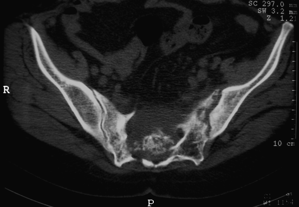
Pelvic axial computed tomography scan showing osteolytic process involving second sacral segment with intracanalar extension mimicking malignant tumor.
Her body temperature was normal. Neurologic examination did not show any focal deficits. Gynecologic, urinary, and rectal examination did not yield positive findings. Laboratory evaluation was normal except for mild inflammation (C-reactive protein [CRP] 9.1 mg/L; and erythrocyte sedimentation rate [ESR] 20 mm/h]. Bone scintigraphy demonstrated non-specific increased activity in the sacrum and left sacroiliac junction. A magnetic resonance imaging (MRI) scan revealed a large sacral fluid collection from S2 to S4, extending into the epidural space and compressing the distal thecal sac. This cystic process manifested as low signal intensity on T1-weighted images and high signal intensity on T2-weighted images with ring-like gadolinium enhancement and presacral extension (Fig. 2). A posterior CT-guided biopsy and aspiration drained about 40 mL of thick green fluid. Histologic analysis showed inflammatory granulations without any malignant tumor. Abscess culture grew Staphylococcus epidermidis, S. hominis, and Streptococcus sanguis. Tests for mycobacteria and mycosis were negative.

Sacral spinal magnetic resonance imaging. Sagittal T1-weighted image with gadolinium (
The patient received a 10-day course of intramuscular gentamicin (160 mg/day). Ciprofloxacin was given intravenously for four weeks (600 mg/day), then orally for eight weeks (1 g/day). Laboratory tests for inflammation were normal three months after treatment initiation. At the same time, the patient's symptoms had resolved completely. She remained symptom-free for 12 months of follow up.
Discussion
Sacral osteomyelitis is a rare infection even in neurosurgical and orthopedic practices [11–14,16,17]. This infection is rarely described postoperatively, but it can have serious local and systemic sequelae [7,8,18]. In gynecology, the most common procedure complicated by SOM is abdominal sacral colpopexy (five cases reported) [4,7–9]. In fact, the sacrum alone is rarely infected; it is more precisely described as lumbosacral spondylodiskitis [11,13–16]. As proposed by Taylor et al., this complication may result in a fistulous tract from the vaginal apex to the sacrum and, eventually, a sacral abscess impinging on the spinal column with diskitis and osteomyelitis [7].
On the other hand, the most common infections associated with total abdominal hysterectomy are nosocomial urinary tract infection, wound infection, and pelvic abscess [1,2,6,10]. Ahmed and Wasti reported only a 0.4% incidence of postoperative pelvic abscess in a series of 827 consecutive abdominal hysterectomies. No SOM was identified [1].
The occurrence of SOM after pelvic surgery often is a late manifestation of disseminated pathogens [7,8,18]. The insidious progression of infection, the permissive surrounding anatomic environment, and the non-specificity of the symptoms may lead to a diagnostic delay, as occurred in our patient [16,18]. This case report demonstrates the need to consider SOM in the differential diagnosis for patients with chronic pelvic or abdominal pain, regardless of a history of hysterectomy. Another unusual element of this case was the absence of neurologic deficit despite the extent of disease.
Plain radiography frequently fails to reveal the true extent of osteolysis. Axial CT scan or, preferably, MRI is necessary for full evaluation of the anatomical characteristic of the osteolysis and its extension into the surrounding structures [14,17,19]. Sacral osteomyelitis can mimic osteolytic malignant tumor, leading to mutilating surgical excision [11,17,19]. Sacral transpedicular percutaneous biopsy with aspiration is a safer minimally invasive option; it may confirm the diagnosis and limit the extent of surgical intervention [20,21]. Antibiotic therapy should be administered for at least 12 weeks and until the CRP and ESR are normalized [15].
Conclusions
Sacral osteomyelitis has not been reported previously after transabdominal hysterectomy. Such infection usually presents late in the postoperative period, and early diagnosis was made difficult by the absence of neurologic findings. Postoperative sacral infection should be considered in patients with histories of hysterectomy or other pelvic surgery to avoid unnecessary examinations and treatments. A CT-guided percutaneous aspiration is a safe minimally invasive option; it may confirm the diagnosis and limit the extent of surgical intervention.
Author Disclosure Statement
No competing financial interests exist for any of the authors.