
Abstract
Abstract
Background:
We present a case of a hydatid cyst located in the left thigh.
Case Report:
A 67-year-old man was admitted to our department with a 10 × 5 × 4 cm mass in the medial compartment of the left thigh. Computed tomography scan indicated possible hydatidosis. Serological testing (indirect hemagglutination) was positive for hydatidosis. The patient was operated seven years ago for liver hydatidosis.
Results:
The patient, after evaluation, underwent surgical excision of the cyst under epidural anesthesia. The cyst was located in the left quadriceps muscle; had a soft, elastic substance; was firmly attached to the muscle fibers; and contained transparent fluid and daughter cysts. Histologic examination confirmed the initial diagnosis. The patient was discharged on the fifth postoperative day.
Conclusions:
The diagnosis of muscular hydatidosis is difficult and the usual diagnostic methods are the serological tests for hydatidosis and imaging (e.g., ultrasound, computed tomography, and magnetic resonance imaging). In every soft tissue mass with benign characteristics the existence of a hydatid cyst should always be considered. Careful surgical excision of the intact cyst is the treatment of choice, but complementary control for liver—or other organ—hydatidosis should be performed.
Case Report
The patient was a 67-year-old man who complained of a smooth, palpable 10 × 5 × 4 cm painless growth on the medial surface of the upper third of the left thigh (Fig. 1). The patient's history included a pericystectomy for hepatic hydatidosis seven years previously and no antihelminthic treatment postoperatively. Serologic testing by indirect hemagglutination assay (enzyme-linked immunosorbent assay) was positive for hydatidosis. Ultrasound examination showed a cystic mass containing septations (Fig. 2). Abdominal computed tomography (CT) showed no evidence of intraabdominal recurrence. The CT scan of the left thigh showed a cystic mass on the upper left thigh, without calcification (Fig. 3). The patient subsequently underwent en bloc excision of the cyst under epidural anesthesia. Through a longitudinal incision on the left thigh, the cyst was removed intact (Figs. 4 and 5). The cyst was elastic and whitish in color, and the contents comprised of a blastic membrane, daughter cysts, and brood capsules in a clear, colorless fluid (Fig. 6). The operative and histologic findings confirmed the initial suspicion of hydatidosis (Echinococcus granulosus). The patient did not receive any antihelminthic treatment pre- or postoperatively, because the cyst was removed intact without any leakage or contamination of the adjacent tissues. The patient had an uneventful postoperative course and was discharged in good condition on the fifth postoperative day.
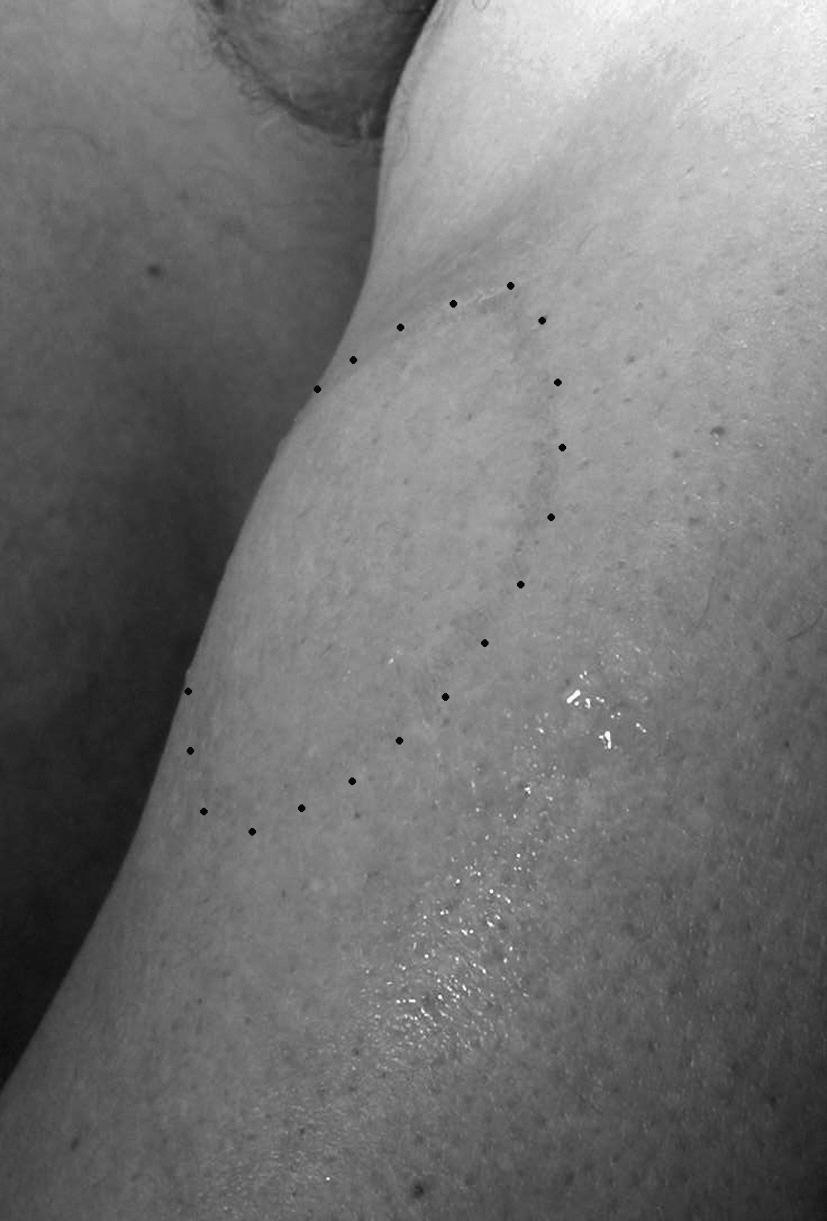
Mass in situ (marked by dots).

Longitudinal ultrasound imaging of the cyst. The arrows indicate the septations inside the cyst.
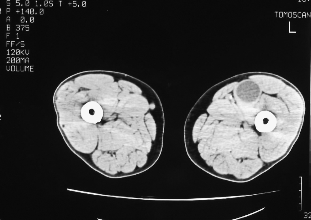
Computed tomography scan of the cyst.

Hydatid cyst in situ intraoperatively.

Hydatid cyst after excision.
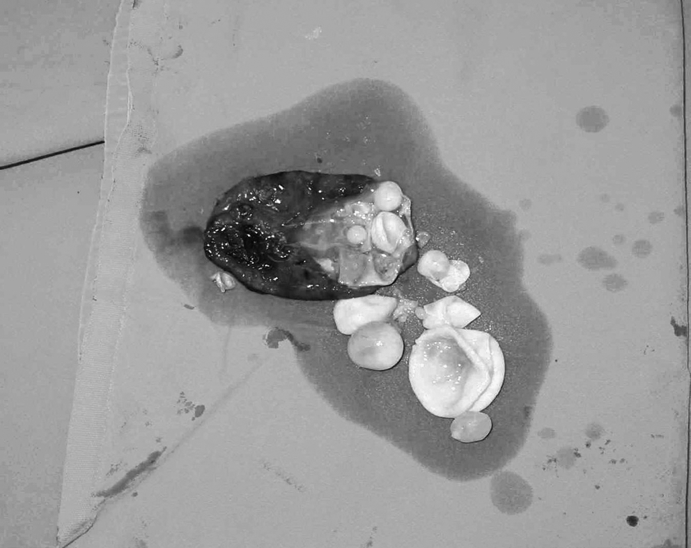
Hydatid cyst opened after excision, showing the blastic membrane, the daughter cysts, and the fluid contained within.
Discussion
The localization of hydatidosis in the soft tissues is considered uncommon, and primary hydatidosis in the skeletal muscles is exceedingly rare, with a prevalence of 4%–5.2% of all cases of hydatidosis [1–3]. Locations that have been described are the rectus abdominis muscle [4], the gluteal muscles [5], and the femoral adductor muscles [6].
The implantation of a hydatid cyst in uncommon locations is mainly primary by the lymphatic vessels of the gastrointestinal tract, the hepatic portal system, and hooked embryo migration; and rarely secondary after a hydatid cyst rupture (spontaneous or iatrogenic) [7] in other organs. In our patient, the pathway of implantation is unclear, as the anatomical site of the first operation (hepatic pericystectomy) was not related to the second site, and the abdominal CT scan showed no abnormal findings, including an intraabdominal recurrence, excluding the possibility of primary spread. Our opinion is that the femoral localization is an independent primary site.
Primary skeletal muscle hydatidosis is rare, because the muscle tissue due to its contractility and high lactate content does not favor the implantation of the hooked embryos.
The diagnosis of muscular hydatidosis is difficult, primarily because of its uncommon location and secondarily, in a hydatid cyst complication, the differential diagnosis includes soft tissue tumor (mainly cystic sarcoma), abscess, lymphatic mass, or obturator hernia [8]. For this reason, preoperative diagnosis or even a suspicion for echinococcosis is essential for the avoidance of an open or transdermal biopsy, or other operative procedures that could facilitate the spread of the disease. In our patient, the history of hepatic hydatidosis raised the possibility of the mass in question to be a hydatid cyst, which was further corroborated by serologic testing.
Clinical findings are characterized by a painless growth of variable size. Diagnosis is achieved primarily by serologic tests and imaging studies (e.g., ultrasound, CT, and magnetic resonance imaging [MRI]). In MRI, the “water-lily sign” appears where shrunk cysts appear at the roof of the mass. In our patient, the imaging studies (ultrasound and CT) were inconclusive (absence of calcification and density of fluid measurement were not helpful). The only diagnostic evidence from imaging workup was the presence of septations within the cyst by ultrasonography.
In our case, the definitive diagnosis was established intraoperatively, because the patient's history, physical examination, positive serologic testing, and imaging studies raised the suspicion of a femoral hydatid, thus avoiding a transdermal aspiration/biopsy.
Mebendazole and albendazole are the only anthelminthics effective against cystic echinococcosis. Albendazole is the drug of choice because its degree of systemic absorption and penetration into hydatid cysts is superior to that of mebendazole. Treatment with albendazole in Echinococcus granulosus infection can result in an apparent cure in as many as 30% of patients, with a further 40%–50% of patients showing objective evidence of response when observed short-term. Albendazole efficacy increases with courses of up to three months in the more common cyst sites. Continuous treatment is preferred and has been administered for periods of up to two years without significant side effects. Albendazole has been demonstrated to be a useful advance in the management of cystic echinococcosis when used as sole treatment or as an adjunct to surgery or other treatments. Praziquantel has recently been suggested, administered additionally once per week at a dose of 40 mg/kg during treatment with albendazole; however, available data are limited [9].
Despite one reported case [10] treated successfully by antihelminthic medications alone, the en bloc resection of the intact cyst remains the standard treatment [11,12]. Although the preoperative and postoperative administration of antihelminthic drugs for hepatic hydatidosis is agreed upon by everyone, it is not clear whether the same protocol should apply for muscular hydatidosis. Some authors suggest the postoperative administration of antihelminthic agents (mainly albendazole/praziquantel or mebendazole for a duration of 14 days) for possible locations elsewhere in the body that are asymptomatic [13–15]. In our case, the patient did not receive any pre- or postoperative antihelminthic treatment and, five years later, had not shown any signs of recurrence or a new primary cyst. In our opinion, the prophylactic administration of antihelminthic medications is not necessary in uncomplicated cases, if an en bloc excision of the intact cyst is achieved.
Conclusion
The diagnosis of muscular femoral echinococcosis is difficult and is usually achieved with serologic hydatidosis tests and imaging studies (e.g., ultrasound, CT, and MRI). The possibility of a hydatid cyst should be considered in any case of a soft tissue mass with benign characteristics. The surgical resection of the intact cyst is the treatment of choice, but a thorough examination for other loci is mandatory. The administration of antihelminthic agents in uncomplicated cases of muscular hydatidosis remains unclear.
Footnotes
Author Disclosure Statement
No competing financial interests exist.