
Abstract
Abstract
Background:
Modern combat- or blast-related injuries are characterized by devastatingly massive zones of injury that violate soft tissue, bone, and neurovascular structures. In our translational research program, we have determined that healing of traumatic combat wounds is dependent on the immune response. Although the majority of combat wounds are not critically colonized with bacteria, there exists a correlation between critical colonization and the concentration of inflammatory cytokines and chemokines measured in wound effluent or patient serum.
Methods:
Patients with penetrating extremity wounds sustained during combat operations were studied prospectively, being followed for 30 days after definitive wound closure. Surgical debridement was repeated every 48–72 h until wound closure at the discretion of the attending surgeon. Serum, wound effluent, and wound bed tissue biopsy were collected at each debridement. Serum and wound effluent were analyzed with a multiplex assay for cytokines, chemokines, and inflammatory proteases, whereas wound tissue was assessed for microbial colonization via quantitative cultures. Correlations between serum and effluent cytokines and chemokines and the degree of tissue colonization were evaluated.
Results:
Samples from 154 debridements in 38 wounds from 25 male patients were investigated. Many of the patients sustained multi-system trauma (mean Injury Severity Score 21±12 points) and were critically ill (mean Acute Physiology and Chronic Health Evaluation II score 7±5 points). Healing failure occurred in 23.7% of wounds. A marked inflammatory profile, including increased serum and wound effluent cytokines and chemokines, was associated with the extent of critical colonization.
Conclusions:
The correlation between systemic and local inflammatory cytokines and quantitative culture suggests that the interplay between the systemic response to injury and the local wound environment is a determinant of outcome. This relationship remains ill defined and requires further investigation in both clinical and pre-clinical studies. A biomarker panel reflective of colonization may provide clinically useful, objective criteria indicating when wound closure is appropriate for successful healing.
In our translational research program, we have determined that healing of traumatic combat wounds is dependent on the immune response and have published a series of papers describing the interplay between a combat casualty's inflammatory response to acute trauma and the clinical ability of those wounds to heal after surgical closure [1–3]. In those publications, we began to describe a dysregulated or improperly compensated immune response that leads to a prolonged pro-inflammatory state in which there is a wound microenvironment conducive to increased cellular infiltration and continued break-down, as opposed to promotion of progression toward wound remodeling and resolution. As bacterial infection of wounds has been and continues to be scrutinized heavily as a cause of healing failure and wound chronicity, we present here an analysis of the association of microbial colonization with the inflammatory response in traumatic acute combat wounds.
Although the majority of combat wounds are not colonized by bacteria, we are interested in the interplay between the systemic response to injury and the local wound environment as a determinant of outcome [4]. Herein, we identify the systemic and local markers of the inflammatory response to combat injury as associated with wound microbial colonization and begin to elicit further the relation to wound healing. Additionally, we describe several inflammatory cytokines that appear to be predictive of wound colonization.
Patients and Methods
Methodology
The study methodology was as reported elsewhere [2,3] but is reiterated here for completeness (see Utz et al. [3]). In brief, this serial, observational study with prospective data collection is in accordance with the ethical standards of the Committee on Human Experimentation as approved by the Institutional Review Board of the National Navy Medical Center (NNMC) and the Naval Medical Research Center (NMRC). Study participants were recruited from wounded U.S. service members evacuated to the National Capital Area from Iraq and Afghanistan and treated at the National Navy Medical Center (NNMC, Bethesda, MD). All service men and women who sustained penetrating injuries to one or more extremities and were without confounding co-morbid conditions, such as immune disorders, connective tissue disorders, or any conditions requiring immunosuppressive agents, were eligible for inclusion, although the final population was exclusively male. The demographic variables are shown in Table 1. Surgical debridement, pulse lavage, and negative-pressure wound treatment were repeated every 48–72 h until surgical closure or coverage, at the discretion of the attending surgeon and in accordance with current institutional standards of practice.
All patients were men.
The volume of two wounds, which showed <103 colony-forming units (cfu)/g, were not available at time of analysis.
APACHE=Acute Physiology and Chronic Health Evaluation; NNMC=National Navy Medical Center; SD=standard deviation.
Sample collection
As described previously [2,3], peripheral venous blood (8 mL) was drawn prior to each surgical debridement and collected in a Red-Top Serum BD Vacutainer® (Becton Dickinson, Franklin Lakes, NJ). Wound effluent samples (≥30 mL) were collected from the vacuum-assisted closure (VAC) canister (without gel pack; Kinetic Concepts, Inc., San Antonio, TX) 2 h after the first surgical debridement and over a 12-h period prior to each subsequent debridement. All serum samples were separated immediately using a centrifuge (Thermo-Electron Corp, Waltham, MA) at 2,500×g for 10 min. Serum supernatant liquids and effluent samples were transferred to individually labeled Cryo-Loc™ polypropylene tubes (Lake Charles Manufacturing, Lake Charles, LA), flash-frozen in liquid nitrogen, and stored at −80°C until analysis.
Cytokine, chemokine, and matrix metalloproteinase analysis
Serum and wound effluent proteins of interest were quantitated using a Luminex® 100 IS xMAP Bead Array Platform (Millipore Corp, Billerica, MA) as described previously [1–3]. Twenty-two cytokines and chemokines (interleukin [IL]-1α, -1β, -2, -3, -4, -5, -6, -7, -8, -10, -12 (p40), -12 (p70), -13, and IL-15; IP (CXCL)-10; eotaxin; interferon [IFN]-γ; granulocyte–macrophage colony-stimulating factor [GM-CSF]; monocyte chemotactic protein [MCP]-1; macrophage inflammatory protein [MIP]-1α; Regulated on Activation, Normal T-Cell Expressed and Secreted [RANTES] (CCL5); and tumor necrosis factor [TNF]α) were quantified using a Beadlyte® Human 22-Plex Multi-Cytokine Detection System (Upstate/Millipore; Cat. No. 48-011) [2]. Five matrix metalloproteinases (MMP-2, -3, -7, -9, and -13) were quantitated using R&D Systems Fluorokine® MAP Human Base Kit, MMP panel (R&D Systems, Minneapolis, MN) according to the manufacturer's instructions and as described [3].
Wound closure and followup
Daily wound examinations were performed, and all patients were followed clinically for 30 days after wound closure or coverage. The mean time to definitive closure or coverage with a skin graft for normal healing was ten days. Impaired healing included delayed closure (definitive closure occurring two standard deviations later than mean normal (>21 days) and dehiscence (spontaneous partial or complete wound disruption after primary closure or >90% skin graft loss) after closure or coverage [2,3].
Quantitative bacteriology
Quantitative cultures of wound tissues were performed as described previously [4]. During each debridement, a 1-cm3 tissue specimen was obtained from the center of the wound bed, weighed, and stored in a sterile 15-mL conical vial. The tissue specimens were transferred to a sterile, disposable tissue grinder, diluted 1:10 (wt/vol) in fastidious broth to a final concentration of 0.1 g of tissue/mL, and ground until homogenized fully. Homogenized tissue specimens (1 and 10 microliters) were inoculated in triplicate on sheep's blood agar and MacConkey plates. Plates were incubated overnight at 37°C. After incubation, colonies were counted, and the number of colony-forming units (cfu)/g of tissue was determined. Phenotypic identification of colonies was accomplished using the Phoenix™ automated bacterial identification system (Becton Dickinson, Sparks, MD).
Statistical analysis
Statistical analysis was performed using SPSS version 16.0 (SPSS, Chicago, IL). The mean concentrations of serum and effluent cytokines, chemokines, and matrix metalloproteinases were compared across degrees of tissue colonization using the Mann-Whitney U test for all biopsies in the dataset. Receiver operating characteristic (ROC) curves were generated using all biopsies by plotting sensitivity vs. 1 – specificity; area-under-the-curve (AUC) values were calculated to assess the predictive value of quantitated cytokines, chemokines, and MMPs as indicators of current critical colonization (>105 colony-form units [cfu]/g of tissue). A p value <0.05 was considered statistically significant. Data are represented as the mean±standard error unless specified otherwise.
Results
Patient and wound characteristics
Thirty-eight extremity war wounds (mean size 259±418 cm3) in 25 patients (mean age 23±5 years) were investigated through sample collection during 154 surgical debridement procedures (Table 1). One hundred seven showed no bacterial growth (<103 cfu/g of tissue) by quantitative culture. Twenty-nine biopsies (18.8%) were critically colonized (>105 cfu/g), of which 16 were from four wounds that ultimately exhibited impaired healing. The bacterial isolates identified in these biopsies were predominantly Acinetobacter (38 biopsies), with smaller numbers of Achromobacter, Escherichia coli, Staphylococcus aureus, Enterobacter, and Enterococcus (Table 2).
Some biopsies had more than one organism that was quantifiable.
Nine wounds (24%) in five patients (20%) demonstrated impaired healing. These problems included five delayed wound closures in three patients (13% of the wounds) and four wound dehiscences in two patients (10.5%). The decision to delay wound closure was secondary to clinical concerns about infection (n=3) or severe systemic illness (n=2).
Many of the patients had sustained multi-system trauma and were critically ill, with a mean (±SD) Injury Severity Score (ISS) of 21±12 and a mean Acute Physiology and Chronic Health Evaluation (APACHE) II score of 7±5 on arrival at the NNMC. A marked inflammatory profile was associated with critical colonization compared with non-detectible colonization (>105 vs. <103 cfu/g of tissue; p<0.05), as exhibited by increased serum concentrations of IL-6, IL-8, IL-10, IP-10, MIP1α, MMP-3, MMP-7, and MMP-13 and increased effluent IL-1β, IL-6, IL-8, IL-10, and MIP-1α concentrations (Fig. 1). Furthermore, the statistical difference between the critically colonized and not critically colonized wounds (>105 vs. <105 cfu/g of tissue; p<0.05) was maintained for each of these analytes except serum IP-10 and MMP-3 and effluent IL-1β, IL-6, and IL-10 (Fig. 2).
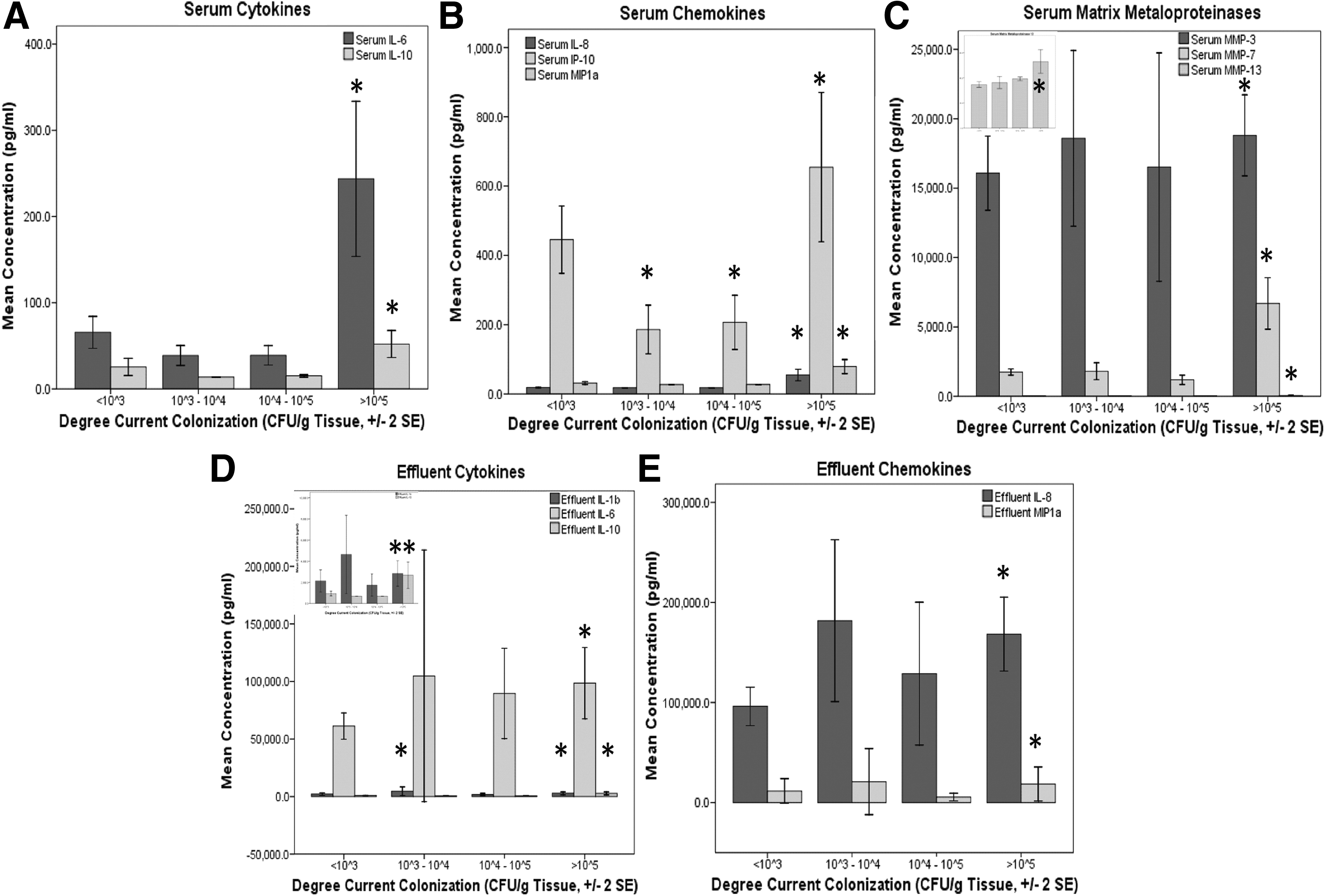
Inflammatory response as associated with current degree of colonization. Bar graphs show the mean protein amount (pg/mL) in the serum (
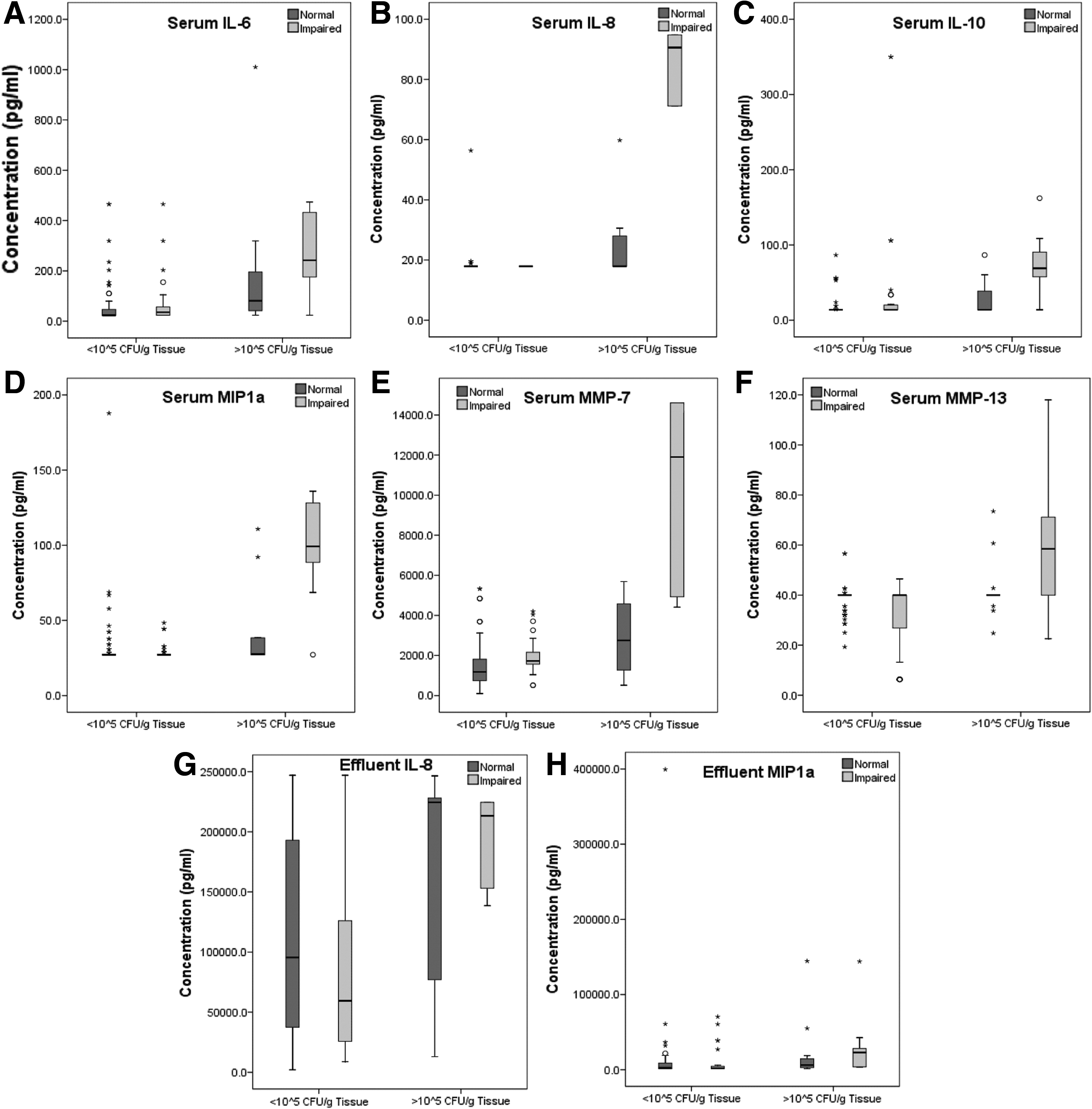
Interplay of inflammatory mediators, wound colonization, and impaired healing. Box plots show serum (
Serum and effluent biomarkers objectively predict critical colonization
Receiver operating characteristic curves were constructed for individual serum and effluent biomarkers. Serum IL-6, IL-8, IL-10, and MMP-7 and effluent IL-6, IL-8, MIP1α were predictive of critical colonization at each debridement prior to wound closure, with AUCs >0.700 (serum: AUCIL-6=0.836, AUCIL-8=0.825, AUCIL-10=0.731, AUCMMP-7=0.797; effluent: AUCIL-6=0.822, AUCIL-8=0.765, AUCMIP1α=0.777) (Table 3; Fig. 3).

Wound colonization biomarkers. Receiver operator characteristic curve for cytokines, chemokines, and matrix metalloproteinases with an area under the curve of >0.700 (see Table 3). Serum and effluent concentrations from each surgical procedure were compared with the respective quantitative culture of biopsy tissue. The positive actual state is “>105 colony-forming units/g of tissue”; i.e., a larger area under the curve indicates greater sensitivity for predicting critical colonization without an increase in false-positive results (or 1 - specificity).
IL=interleukin; MIP=macrophage inflammatory protein; MMP=matrix metalloproteinase.
Discussion
There is a long-standing belief that a direct correlation exists between bacterial counts and wound healing in traumatic (as exemplified by combat-derived injuries from the Vietnam era) and chronic [5,6] wounds. Studies have suggested limits of <106 bacteria/mL for pressure ulcer healing [5], <105 for 94% skin graft survival (which dropped to only 19% when bacterial counts were >105) [7], and, in a delayed wound closure study with use of topical antibiotics [8], <105 cfu/g for uncomplicated healing vs. failed closure if >105 cfu/g were present. The authors of that last study applied their findings in a prospective evaluation of wound closure with quantitative cultures and achieved a 96% success rate for wounds indicated for closure [9]. In this report, we extend these previous studies by linking the microbial status of the combat wound with the inflammatory and remodeling response by measurement of serum and effluent cytokines, chemokines, and proteases.
Contributing to the understanding of the intimate link between wound healing and bacterial colonization, we have associated components of the inflammatory response with high-energy, acute trauma as part of this triad. Although the current data set does not provide direct prediction of healing outcome from the quantitative bacteriology values (see Fig. 2), there is an association of the degree of tissue colonization with the quantifiable serum or effluent concentrations of IL-1β, IL-6, IL-8, IL-10, IP-10, MIP1α, MMP-3, MMP-7, and MMP-13 (see Figs. 1 and 2). Informatively, with the exception of IL-1β, IL-10, and MMP-13, each of these biomarkers is associated independently with wound outcome [2,3].
The correlation between the systemic and local inflammatory cytokines and quantitative cultures suggests that the interplay between the systemic response to injury and the local wound environment is a determinant of outcome. It is not an unreasonable hypothesis that the persistence of bacteria alters the critical balance of cytokines, chemokines, and matrix metalloproteinases (or vice versa) that coordinate the progression through the healing stages of acute wounds [10–12]. Severe trauma is capable of inducing systemic inflammatory response syndrome, in which the inflammatory process can become dysregulated further and result in remote organ failure [2,13]. Conversely, dysregulated post-injury inflammation may result in a maladaptive compensatory anti-inflammatory response syndrome with a higher risk of immunosuppression-associated infection. Acute wound failures are likely a consequence of a maladaptive inflammatory phase with failure to progress into the subsequent phases required for normal healing [14,15]. In contrast to normal healing, chronic wounds exist in a state of continual inflammation, as might be seen with frank infection or the presence of large numbers of bacteria [15]. Wound persistence, non-healing, and progression often are attributed to infection of the surface and are treated with long courses of antimicrobial agents. A wound surface can represent an ecological system of complex microbial communities interacting with the host. Surprisingly, little is known about how these interactions affect disease progression. Although studies such as these begin to examine this interplay between the host and the bioburden, the relation remains ill defined and requires further investigation in both clinical and pre-clinical studies. To that end, we are developing an animal model that combines systemic insult, local wounds, and bioburden to address such questions from a mechanistic viewpoint.
As our understanding of this response develops, a biomarker panel reflective of colonization may provide clinically useful, objective criteria indicating when wound closure is appropriate for successful healing. Additionally, and perhaps more evident here, a personalized classifier of the current degree of wound colonization based on an individual patient's inflammatory response may allow determination of appropriate antibiotic usage. The multiplex technologies and rapid quantification techniques that will enable a timely, accurate, and sensitive assay for use by today's trauma surgeons are becoming ubiquitous and familiar in modern trauma centers. It is our goal to translate basic science research in military medicine to forward deployment in military medical facilities and ultimately to utilization in the general population for the advancement of trauma care and wound healing.
Footnotes
Acknowledgments
The multidisciplinary care of these patients would not have been possible without the dedicated efforts of everyone at Walter Reed Army Medical Center (WRAMC), NNMC, and Naval Medical Research Center (NMRC). Both civilian and military personnel have rendered skilled and compassionate care for these casualties. All of our efforts are dedicated to those who have been placed in harm's way for the good of our nation. We further recognize the diligent work of Fred Gage, our clinical research coordinator, for the care he has shown for both our nation's patients and the precious samples they have provided; of Edward Utz, MD, dedicated to the mission with his collection and processing of the MMP data; and of Nancy Porterfield, BS, our research assistant with a knack for data analysis and data base curation in support of this study. We also are grateful for the dedicated support of this study by Stephanie Sincock, PhD, and Matt Kasper, PhD, of the microbiology laboratory of the NNMC.
This effort was supported in part by the U.S. Navy Bureau of Medicine and Surgery under the Medical Development Program (PE 0604771 N) and an Alpha Omega Alpha Carolyn L. Kuckein Student Research Fellowship.
We are military service members or employees of the U.S. Government. This work was prepared as part of our official duties. Title 17 U.S.C. 105 provides the “Copyright protection under this title is not available for any work of the United States Government.” Title 17 U.S.C. 101 defines a U.S. Government work as a work prepared by a military service member or employee of the U.S. Government as part of that person's official duties.
This study was approved by the National Naval Medical Center Institutional Review Board in compliance with all Federal regulations governing the protection of human subjects. The IRB Protocol number and title is NNMC.2005.0069 “The Use of Vacuum-Assisted Wound Closure Device in Treating Extremity Wounds.”
We certify that all individuals who qualify as authors have been listed; each has participated in the conception and design of this work, the analysis of the data (when applicable), the writing of the document, and the approval of the submission of this version; that the document represents valid work; that if we used information derived from another source, we obtained all necessary approvals to use it and made appropriate acknowledgements in the document; and that each author takes public responsibility for it.
None of the authors has any financial interest in the results of this study.
Author Disclosure Statement
No conflicting financial interests exist.