
Abstract
Abstract
Background:
Thyroidectomy is rarely complicated by a surgical site infection (SSI). Despite its low incidence, post-thyroidectomy SSI is especially concerning because of its proximity to vital head and neck structures and the very real potential for airway compromise and death. Severe SSIs frequently are caused by Group A Streptococcus (GAS) because of its potential for developing into necrotizing fascitis. No description of the surgical approach to a necrotizing soft-tissue infection after thyroid resection is available in the current literature.
Methods:
Case report and review of the pertinent English-language literature.
Results:
A 47-year-old male underwent a right thyroid lobectomy and isthmusectomy for a follicular neoplasm. On post-operative day 2, the patient presented to the emergency department with persistent pain, rapid onset of swelling, and airway compromise shown on computed tomography scan. Emergency incision and drainage revealed a severe soft tissue infection. Because of subsequent worsening erythema and soft-tissue swelling, the patient had to be re-explored. The infection, later identified as caused by GAS, might have been transmitted from the patient's daughter.
Conclusion:
To our knowledge, this is the first case reported of exposure to a family member with GAS pharyngitis. Successful treatment requires an appropriately high level of suspicion followed by emergent operative debridement and systemic antibiotics.
Severe SSIs frequently are associated with Group A Streptococcus (GAS) because of the ability of these infections to evolve into necrotizing fascitis. Few cases of post-thyroidectomy GAS SSI have been reported in the literature (Table 1) [6–10]. Many of these cases were complicated by septic shock and death, underlining the importance of early identification and proper treatment of GAS SSI post-thyroidectomy. No description of the surgical approach to a necrotizing soft-tissue infection after thyroid resection was identified in the current literature.
CDC=U.S. Centers for Disease Control and Prevention.
Case Report
A 47-year-old male was referred to the surgical service at the Providence Veterans Affairs (VA) Medical Center because of an incidental finding of a right thyroid nodule on a magnetic resonance imaging scan performed for back pain. A surgeon-performed ultrasound scan confirmed a 1.6-cm hypoechoic right thyroid nodule with regular borders. Ultrasound-guided fine-needle aspiration of the nodule was consistent with follicular neoplasm. The contralateral lobe was normal, and the operative plan was to perform a right thyroid lobectomy and isthmusectomy.
His medical history included bipolar disorder, hyperlipidemia, previous alcohol dependence, osteoarthritis, prior tibia fracture, and attention-deficit hyperactivity disorder. The surgical history included laparoscopic cholecystectomy, left rotator cuff repair, remote incision and drainage of a right buttock abscess, and right knee arthroscopy with partial medial meniscectomy. Medications consisted of valproic acid, quetiapine, fluoxetine, simvastatin, and omeprazole. The patient denied recent smoking or alcohol use.
A right thyroid lobectomy and isthmusectomy was performed with standard sterile technique. The skin was prepared with 2% chlorhexidine gluconate and 70% isopropyl alcohol (ChloraPrep®; CareFusion, San Diego, CA), covering the area from the chin to the nipples and between the shoulders. No pre-operative antibiotics were administered. A 4-cm Kocher incision was made in a natural skin crease. Bovie cautery was used to divide the platysma and subsequently to create subplatysmal flaps. The thyroid vessels were divided with the Ethicon Harmonic Scalpel (SomaTechnology, Bloomfield, CT) or ligated with silk ties. The isthmus was divided at its junction with the left lobe using the harmonic scalpel. The strap muscles and platysma were closed with interrupted 3-0 polyglactin 910 (Vicryl) suture (Ethicon, Inc., a division of Johnson & Johnson, Piscataway, NJ), and the skin was closed with Dermabond® (Ethicon) prior to extubation. Typically, drains are not placed during routine thyroidectomies, and there was nothing about this case that warranted drainage. The operation was uncomplicated. There were no identified breaches in sterile technique. The estimated blood loss was 10 mL, and he received 1,500 mL of lactated Ringer's solution.
On post-operative Day 0, the patient reported moderate pain at the incision site and moderate odynophagia. Examination revealed no erythema, swelling, or hoarseness. The patient was given nothing by mouth overnight. The next morning, his pain and odynophagia had improved. He tolerated a solid diet and was discharged home. Physical examination at the time of discharge revealed mild swelling at the incision site with minimal erythema, which were considered normal postoperative findings. His temperature was 98.7°F, pulse 91 bpm, and blood pressure 117/78 mm Hg. His white blood cell (WBC) count of 17,000/mm3 was attributed to post-operative leukocytosis.
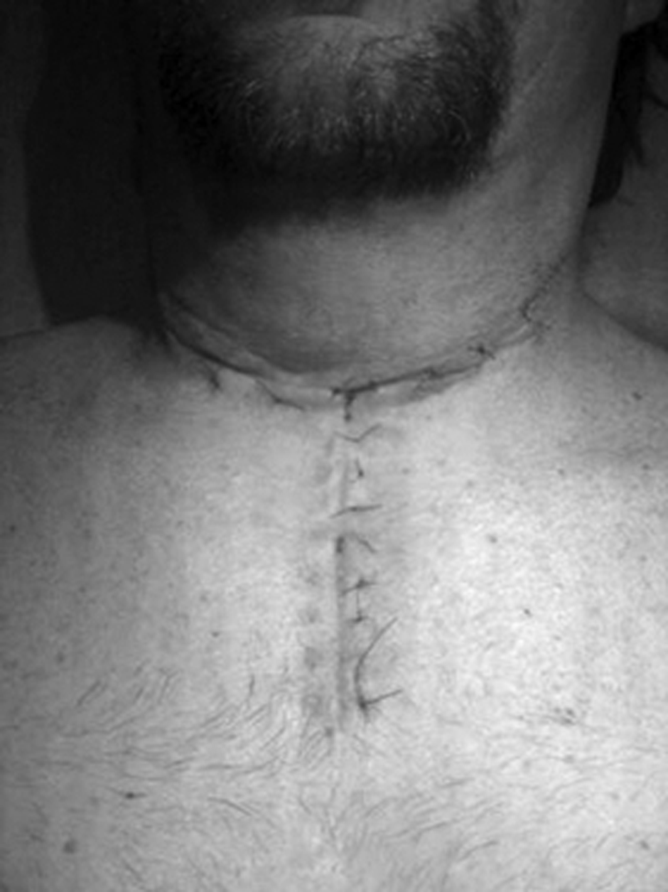
At 9:30

Appearance of surgical site on post-operative day 2. Black line designates erythemutous indurated region.
A computed tomography (CT) scan of the neck was ordered prior to the notification of the surgery team, and it revealed diffuse soft-tissue swelling with fluid accumulation and gas tracking along the superficial and deep layers of the cervical fascia bilaterally. There was substantial airway compromise (Fig. 2).

Computed tomography scan on post-operative day 2.
The patient was taken to the operating room (OR) urgently to establish a secure airway in a controlled environment and for wound exploration. He was intubated without difficulty in the OR. The original incision was opened and extended laterally on both sides. Exploration revealed copious amounts of thin odorless fluid tracking along the previously dissected muscle planes. The muscle tissues were pale, and viability was uncertain. All Vicryl sutures were removed, and the strap muscles were separated. Murky fluid was drained from the tissue planes between the strap muscles and the sternocleidomastoid, as well as the paratracheal regions bilaterally. There was no extension into the mediastinum. Necrotic paratracheal tissue was debrided from the dissected right thyroid bed. There was no evidence of tracheoesophageal injury. Esophagoscopy and direct laryngoscopy revealed no evidence of mucosal injury in either structure. Penrose drains were placed bilaterally in the paratracheal spaces and brought through the incision. The medial portion of the incision was left open and packed with gauze. The lateral portions were closed loosely with 3-0 nylon suture.
Intraoperatively, the patient was hypotensive and required intravenous phenylephrine, which was withdrawn quickly post-operatively. A gram stain of the fluid showed gram-positive cocci and numerous polymononuclear leukocytes.
Post-operatively, the patient was admitted to the surgical intensive care unit. Empiric vancomycin and piperacillin-tazobactam were started; 24 h later, cultures yielded beta-hemolytic GAS and coagulase-negative Staphylococcus, and the antibiotics were changed to penicillin, clindamycin, and vancomycin.
The day after initial debridement, the maximum temperature was 100.2°F, the pulse ranged from 80 to 110 bpm, and the blood pressure ranged from 80 to 120s/60 to 70s mm Hg. Urine output was adequate with aggressive fluid resuscitation. His serum lactate concentration had decreased to 2.6 mg/dL. The WBC count was 22,200/mm3 with 53% bands and 42% neutrophils. The creatinine concentration had normalized to 0.96 mg/dL.
Unfortunately, the patient's neck erythema had spread circumferentially, and the soft-tissue swelling worsened. Because of heightened concern about untreated necrotizing fasciitis, he was returned to the OR for re-exploration. Examination showed no crepitus. A head and neck ultrasound scan was performed under anesthesia to rule out undrained collections. It demonstrated severely edematous tissue without obvious fluid collections. The Kocher incision was extended in a cruciate fashion onto the sternum to evaluate the viability of the pectoralis muscles and assess for undrained fluid (Fig. 3). There was excellent tissue perfusion, no wound tracking, and no undrained collections. All muscles and soft tissues were viable. The wound was irrigated copiously and packed open.

Postoperative day 4. Extended open unpacked incision.
Antibiotic coverage was narrowed to high-dose penicillin. The patient was extubated without difficulty the following day, continued to improve, and eventually was discharged from the hospital on intravenous penicillin with a vacuum-assisted dressing in place.
At 22 days post-thyroidectomy, the patient's swelling and erythema had nearly resolved, and a delayed primary closure was performed. The following day, he developed obvious cellulitis. Vancomycin and high-dose penicillin were started immediately, and over the next 24 h, there was noticeable improvement. Over the next few days, the swelling abated, and he was discharged on two weeks of intravenous vancomycin. His incision continued to heal well, and he returned to normal activity (Fig. 4).
Healed incision weeks after surgery.
The potential cause of such a life-threatening post-operative infection in an otherwise-routine thyroidectomy was investigated. No health care workers involved in his care were known to have, or had symptoms of, streptococcal infections. However, his daughter had been found to have streptococcal pharyngitis within the week prior to his original thyroidectomy. In fact, antibiotics were begun within 24 h of her symptoms. The patient himself had no symptoms of a pharyngeal GAS infection and denied sore throat prior to surgery. No throat cultures were obtained from the patient, family, or involved health care workers. Of note, the patient's preoperative nares screening culture was negative for methicillin-resistant Staphylococcus aureus (MRSA).
Discussion
This patient's course was complicated by a severe soft-tissue GAS infection with sepsis. GAS causes approximately 9,000–11,500 invasive soft-tissue infections per year in the United States, and rates have been stable for the last seven years [11]. GAS is isolated from 3% of post-partum infections and 1% of surgical-site infections [7]. Although rare, the severity of such infections requires heightened suspicion and a low threshold to begin appropriate antibiotics and debride infected tissue aggressively.
As is routine in our institution, pre-operative antibiotics were not given to this patient. Current guidelines cite no benefit for prophylactic antibiotics in clean surgery such as thyroidectomy, for which the rate of infection is<5% [12]. In a trial of prophylactic antibiotics in thyroidectomy for goiter or thyroid carcinoma, 500 patients were randomized to either 3 g of ampicillin-sulbactam 30 min before surgery or no pre-operative antibiotic. Of the three patients who developed an SSI, two had received prophylactic antibiotic [13].
Some surgeons do use prophylactic antibiotics for thyroidectomy. An Internet-based survey of all members of the American and International Associations for Endocrine Surgeons showed that of 275 endocrine surgeons who responded, 62% “almost never” used prophylactic antibiotics, 26.2% administered prophylactic antibiotic “almost always,” and 11.4% reported that they varied their prophylactic antibiotic use [14]. In a retrospective study of 14,934 thyroid operations in Italy, 50% of the surgeons administered antibiotic prophylaxis, 17% provided “antibiotic therapy,” and 33% did not use any antibiotics; the incidence of infection was not significantly different in these groups [1].
Group A Streptococcus frequently colonizes various sites in the body, including the skin, pharynx, vagina, and anus, asymptomatically [13]. Rates of asymptomatic GAS colonization in adults range from 2% to 8% [15,16]. Our patient was tested for MRSA carriage in his nares, which was negative. However, he was not tested for GAS carriage, as this is not routine practice.
The patient had a family member with recent streptococcal pharyngeal infection. Although his daughter was treated properly in the week prior to his surgery, this exposure may have led to subsequent pharyngeal GAS carriage in our patient. Pharyngeal carriage by either the patient or a healthcare team member is a possible source of SSI. Other characteristics that increase the risk of invasive GAS soft-tissue infections are diabetes mellitus, alcohol abuse, cancer, and cardiac or pulmonary disease [17].
The U.S. Centers for Disease Control and Prevention (CDC) recommends that when a patient has tested positive for a GAS SSI, other possible cases within the same healthcare institution be sought diligently [8]. Because case reports describe the clustering of GAS SSIs, early identification of similar cases can greatly decrease morbidity and mortality rates. Linkage of multiple infections can be confirmed by strain testing using emm genotyping for M protein, serologic, or other molecular methods [8]. In our case, there were no other GAS SSIs at our institution that were temporally related to this case.
The testing of involved health care workers (HCW) is reasonable when unexplained infections occur and is recommended by the CDC [8]. In a report of two cases of post-thyroidectomy GAS SSI and one case of post-parathyroidectomy GAS SSI within seven days at the same institution in California, 1 of 41 tested HCWs was GAS positive. However, the orderly who tested positive for GAS carriage was colonized with a different strain and could not be linked to the SSIs. One surgeon who was present in all three cases initiated voluntary prophylactic antibiotic treatment before testing could be administered [8]. In two other post-thryoidectomy GAS SSI cases in France, no HCW was found to carry GAS [7]. The inability to identify the source of infection among surgical staff in this report may reflect the fact that cultures were obtained only from the throat of involved staff; however, GAS carriage on the rectum has been linked to SSI, and rectal or vaginal carriage by the surgical or anesthesia staff may not have been ruled out in these cases.
There have not been any cases reported in the literature citing exposure to a family member with GAS infection as a source of GAS SSI. The severity of GAS SSIs evident in case reports such as ours illustrates the need for prompt identification and treatment of such infections. It would not be difficult to ask patients about pharyngitis or recent GAS exposure before head and neck endocrine surgery. If a positive response is elicited, it should be followed up by pharyngeal culture for GAS or with prophylactic antibiotics. Physicians should be aware of the signs of severe GAS infection, which include rapidly increasing severe pain and swelling, fever, erythema, crepitus, ecchymosis, drainage, and exquisite tenderness [11]. Any suspected invasive GAS infection should be treated with high-dose penicillin and clindamycin [11]. Immediate and aggressive debridement is mandatory, as failure to pursue aggressive surgical treatment may result in the patient's death. The greatest challenge for the treatment of these infections remains the rapid progression and the need for quicker recognition and diagnosis by surgeons.
This case report illustrates the importance of surgeon–patient communication in the peri-operative setting. Previous studies have shown difficulty in improving patients' knowledge and recall of SSI symptoms, even with take-home educational documents [18]. Potentially dangerous home exposures, such as pharyngeal GAS infection, should be explained pre-operatively and post-operatively. Patients should be able to recognize symptoms of SSI compared with normal wound appearance and healing. As in this case report, patients should be encouraged to return to care immediately for evaluation of any signs of infection. Further research should be directed at efficient and effective dissemination of this information to patients with the ultimate goal of decreasing SSI morbidity and mortality rates.
Footnotes
Author Disclosure Statement
The authors have no conflicts of interest to disclose.