
Abstract
Abstract
Background:
Phlegmonous gastritis is a rare and highly lethal primary bacterial infection of the stomach. The pathogenesis of this disease is understood poorly and no detailed description of its associated findings on computed tomography has been reported.
Methods:
Case report and literature review.
Case Report:
The authors describe an 84-year-old male with phlegmonous gastritis presenting as an abdominal catastrophe with portal venous pneumatosis observed on computed tomography.
Conclusion:
The association of portal venous air and related computed tomographic findings suggesting compromise of the gastric wall should be regarded with suspicion, and the possibility of phlegmonous gastritis should be entertained. Broad-spectrum antibiotic coverage should be instituted. Gram stain of the tissues of the stomach wall may help direct antibiotic therapy toward streptococcal infections as opposed to polymicrobial processes.
Case History
An 84-year-old male presented to the emergency department complaining of intermittent nausea and vomiting over the preceding month, followed by severe nausea and vomiting over the preceding 3 d. He began to experience retching and hematemesis with upper abdominal pain on the day of presentation. Past medical history was remarkable for colon cancer treated by partial colectomy more than five years before his presentation. He denied any history of peptic ulcer disease but reported a history of hypertension, asthma, chronic obstructive pulmonary disease, mild chronic kidney disease, and prostate cancer treated with radiation therapy. He denied a history of myocardial infarction and of congestive heart failure. His surgical history was further remarkable for a ventral hernia repair after his colectomy. The patient's medications included atenolol, diltiazem, aspirin, and rosuvastatin. He took lorazepam and sildenafil as needed. He was allergic to ciprofloxacin. He lived at home with his wife and was independent and highly functional at baseline.
On physical examination, and patient was confused and appeared acutely ill. His temperature was 98.3°F and his heart rate was 90 beats/min and regular. His blood pressure was 116/68 mm Hg, respirations 16 breaths/min, and oxygen saturation 95% on 28% O2. The patient's lungs were clear. His abdomen showed moderate tenderness in the left upper quadrant, without involuntary guarding or rebound. A nasogastric tube was placed and drained grossly bloody fluid. The patient's extremities were warm and without edema.
Laboratory studies were remarkable for a white blood cell count of 26.1×10³/mcL with 94% neutrophils. The serum lactate concentration was 1.4 mmol/L. Because the serum creatinine concentration was 1.8 mg/dL, computed tomography (CT) of the abdomen and pelvis was done without intravenous contrast. This showed evidence of subserosal and intramural gastric pneumatosis at the lateral fundus. There was extensive portal venous air (Fig. 1), which was described by the radiology service as suggesting air dissecting through a Mallory–Weiss tear, with possible extension to contiguous veins. There was also edema and a question of mucosal air along the lesser curve. There was radiologic suggestion of an ileus but no evidence of intra-peritoneal free air or fluid. Although the patient's clinical symptoms appeared relatively benign, the suggestion of a gastro-esophageal perforation, along with the presence of portal venous air on CT and his marked leukocytosis, was believed to mandate exploratory laparotomy. He was therefore taken emergently to the operating room.
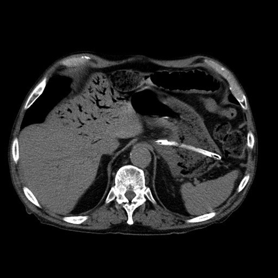
Computed tomographic image of a transverse section of the abdomen showing portal venous air.
At operation there was no evidence of diffuse peritonitis. The small and large intestines appeared unremarkable except for the prior colectomy. The anterior stomach appeared normal but there was extensive edema within the hepato-gastric ligament and in the fat pad over the esophageal hiatus. There was no overt evidence of perforation. The lesser sac was opened and the posterior stomach was noted to have a bluish appearance suggestive of possible venous congestion from ischemia. The stomach was therefore explored through a long anterior gastrotomy to inspect for ulcers or other evidence of perforation. Exploration revealed a large portion of the gastric mucosa to be friable and necrotic-appearing (Fig. 2A). On bi-manual palpation, the gastric wall of the entire upper one-half of the stomach was felt to be thinned, and the only viable mucosa in the upper stomach appeared to be a 1–2-cm cuff around the esophagus. A near-total gastrectomy was performed using a Roux-en-Y reconstruction to the gastro-esophageal cuff. A feeding jejunostomy tube was also placed.

Direct views of the diseased stomach. (
On gross pathologic examination the serosa of the stomach was smooth and glistening except on the posterior aspect proximally, where it was dusky and erythematous (Fig. 2B). A 19×9 cm area of the posterior stomach wall proximally was dusky, friable, erythematous, and grossly necrotic-appearing, with fibrinous debris, ulceration, and necrosis (Fig. 2C). The remainder of the stomach mucosa was pink and grossly unremarkable, and no other lesions were identified.
Microscopic sections revealed areas of ulcerations and fibrinopurulent exudates, although most of the mucosa was intact and viable. The submucosal layer was thickened and edematous, and there was conspicuous neutrophil-rich transmural acute and chronic inflammation, with patchy necrosis of the stomach wall (Fig. 3). In addition, polymicrobial bacterial aggregates were present, with tissue Gram stain revealing both gram-positive and gram-negative organisms (Fig. 4).
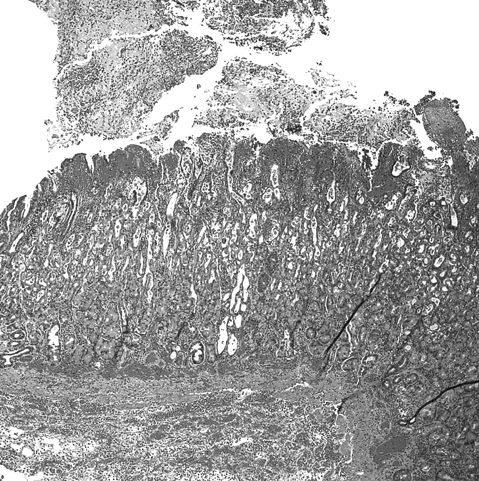
Histologic sections of the stomach demonstrate oxyntic mucosa with overlying fibrinopurulent exudate. The mucosa itself is intact, and there is transmural acute and chronic inflammation, as well as hemorrhage (hematoxylin and eosin stain, ×100).

Within the gastric muscular layer, polymicrobial transmural aggregates of mixed bacterial populations are present. These are composed of cocci and bacilli, including both gram-positive and gram-negative bacteria (tissue gram stain, ×400).
The patient's subsequent hospital course was unremarkable. Postoperatively, he was admitted to a surgical intensive care unit. He was given an empiric course of piperacillin–tazobactam because of concern for his portal pylephlebitis. His leukocytosis resolved rapidly and he was extubated on post-operative day 1. His renal function improved progressively. He was never oliguric and his serum creatinine concentration returned rapidly to within the normal range. His atrial fibrillation was treated with a diltiazem infusion and resolved eventually. Feedings were initiated through his jejunostomy tube and he was transferred to a surgical floor. The patient was discharged to a rehabilitation facility on post-operative day 12. He progressed to a post-gastrectomy diet and the jejunostomy tube was removed two months after his surgery.
Discussion
Phlegmonous gastritis can present as a localized or a diffuse infection of the stomach. Review of the available reports suggest that it is caused by Streptococcus species alone in about two thirds of cases and is polymicrobial in the remaining one third. Reported isolates include Staphylococcus, Escherichia coli, Haemophilus influenzae, Proteus, and Clostridia [1]. From that perspective, phlegmonous gastritis may be considered to behave as a necrotizing mucositis of the stomach. Predisposing risk factors are thought to include mucosal injury, alcoholism, achlorhydria, debilitation, and immune compromise, although none of those factors were present in this case.
Hepatic portal venous gas (HPVG) was first reported in 1955 as subcapsular radiolucencies seen in the peripheral liver of neonates dying after abdominal catastrophes [3,4]. Such gas can be noted on many types of imaging including plain radiography, ultrasonography, and CT. But with the increased use of CT the incidence of HPVG as a finding has increased in many conditions. In adults, HPVG has been associated with a variety of sources of intra-abdominal pathology. The most commonly associated pathology is ischemic enterocolitis, which may be responsible for up to 75% of cases of HPVG in some series [3].
The etiology of HPVG is also understood poorly. It may stem from gas-forming organisms that escape the gut lumen to invade the bowel wall. This appears to have been what happened in the case described here, with the process being a polymicrobial necrotizing mucositis rather than the more common streptococcal mucositis. On the other hand, HPVG may also result from a non-infectious dissection of air through a mucosal injury, with this latter process typically being a benign finding. Thus, although portal venous air must always be considered a potentially ominous finding, the mortality attributed to it varies widely. Known associated infective pathologies include ischemic enterocolitis, diverticulitis, and other intra-abdominal suppurative diseases.
Notwithstanding its reputation as a “bad actor,” HPVG has also been seen as a benign finding in pediatric pyloric stenosis [5,6]. There have also been reports of benign isolated HPVG after blunt abdominal trauma that did not require operation [7]. Portal venous gas can be seen immediately after liver transplantation and after chemotherapy [8,9]. There has also been a report of gastric pneumatosis with associated HPVG that developed in a young woman with neurofibromatosis immediately after a cesarean section. That patient was successfully treated non-operatively with broad-spectrum antibiotics and nasogastric decompression [10].
The severity of conditions associated with HPVG therefore ranges from rapidly lethal abdominal disasters to benign post-procedural findings. Since the prognosis and need for surgery in HVPG relates to its source and the overall health of the patient, rather than to any direct effect of portal venous gas per se, evidence-based guidelines have now been created proposing management of HPVG that focuses on treatment of the underlying pathology [11].
Conclusion
We describe a case of phlegmonous gastritis presenting as an acute abdominal emergency with portal venous pneumatosis seen on CT, in association with signs of gastric mucosal compromise. The patient recovered fully after gastrectomy and broad-spectrum antibiotic therapy. The presence of portal venous air and related CT findings suggesting compromise of the gastric wall should be regarded with suspicion, and the possibility of phlegmonous gastritis should be entertained. Broad-spectrum antibiotic coverage should be instituted much as one might for a necrotizing fasciitis. Gram stain of the tissues of the stomach wall may help in directing antibiotic therapy toward streptococcal infections as opposed to polymicrobial processes. In addition, when operating in the presence of portal venous gas, subtle findings of edema or congestion in the perigastric tissues should suggest consideration of exploratory gastrotomy to evaluate the gastric mucosa visually for suppuration, ulceration, and necrosis.
Footnotes
Author Disclosure Statement
No competing financial interests exist.