
Abstract
Abstract
Background:
Numerous studies have described various complications after endoprosthetic reconstructive operations. However, there are limited reports that focus specifically on deep infections (e.g., deep incisional surgical site infections), which remain one of the most dreaded complications of these operations, with rates ranging from 10% to 17%. Thus, this study was undertaken to determine the deep infection rates and to analyze possible risk factors, clinico-pathologic characteristics, and treatment modalities of endoprosthetic infections.
Methods:
We reviewed retrospectively the records of 105 consecutive patients who underwent endoprosthesis replacements from January 2007 to September 2011, with a minimal follow-up period of 32 mo. Comparison was made between patients with and without endoprosthetic infections.
Results:
Thirteen of the 150 patients (12.38%) who underwent endoprosthetic operations developed deep infections. Ninety-seven (92.4%) patients presented with a primary bone/soft tissue tumor, 5 (4.8%) with bone metastasis, and 3 (2.9%) with non-tumor conditions. Distal femoral was the most common implant location (42%). The majority of the infections (6/13) occurred within 3 mo post-operation. An elevated C-reactive protein concentration or erythrocyte sedimentation rate were present consistently in all patients at time of diagnosis, whereas clinical presentations and leukocytosis were inconsistent in determining infection. Staphylococcus aureus and coagulase-negative staphylococci (CoNS) were the most common organisms isolated, with high numbers demonstrating methicillin-resistance. Overall, multi-drug resistance was noted in 52.6% of the isolated strains. Four risk factors were associated independently with deep infection by multivariable analysis (p<0.05) and these were proximal tibia endoprosthesis, pelvic endoprosthesis, pre-operative duration of hospitalization of more than 48 h, and additional surgical procedures performed after the initial endoprosthetic insertion. Overall, infection was eradicated successfully in 53.8% (7/13) of the patients. Two-stage revision successfully treated the infection in 80% (4/5) of the patients, whereas surgical debridement without a change of implant was successful in only 42.8% (3/7) of patients. Amputation was performed in three patients.
Conclusions:
Patients undergoing endoprosthetic replacement for various orthopedic oncologic conditions have high infection rates. The present study allows early identification of such patients in view of the high morbidity associated with this condition. This report also highlights the high rate of multi-drug–resistant infections, especially methicillin-resistant strains of S. aureus and CoNS encountered, which complicates further the management of these patients.
I
Endoprosthesis offers various advantages over other reconstructive methods. They are readily available, reliable, and durable; allow rapid restoration of weight-bearing function and can be sized appropriately to the patient [3,4]. Although the benefits are indisputable, implantation of these prostheses carries the risk of various complications such as infection, structural breakage, soft tissue failure, and aseptic loosening, which may ultimately result in implant failure [2,5]. Deep infection (e.g., deep incisional surgical site infection) remains one of the most devastating complications in endoprosthetic reconstruction, with reported rates ranging from 10% to 17% [2,4,6,7]. The lengthy and complex operations compounded with the immunocompromised status of patients favor the higher rates [4,6]. These high rates are in contrast with other implant-related orthopedic infections that report lower infection rates ranging from 0.39% to 0.98% [8,9]. Endoprosthetic infections have various health-related and socioeconomic repercussions on patients that include the need for multiple surgical procedures, a long period of disability, long-term hospitalization, weeks-to-months of intravenous antibiotics, a delay in adjuvant therapy, and an escalating treatment cost to the patient and health care system [4,6,10]. Unfortunately, 19% to 51% of patients with deep infections may ultimately result in limb salvage failure or amputation [2,6,11].
Whereas complications after endoprosthetic reconstructions have been described extensively, there are few studies that specifically focus on deep infections involving tumor endoprostheses. Furthermore, reports have not been well characterized on the clinical presentations at the outset of these infections, microbiologic culture results, treatment modalities, and use of antibiotics that influence the outcome of these infections.
We review all patients who underwent endoprosthetic reconstructions to determine the resulting deep infection rates and to identify the risk factors for these infections. We also report our experience with the causative organisms, clinical characteristics, and treatment modalities of these patients.
Patients and Methods
We performed a retrospective study of all patients who underwent endoprosthetic reconstructions over a period of 57 mo, from January 2007 to September 2011. All of the operations were performed at a single Orthopaedic Oncology Center at University Malaya Medical Center, which serves as the tertiary referral center for orthopedic bone and soft tissue tumors. The study was approved by the University Malaya Medical Center (UMMC) Medical Ethics Committee (MEC ID No: 201402-0752). Informed consent was not obtained from the patients because this was a retrospective study.
All the endoprosthetic reconstructions were performed by the same orthopedic surgical oncologist throughout the study period. Generally, two types of endoprosthesis were used in these patients: Modular endoprosthesis (Fig. 1) for adult patients and growing or lengthening prosthesis (Fig. 2) for children who were still growing. Limb-lengthening procedures are done when there is a limb length discrepancy of more than 1 cm. The limb is lengthened to a maximum of 1 cm each time, to prevent damage to the neurovascular structures because of stretching. The growing prosthesis is lengthened mechanically with a screwdriver, under strict aseptic techniques in the operating theater under image intensifier control.
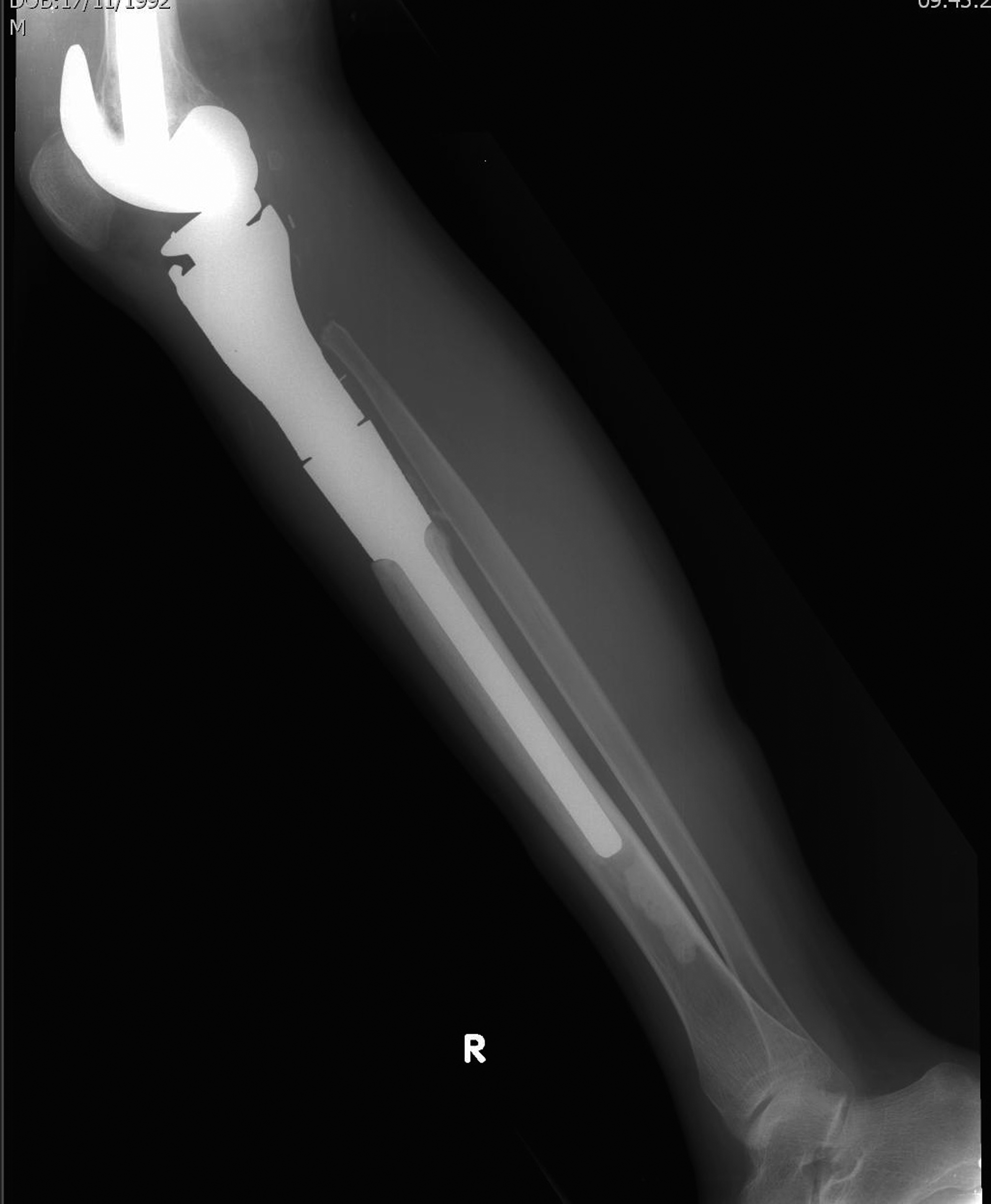
Radiograph of modular proximal tibia endoprosthesis.
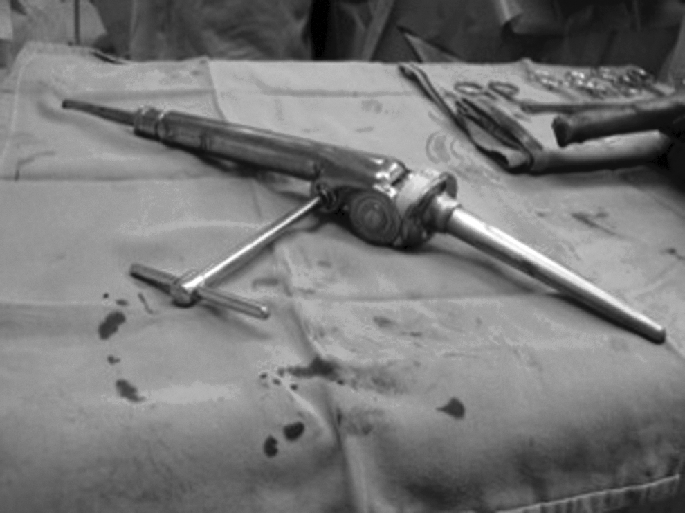
A distal femur-growing prosthesis with the lengthening screwdriver in situ.
Data were collected by reviewing the surgical case notes, histopathology reports, microbiology, and hematology laboratory results. Patients were followed up for at least 32 mo, except in cases in which death occurred before this period. On the basis of the institutional protocol, all patients were given prophylactic intravenous cefuroxime 30 min before incision and this was continued for 5 d post-operatively. Patients who were allergic to cefuroxime were given intravenous vancomycin. The pre-operative skin preparation agents used were 10% povidone-iodine and 70% alcohol. Drainage tubes were used routinely and remained in place for an average of 5 d post-operation or until the drainage was less than 50 mL/d. Pre-operatively, all patients had normal leukocyte counts with no clinical evidence of infection.
Data retrieved included demographic data, histopathologic diagnosis, site and origin of tumor (primary or metastasis), concurrent non-neoplastic medical comorbidities, administration of chemotherapy or radiotherapy, American Society of Anesthesiologists (ASA) score, type of excision, type and site of prosthesis, pre-operative length of hospitalization before operation, length of operation, estimated intra-operative blood loss during operation, and blood products transfused intra-operatively.
If a patient developed endoprosthetic infection, the time frame between endoprosthetic insertion and development of deep infection was recorded. When additional operations were performed after the initial operation, the interval between the additional operations and deep infection and the types of additional operations involved was determined. Other variables determined were the initial clinical presentations, inflammatory markers (erythrocyte sedimentation rate [ESR] and C-reactive protein concentration [CRP]), leukocyte counts at the time of infection, microbiologic culture results, types and number of surgical interventions required to control infection, antibiotics administered, and the final outcome of the infected endoprosthesis.
Comparison was made between patients with and without endoprosthetic infections. Data were analyzed using the Statistical Package for Social Sciences (SPSS version 20.0, IBM, Armonk, NY). The χ2 test or Fisher exact test and Student t test or Mann-Whitney U test were used to compare categorical and continuous variables between the two groups, respectively. The odds ratio (OR) and its 95% confidence intervals (CI) were calculated. The p value<0.05 (two-tailed) was taken as the level of significance. Those predictors that were statistically significant were entered together into stepwise logistic regression model, to evaluate independent risk factors for infection.
Definition
Deep infection was diagnosed based on the U.S. Centers for Disease Control and Prevention (CDC) guidelines [12] with the following modifications: Organ-space infections and deep incisional infections were managed together as “deep infections” and infections occurring more than 12 mo after the initial tumor endoprosthesis operation were also considered [6,7,11]. All tissue samples for microbiologic cultures were taken intra-operatively and were obtained close to the surface of the prosthesis and from contiguous inflamed tissue. We excluded patients with superficial incisional surgical site infections who did not progress to deep infections. The duration of the operation was defined as the time between incision and closure. Persistent infection was defined as the failure to eradicate the infection completely, as suggested by persistent clinical and laboratory evidence of infection necessitating repeated courses of antibiotics. Infections were categorized based on the interval between the initial operation and development of infection, using the classification proposed by Coventry [13] and refined by Fitzgerald et al. [14]: early (less than 3 mo after operation), delayed (3 to 24 mo after operation), or late (more than 24 mo after operation). Prolonged operation was defined as operation lasting longer than 2.5 h [8]. As described by Hardes et al. [10], we regarded infections as cleared based on the absence of clinical evidence of inflammation and normalization of CRP with the additional criteria of negative microbiologic cultures.
Results
A total of 105 patients underwent endoprosthetic reconstructive operations after tumor excision and were followed up for a minimum period of 32 mo. Six patients died before this follow-up period (range, 8–28 mo); five died of advanced disease and one succumbed to upper gastrointestinal bleed.
Patient characteristics
There were 59 (56.2%) male and 46 (43.8%) female patients with a median age of 25 y (range, 7–89 y). A majority of the patients were Malays (51.4%) followed by Chinese (36.2%), and Indians (12.4%), which parallels closely the ethnic makeup of the Malaysian population. There were 25 (23.8%) pediatric patients (younger than 16 y). Ninety-seven (92.4%) patients presented with primary bone/soft tissue tumors, five (4.8%) with bone metastasis, and three (2.9%) with non-tumor conditions (Table 1). Distal femoral was the most common implant location (41.9%), followed by proximal tibia (21%) and proximal femoral (16.2%; Table 2).
PVNS=pigmented villonodular synovitis.
Risk factors for infection
On the basis of the modified CDC case-definition criteria for surgical site infections (SSI), deep endoprosthetic infections were diagnosed in 13 of 105 cases, representing an overall incidence of 12.38%. The infection rates according to the sites of endoprosthetic implantation are listed in Table 2. Except for one patient who developed deep infection after revision operation, all other infections were associated with primary replacement operations. The interval between the initial operation to the diagnosis of deep infection ranged from 9 d to 63 mo (Table 3). Of the 13 infected endoprosthesis; majority cases (n=6) occurred within 3 mo of the primary operation, four cases occurred between 3 mo and 2 y, and three cases occurred after 2 y.
Acute antibiotics administered for all cases; maintenance antibiotics administered for cases of debridement with prosthetic retention or failed two-stage revision.
AKA=above-knee amputation; D=debridement; F=flap; 2SR=two-stage revision; WE=wide excision; ME=marginal excision; CS=cement spacer; E=expandable; C=conventional; SSG=superficial skin graft; WBC=white blood cell count; CRP=C-reactive protein concentration; ESR=erythrocyte sedimentation rate; ESBL=extended spectrum β-lactamase; MDR=multi-drug–resistance; MRSA=methicillin-resistant Staphylococcus aureus; MRSE=methicillin-resistant Staphylococcus epidermis; HPE=histopathological examination.
By univariate analysis (Table 4), patients who underwent additional procedures after implantation of the original endoprosthesis were at 13 times increased risk of developing endoprosthetic infections (p<0.001), favoring the role of subsequent surgical intervention as a determinant of infection. There were 18 patients who had undergone additional surgical interventions after the initial operation. These interventions were lengthening procedures (n=14), post-operative wound exploration for bleeding (n=1), arthrotomy washout for knee hematoma (n=1), re-exploration of flap (n=1), and revision of endoprosthesis (n=1).
Fisher exact test.
Chi-square test
and b expressed as number (%)
n=105 for all patients except; surgery >2.5 h (n=97) and tumor origin (n=102; 97 primary and 5 metastasis).
OR=odds ratio; CI=confidence interval; ASA=American Society of Anesthesiologists.
Other factors associated with a significantly higher risk of endoprosthetic infection by univariate analysis were the presence of comorbidity (p=0.02), proximal tibia endoprosthesis (p=0.028), pelvic endoprosthesis (p=0.006), and pre-operative length of hospitalization of more than 48 h (0.004). By contrast, distal femoral endoprosthesis was protective of infection (p=0.038). When the amount of blood transfused was examined, transfusion of two or more units of allogenic packed cells emerged as a predictor for infection (p=0.016), whereas one unit or less did not appear to increase infection risk. The median (range) duration of hospitalization prior to operation was significantly longer in the infected group compared with the non-infected group: 48 h (24–480 h) versus 24 h (20–700 h); p<0.0001. The mean (standard deviation [SD]) estimated blood loss was not higher in the group with infected endoprosthesis, 1,573 mL (1,425 mL) versus 896 mL (647 mL), p=0.118.
Four risk factors were associated independently with deep infection by multivariable analysis; these were proximal tibia endoprosthesis (OR 6.268; 95% CI 1.170–33.596; p=0.032), pelvic endoprosthesis (OR 90.324; 95% CI 5.268–1548.773; p=0.002), pre-operative duration of hospitalization of >48 h (OR 6.449; 95% CI 1.018–40.852; p=0.048) and additional surgical procedures after the original endoprosthetic insertion (OR 11.672; 95% CI 2.309–58.996; p=0.003). In contrast, co-morbidity and transfusion of more than two units of blood were not independent predictors for endoprosthetic infection.
Prolonged primary operation (>2.5 h) was not included in the logistic regression model, because it would have constrained the use of the model since we had excluded eight patients who developed infections after additional operations after primary constructive operations. In the remaining 97 patients, 80% (4/5) of the infected patients underwent operations that took longer than 2.5 h compared with 16.3% (15/92) in patients without infection (p=0.005). The duration of operation was significantly longer for patients with an infected endoprosthesis. The median (range) duration of operation was 220 min (100–240 min) in the infected group compared with 95 min (60–320 min) in the non-infected group (p=0.004).
Clinical manifestation and laboratory results
The diagnosis of deep infection was based on clinical symptoms, elevation of inflammatory markers, and microbiology results. The clinical presentation that consistently appeared in most patients was pain/tenderness, which appeared in 11 patients (Table 3). Swelling was noted in 10 patients and discharge in nine patients. However, other inflammatory manifestations such as fever at presentation and localized redness were not as common and were observed in only seven and six patients, respectively. Three patients presented with wound dehiscence and one had a draining sinus. None of the cases presented with loosening of the endoprosthesis. However, an elevation of CRP (normal <0.8 mg/dL) and ESR was consistently present in all patients at the time of diagnosis. C-reactive protein concentrations ranged from 1.1 to 10.6 mg/dL. In contrast, leukocytosis was demonstrated in four patients only, with all the other patients demonstrating normal white cell counts.
Culture and microbiology
In all 13 patients, microbiologic cultures of intra-operative peri-prosthetic tissue with or without pus were positive (Table 3). Eight patients (61.5%) had only one organism isolated, whereas in five (38.5%) patients more than one organism was isolated. The most common organisms isolated were Staphylococcus aureus (n=5, of which three were methicillin-resistant) and coagulase-negative staphylococci (CoNS; n=4, of which three were methicillin-resistant). The other organisms isolated were Klebsiella pneumoniae (n=3, of which two were extended spectrum β-lactamase [ESBL] producers), Pseudomonas aeruginosa (n=3), Enterococcus faecalis (n=2), multi-drug–resistant (MDR) Acinetobacter calcoaceticus (n=1), and Stenotrophomonas maltophilia (n=1). Overall, multi-drug resistance was noted in 52.6% of the isolated strains. Whereas staphylococcal species were associated both with monomicrobial and polymicrobial infections, gram-negative organisms and E. faecalis were more prominent in the polymicrobial infections. Almost 80% of the isolated microorganisms (15/19) were not susceptible to cefuroxime, which was the standard prophylactic antibiotic administered.
Treatment
Various modalities of treatment were used for endoprosthetic infections and the selection depended on the site of endoprosthesis, soft tissue coverage, onset of infection and the clinical condition of the patient. In our center, the standard protocol for early infection (<1 mo) is wound debridement with or without local or free flap for soft tissue coverage, combined with 3–6 mo of antibiotics depending on the clinical picture and inflammatory markers. Infections manifesting after 1 mo are dealt with two-stage revision operation. This involves removal of the implant and insertion of an antibiotic-impregnated cement spacer, consisting of vancomycin, gentamicin, or a combination of both. If a free muscle transfer is necessary, this is usually performed at this time, to ensure adequate muscle coverage during the second stage procedure. Intravenous antibiotics are administered for a minimal period of 6 wk and the choice of antibiotic is based on the microbiology culture report of the deep intra-operative tissue obtained during primary debridement. Upon completion of 6 wk of antibiotics, a second stage revision will be performed only if the inflammatory markers such as ESR and CRP have normalized and the cultures taken from aspiration of the peri-prosthetic cavity are negative. If these markers remain persistently high or the aspirated cultures demonstrate growth of organism/s, repeated debridements and a change of cement spacer is performed. This will be followed by another 6-wk course of intravenous antibiotics, after which cultures of aspiration and inflammatory markers are repeated again. During the second stage revision, the cement spacer is removed, the scar tissue is meticulously excised, and a new prosthesis is inserted. Post-operatively, the patient is usually covered with the similar antibiotics for another 2 wk until the sutures are removed. Two-stage revision was performed in five patients and was effective in controlling infection in four patients (Table 3); patient number 13 had intermittent discharge and is currently on intermittent chronic antibiotic suppression (Table 3).
Surgical debridement with or without soft tissue flap was done in seven patients. Three of the seven patients (two of whom had an early infection) had pelvic endoprosthesis. Two-staged revision was not done in these patients because it would entail a hindquarter amputation, therefore, these patients were managed with repeated debridement during acute flare-ups and are currently on long-term antibiotic suppression. In three other patients, infection occurred within 4 wk of implant insertion and these early post-operative infections were treated with debridement and intravenous antibiotics. In another patient, a two-stage revision was not possible because of poor quality of soft tissue, resulting from radiotherapy. This patient was treated with repeated debridement and antibiotics.
Overall, surgical debridement was successful in eradicating infection in only three of the seven patients. Two patients are living with subtle infections manifesting as intermittent discharge and are currently being treated with intermittent antibiotics. These patients are performing well functionally and are living with the chronic recurrent infection with antibiotic suppression. Two of the seven patients had eventual amputation because of failure to control infection. None of the patients had amputation as a primary surgical procedure. In patient number 5 (Table 3), a two-stage revision was attempted but failed at the first stage because the infection was unmanageable despite placement of a tobramycin and vancomycin cement spacer. An amputation was eventually performed upon request of the patient.
Discussion
Since the mid-1980s, there has been an escalation in endoprosthetic operations for management of patients with musculoskeletal tumors. However, these operations are often accompanied by infections, which remain among the most important causes of morbidity in these patients. An immunocompromised state caused by pre-operative chemotherapy and underlying malignant disease, long and complex surgical procedures, voluminous dead space created after removal of bone and surrounding tissue, and the presence of large wounds that often lack adequate soft tissue coverage contribute to an increased risk of infection [4,6]. Identification of patients at high risk for endoprosthetic infection is necessary, so that surgeons could maintain a high index of suspicion in these groups of patients and take adequate preventive measures, wherever possible.
In the present study, the overall rate of post-operative deep infection was 12.38%. In a systematic review of patients with primary bony malignant tumors treated with endoprosthetic reconstruction, the overall infection rate was reported as 10% [4], whereas Jeys et al. [6] reported an overall rate of 11%. In another study specifically confined to endoprosthesis around the knee, a higher rate of 17% was observed [7].
In our study, proximal tibia and pelvic endoprosthesis were identified as strong risk factors for endoprosthetic infections. Similarly, proximal tibia and pelvis were also predictors for infection in a study involving a much larger cohort of patients [6]. This is not an unexpected finding, because operations involving pelvic tumors are challenging and require long operating times because of the complex anatomy, large bone and soft tissue defects after tumor resection, and difficulty in achieving wide surgical margins. On the other hand, proximal tibia is susceptible to infection because of the difficulty in achieving good soft tissue coverage [10].
Additional surgical procedures performed after insertion of the initial endoprosthesis contributed significantly to infections, with similar observations noted in other series [2,6]. Longer pre-operative hospital stay was an independent predictor of infections, concurring with other reports [15,16]. Pre-operative stay of longer than 2 d was found to be associated with a greater risk of deep infection after primary total hip arthroplasties [16]. The length of pre-operative stay is a probable indicator for illness severity and comorbid conditions requiring diagnostic workup and therapy before the operation [12]. Longer hospital stay and additional procedures will invariably expose patients to nosocomial organisms and may be partly responsible for the large number of resistant organisms reported in our series.
In our study, the duration of operation of more than 2.5 h led to a higher risk of endoprosthetic infection. This concurs with Peersman et al. [8], who found that an operating time for knee replacement of longer than 2.5 h increased the incidence of infection and predicted those patients at risk. In another series involving hip arthroplasties, a higher risk of SSI was noted in operations lasting longer than 2 h [15]. Longer operation duration may be a reflection of complex nature of an operation and is likely to be associated with greater blood loss, with an associated intra-operative “drain-out” of prophylactic antibiotics, and a higher risk of intra-operative contamination.
A trend toward an increased development of endoprosthetic infection was observed in patients who received two or more units of packed cells, although it did not reach statistical significance by multivariable analysis. Nevertheless, a strong association between allogeneic blood transfusions and post-operative peri-prosthetic joint infections [17] and surgical site infections [18] have been reported in the literature; this appears to be related to the immunomodulatory effects of the transfusion.
By contrast to other reports [6], our study did not find radiotherapy predicted endoprosthetic infections, although the rates were four times higher in these patients. One possible reason could be that the number of patients exposed to radiotherapy in our study was relatively small. Similarly, patients undergoing chemotherapy did not appear to be at increased risk of infection, concurring with other reports [6,7].
Clinically, the diagnosis of an endoprosthetic infection may be challenging because symptoms are highly variable. Pain, swelling, and discharge were the most common symptoms, whereas other symptoms were inconsistently present. Consistent with other reports [19], our findings demonstrate the importance of elevated inflammatory markers in supporting the diagnosis of deep infections. All the patients in this study had elevated CRP and ESR, suggesting that a normal ESR along with a normal CRP would point unfavorable to an infection. In contrast, white blood cell count was less useful in the diagnosis of infection, in agreement with other reports [19]. This becomes especially important as the presence of an immunocompromised condition may camouflage an infectious process.
In addition to the clinical-pathologic factors described above, the causative bacteria and their pathogenic properties need to be considered to further appreciate the pathogenesis of endoprosthetic infections. Staphylococcus aureus and CoNS were the most common infecting organisms with similar trends observed in several other studies [4,7,10,11]. All the organisms implicated in the present study, which includes S. aureus, P. aeruginosa, Enterococcus spp., A. calcoaceticus, K. pneumoniae, and S. maltophilia, are able to form biofilms and have an increased resistance to the local host defenses and to antibiotics [20–22]. Moreover, the recovery of several multi-drug–resistant organisms in our series, which included methicillin-resistant S. aureus (MRSA), methicillin-resistant coagulase-negative staphylococci (MRCNS), multi-drug–resistant Acinetobacter, and ESBL Klebsiella, could have complicated treatment further. The majority of the infections were most likely acquired exogenously during operations or in the post-operative phase in case of wound healing disturbances. In three patients, infections manifested more than 2 y after the initial implant insertion and were most likely introduced at the time of additional procedures.
Overall, infections were successfully eradicated in 53.8% (7/13) of the patients. Two-stage revision was the most successful modality in eradicating infection and successfully controlled infection in four of five patients (80%). The superiority of two-stage revision over other limb salvage modalities has been well documented [6,11]. By contrast, local surgical debridement was successful in only three of seven patients (42.8%); all three patients had early post-operative infections. In comparison, the rates of eradication of deep infection after local surgical debridement in other studies were reported to be less than 10% [6,11]. Evidence supports debridement with prosthetic retention provided the following criteria are met; acute onset infection (14–28 d), clear-cut diagnosis based on histopathology and microbiology, stable implant, and susceptibility of the micro-organism to an effective orally available antimicrobial agent [23]. In our series, three (23%) patients had eventual amputation with reported rates from other studies ranging from 37% to 51% [2,6]. Consistent with other studies [10], our study emphasized a high risk of amputation in areas with poor soft tissue conditions, frequently found at the proximal tibia, as all our cases of amputations involved tibia.
As for prophylactic antibiotic, our current practice is to administer intravenous cefuroxime, with the pre-operative dose given at least 30 min before skin incision and continued for 5 d post-operatively, until the drainage tube is removed. We remove the drainage tubes on day 5 post-operatively to minimize the collection of hematoma within the operative site, which can predispose to infection. Studies show that wide variations exist among orthopedic oncologists on prophylactic antibiotic regimen practices in tumor operation [4,24], ranging from none to 24 h to 7 d or until surgical drains were removed [24]. However, these studies do not suggest that long-term antibiotic prophylaxis is more effective at minimizing infection risks in patients undergoing endoprosthetic reconstruction [4,24].
The strength of this study is that it involved a relatively large group of patients undergoing endoprosthetic operation at a single institution. All patients were subjected to a similar management protocol and the same surgeon performed the operations. However, there are several limitations to this current study. All retrospective studies depend on the obtainability, accuracy, and completeness of the medical data. Therefore, certain factors such as the role of invasive devises and antibiotics administered during repeated hospital stay and its impact on development of subsequent infections and antibiotic resistance were difficult to analyze. Second, because of the relatively low number of endoprosthetic infection cases, our study may be powered inadequately to examine the influence of certain characteristics. Moreover, six patients died before the 32-mo follow-up period; with longer follow-up, the rate of infection may have been higher.
As the number of endoprosthetic reconstructive operations increases, the number of endoprosthetic infections will inevitably increase. Because these infections are difficult to treat and eradicate once they become established, measures to minimize or prevent these infections are of utmost importance. Where possible, we should minimize the duration of pre-operative hospitalization by carrying out all the pre-operative investigations and assessments on an outpatient basis and admitting these patients 1 d prior to operation. This report also highlights the high rates of multi-drug–resistant organisms (MDROs) especially MRSAs and MRCNs. Daily bathing with chlorhexidine-impregnated washcloths significantly diminishes the risks of acquiring MDROs and developing hospital-acquired blood stream infections [25], although there are still gaps to be filled regarding its impact on SSIs [26]. These measures would help reduce the risk of colonization of patient's skin with MDROs. We may consider including vancomycin as a prophylactic antibiotic, as it has been recommended in institutions with high prevalence of MRSA and MRCNS [27] and for patients with known or high risk for MRSA [28]. Because vancomycin requires a long administration time, the dose should begin 120 min before skin incision [28].
In order to formulate strategies to adopt these recommendations, continuous surveillance is needed to determine the antibiotic resistance trends of organisms implicated in SSIs after orthopedic implant operations in our institution. The resistance patterns of organisms causing SSIs and in some cases procedure-specific resistance pattern, should take precedence over hospital-wide antibiograms [28].
The high infection rates in operations with prolonged operative time, for example pelvic operations, are attributed to extensive soft tissue dissection, long operating times, and excessive blood loss during operation. Therefore, if the duration of the operation exceeds two half-lives of the prophylactic antibiotic or there is excessive blood loss, intra-operative re-dosing is needed to ensure adequate serum and tissue concentrations of the antibiotics [28].
Conclusion
Our study found that proximal tibia endoprosthesis, pelvic endoprosthesis, pre-operative duration of hospitalization of longer than 48 h and additional procedures after original endoprosthetic insertion were independently associated with an increased risk of endoprosthetic infection. Early identification of patients at high risk, and taking measures to minimize long hospital stays and multiple surgical procedures wherever possible, are important in view of the high morbidity associated with these infections. We advocate currently the use of two-stage revision, as our study has demonstrated a high rate of eradication of infection using this technique. In addition, we hope to reduce infection rates further in the future with the use of silver-coated implants [29] and iodine-coated implants [30]. These coated implants show promise in reducing infection rates. In view of emerging antibiotic resistance, continuous surveillance of the organisms and the antibiotic sensitivity patterns becomes absolutely crucial. On the basis of a larger scale study, existing preventative strategies, in particular empirical and prophylactic antibiotic guidelines, should be optimized to the local susceptibility patterns and ideally should provide methicillin-resistant staphylococcus coverage.
Footnotes
Author Disclosure Statement
No competing financial interests exist.