
Abstract
Abstract
Background:
Metal implants are used frequently in orthopedic procedures and the occurrence of subclinical low-virulence infection is difficult to diagnose. The objective of this study was to examine the hypothesis that peri-prosthetic subclinical infections may be diagnosed effectively in a murine model system using scintigraphic imaging with 99mTc-labeled ceftizoxime.
Methods:
A sample population of 3-mo old Wistar rats (mean weight 327 g) was divided randomly into a control group (n=6), which received sterile implants, and an experimental group (n=6), which received implants contaminated with Staphylococcus aureus strain ATCC6538-P. Animals were anesthetized and femoral titanium implants were fixed beneath muscle tissue in left hind limbs. Three weeks after surgery, animals were injected with 99mTc-ceftizoxime solution (62.9 MBq) and scintigraphic images were obtained at 3.5 and 6.5 h after tracer injection.
Results:
According to the scintigraphic images, the radiopharmaceutical showed affinity for the operated thigh areas of experimental animals but not for those of the control group. There was no difference between the control and experimental groups regarding the amount of radioactivity in the regions of interest measured at 3.5 h after injection of radiolabeled antibiotic, but the between-group difference determined at 6.5 h after treatment was statistically significant (p=0.026). Moreover, the level of radioactivity recorded in resected thigh tissues derived from experimental animals was greater than that of the control group (p=0.035).
Conclusion:
99mTc-ceftizoxime scintigraphy can localize preferentially periprosthetic-infected areas adjacent to metal implants in a murine model. Furthermore, the radiolabeled antibiotic appears to be capable of detecting alterations in the micro-environment close to the implant and of reaching the bacteria attached to the implant surface.
M
In many cases, an unsatisfactory clinical outcome may be the result of a subclinical low-virulence infection that is difficult to diagnose. Several studies have shown that the formation of biofilm, a membrane comprising multiple layers of bacteria and a matrix of extracellular polysaccharides, hampers host defenses, blocks the effect of antimicrobial agents [3,4], and limits the sensitivity/specificity of the microbiologic methods used normally for the identification of pathogens [6–10].
Scintigraphic imaging offers a straightforward and relatively low-cost technique for the diagnosis of implant-related infections. However, although scintigraphy offers high sensitivity in localizing potential infectious foci, the specificity of the method is low in that it cannot always differentiate between an inflamed, aseptically loosened prosthesis and an infected prosthesis. One tactic for improving the specificity of the technique involves the use of radiolabeled antibiotics that can be taken up by the infecting bacteria [11–13]. Although a number of antibiotics have been considered for application in this type of scintigraphic imaging, the results obtained are somewhat conflicting [14,15]. Gomes-Barreto et al. [16] described the labeling of ceftizoxime, a third-generation cephalosporin that is active against gram-positive and gram-negative bacteria. Studies with model systems have demonstrated that the scintigraphic detection of infectious processes in soft tissues and bones using radiolabeled ceftizoxime was satisfactory [16,17].
In the present study, we examine the hypothesis that implant-associated subclinical infections may be diagnosed using radiolabeled antibiotic and scintigraphic imaging. For this purpose, we used a murine system to model the presence of subclinical infection and we assessed the effectiveness of ceftizoxime labeled with a metastable isomer of technetium-99 (99mTc-ceftizoxime) in the detection of infected foci formed on titanium implants.
Materials and Methods
Details of the project were submitted to and approved by the Committee of Ethics in Animal Research of the Universidade Federal de Minas Gerais (UFMG: protocol no. 150/2011). The experiments were conducted in the Laboratory of Experimental Surgery and the Radioisotope Laboratory of the Faculty of Medicine at UFMG. All of the procedures described were performed in accordance with the guidelines issued by Conselho Nacional de Controle e Experimentação Animal/Ministério da Ciência e Tecnologia.
Animals
Three-month-old Wistar rats (mean weight 327 g; range, 290–350 g) were maintained at the Central Animal Facility, UFMG, in wire-mesh cages under controlled temperature and ventilation, a natural day/night cycle, and with free access to chow and water. Minitab® version 14.1 software (Minitab Inc., State College, PA) was used to calculate the size of the sample population required for the analysis of variance (ANOVA) of independent samples with statistical significance (α) set at 0.05, power of the study>0.09, and maximum deviation between results equivalent to one-half of the mean value. On the basis of a calculated minimum requirement of six animals per group, the experiment was conducted using a sample population of 12 rats (in order to allow for possible losses) divided randomly into a control group (n=6), who received sterile implants, and an experimental group (n=6), who received bacteria-contaminated implants.
Preparation of implants
Cylindrical titanium implants (1.5 mm length×2.0 mm diameter) were manufactured by Baumer (Mogi Mirim, SP, Brazil) and sterilized with ethylene oxide. Suspension cultures containing 1×10⁹ colony-forming units (CFU)/mL of the susceptible Staphylococcus aureus strain ATCC6538-P (American Type Culture Collection, Rockville, MD) were prepared from cultures grown on blood agar plates. The implants that were to be fixed in animals of the experimental group were immersed in the bacterial suspension for 5 min immediately prior to insertion.
Preparation of radiopharmaceutical 99mTc-ceftizoxime
Ceftizoxime (Cefizox®, SmithKline Beecham, Rio de Janeiro, Brazil) was labeled with 99mT according to the method described by Diniz et al. [17]. Ceftizoxime was dissolved in 1.2 mL of a solution containing sodium pertechnetate (Na99mTcO4; 481 MBq) and heated to 100°C for 10 min. The reaction mixture was then cooled under running tap water for 5 min and the radiopharmaceutical collected after vacuum filtration through a 0.22 mcm cellulose membrane.
Surgical procedure
Animals were sedated and anesthetized by an intra-peritoneal injection of xylazine hydrochloride (40 mg/kg) and ketamine hydrochloride (5 mg/kg). The left hind limb was shaved, disinfected with polyvinylpyrrolidone-iodine (povidone) antiseptic solution followed by aqueous alcohol, and covered with a sterile plastic drape. A surgical incision was made under aseptic conditions in the lateral impact of the thigh, and the posterior parosteal region of the femur was exposed. The metal implant was placed in position and covered with muscle tissue to avoid contact with the skin incision. The incision was closed with 4.0 nylon monofilament sutures and the animal was returned to its cage and maintained at the Central Animal Facility under the conditions described above. Animals were inspected regularly for clinical signs of local and systemic infection throughout a 3-wk post-operative period, after which they were referred for scintigraphic study.
Scintigraphic imaging analysis
Three weeks after surgery, the animals were transferred to the Radioisotope Laboratory at UFMG, anaesthetized as described above, and injected in the tail vein with 0.1 mL of 99mTc-ceftizoxime solution (62.9 MBq). Scintigraphic images were obtained at 3.5 h and 6.5 h after injection of tracer by placing the animals in the supine position under a gamma-ray camera equipped with a low-energy collimator (Nuclide™ TH 22, Mediso, Hungary). Static planar images (resolution 256×256 pixels) were acquired over a 10-min period and the levels of radioactivity within the regions of interest (ROIs) in the thigh (target) and in the contralateral (non-target or background) areas of control and experimental animals were determined. The uptake of 99mTc-ceftizoxime was expressed in the form target/non-target ratio.
After the gamma images had been obtained, the rats were euthanized with an overdose of anesthetic and the implants were removed surgically under aseptic conditions and submitted to microbiologic analysis. The amount of radiopharmaceutical taken up by the thigh tissues was determined by weighing the resected tissues and measuring the radioactivity using a Wallac 1470 Wizard Gamma Multi-well Counter (Perkin Elmer, Sao Paulo, Brazil). Radioactivity was expressed as cpm/g of tissue.
Microbiologic analysis
Implants removed from the animals were placed immediately into trypticase soy broth (MBio®; Belo Horizonte, Brazil) and incubated at 37°C for 5 d, after which bacterial growth was assessed. The results obtained from scintigraphic imaging were excluded from the study when either the culture of an implant removed from a control animal was positive for the presence of bacteria or the culture of an implant taken from an experimental animal was negative for the presence of bacteria. In such cases, the animals were replaced and the substitute was submitted to the appropriate experimental procedure described above.
Statistical analysis
Statistical analyses were carried out with the aid of SPSS® software for Windows, version 12.0 (IBM Corporation, Armonk, NY). Results were expressed as mean values±standard deviations, and between-group comparisons were performed using the non-parametric Kruskal-Wallis test with the level of significance set at 95% (p≤0.05).
Results
Two of the animals had to be replaced during the experiment because the implant removed from one member of the control group showed signs of bacterial growth during the microbiologic analysis, and one animal died during administration of anesthetic. None of the animals exhibited signs of local or systemic infection after implant surgery.
According to the scintigraphic images, the radiopharmaceutical showed affinity for the operated thigh areas of experimental animals but not for those of the control group (Fig. 1).
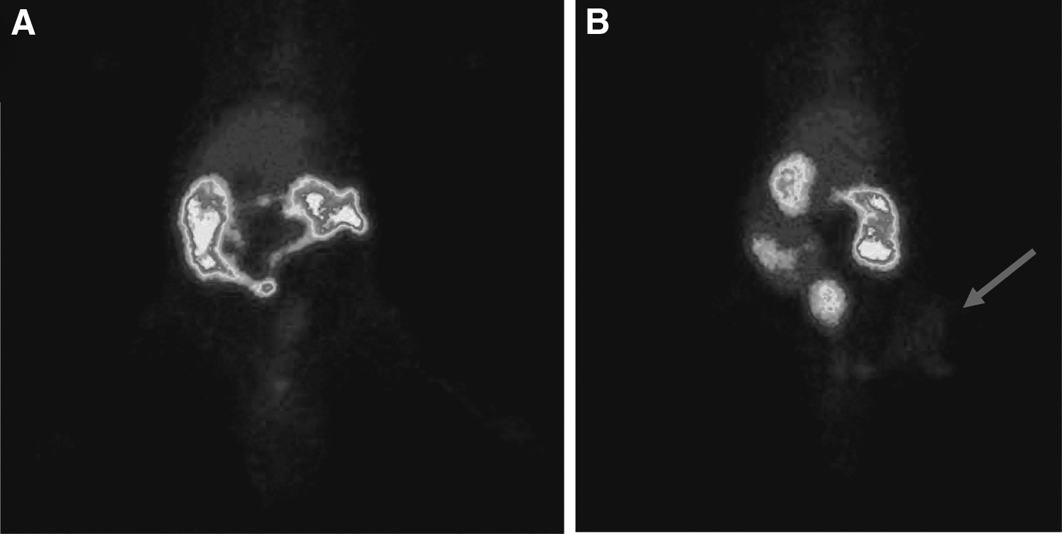
Scintigraphic images obtained at 6.5 h after injection of 99mTc-ceftizoxime showing: (
The results presented in Table 1 demonstrate that although there was no difference between the control and experimental groups regarding the amount of radioactivity in the ROIs recorded at 3.5 h after injection of radiopharmaceutical, the between-group difference determined at 6.5 h after treatment was statistically significant (Fig. 2). Moreover, the level of radioactivity in thigh tissue, measured after the euthanasia of animals and the removal of implants, was significantly higher in animals of the experimental group in comparison with the control group (Table 1).
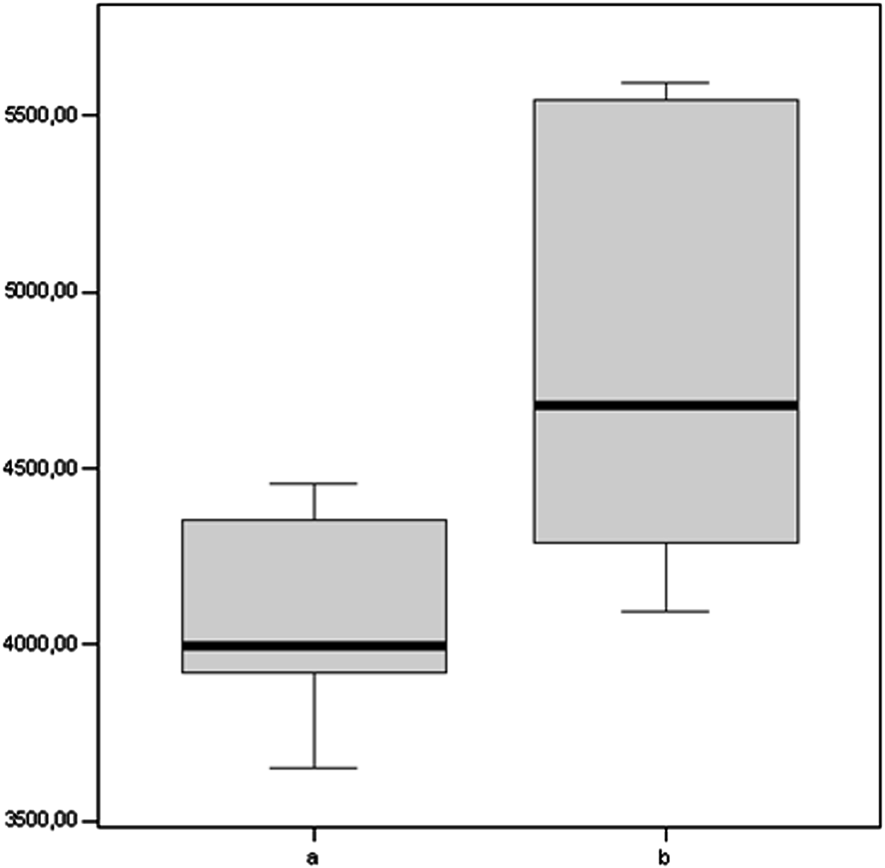
Box plot showing the levels of radioactivity at 6.5 h after injection of 99mTc-ceftizoxime present in regions of interest of: (
Values shown are mean±standard deviation.
Animals received sterile implants (n=6).
Animals received implants contaminated with Staphylococcus aureus ATCC6538-P (n=6).
Between-group difference statistically significant according to the Kruskal-Wallis test (p≤0.05).
Total counts per pixel in 10 min static images.
ROI=regions of interest.
Discussion
The use of temporary or permanent orthopedic implants has become a common practice in modern medicine [18], but the their permanence may be limited by infection [4]. Although the incidence of implant infection is relatively low, the recent increase in the number of patients submitted to prosthetic surgery has been accompanied by an increase in the occurrence of infection-associated problems and, consequently, greater risk of morbidity or mortality [19].
An implant acts as an extraneous body that modifies the local micro-environment, and facilitates bacterial contamination either directly or via hematogenous dissemination. Typically, the surface of the biomaterial becomes colonized by bacteria, thereby creating a biofilm that is highly resistant to the host defenses and to antibiotics. The presence of a bacterial biofilm on medical implants is difficult to diagnose and treat by virtue of its subclinical evolution and the absence of clinical manifestations of infection. However, the progress of infection culminates in osteomyelitis, bone loss, loosening of implant, and failure of the surgery [20].
The development of a biofilm impedes the detection of infection by conventional histologic and microbiologic methods, thus giving rise to an underestimation of the true incidence of subclinical infection. In view of these difficulties, considerable effort has been devoted to the development of new approaches, such as immunofluorescence and polymerase chain reaction, for the diagnosis of implant-associated infections. However, these techniques are of limited clinical application [7], and no effective, uncomplicated and low-cost method for diagnosing subclinical peri-prosthetic infections is currently available.
In the present study, we used a murine model to simulate an implant-related infection produced by a low virulence pathogen. The surgical procedure, which was simple and associated with low morbidity, involved the placement of a small titanium cylinder in the left hind limb of a rat. Titanium was used as the implant in the model system because its strength, lightness, and biocompatibility render it the material of choice for the manufacture of prostheses for application in the human body. The choice of Staphylococcus aureus ATCC6538-P as the contaminating bacterium on implants fixed in the experimental group of animals was based on its broad susceptibility to antibiotics and its affinity for metallic implants [22,23]. A 3-wk period was allowed between surgery and scintigraphic assessment because infections at this post-operative stage are considered sub-acute rather than acute.
Gamma scintigraphy is a non-invasive and sensitive technique that allows in vivo imaging of gamma-emitting radioisotopes. Most researchers agree that bone scintigraphy cannot identify unambiguously a periprosthetic infection because the significance of a scintigraphic “hot spot” could be associated with an inflamed, aseptically loosened implant or an infected prosthesis [11]. Various techniques have been described for improving the specificity of scintigraphy, including complementary imaging with gallium 67 citrate, indium-111–labeled leukocytes, and radiolabeled antibiotics. However, despite the substantial enhancements obtained using these approaches, none of the methods are 100% efficient in determining the presence of subclinical infections.
Fluoroquinolone antibiotics, which include ciprofloxacin, sparfloxacin, and levofloxacin, are the most commonly scintigraphic markers of infection used. However, the results obtained with these antibiotics are contradictory and exhibit low reproducibility [23]. Moreover, Sarda et al. [24] and Larikka et al. [25] have reported that scintigraphic imaging with 99mTc-ciprofloxacin cannot distinguish aseptic processes from osteomyelitis, and suggested that this limitation was because of pharmacodynamic characteristics, such as low molecular weight and low concentration of the agent used.
The third-generation semi-synthetic cephalosporin, ceftizoxime, used in the present study is β-lactamase–stable and active against aerobic and anaerobic gram-positive and gram-negative organisms. Gomes-Barreto et al. [16] demonstrated previously the reliability of scintigraphic imaging with 99mTc-ceftizoxime for the detection of infections in soft tissues and bone using model systems. The present study has shown that 99mTc- ceftizoxime scintigraphy can localize preferentially peri-prosthetic–infected areas adjacent to metal implants. The efficacy of the agent was particularly noticeable in the images acquired 6.5 h after injection of the radiopharmaceutical, suggesting that the labeled antibiotic is capable of detecting alterations in the micro-environment close to the implant and of reaching the bacteria attached to the implant surface. Radioactivity counting in tissues adjacent to the implant revealed that radiation remained even after the bacteria-contaminated implant had been removed, implying that the agent may be used as a marker of the evolution of the infectious process in the absence of the biomaterial.
Differentiating infection from aseptic inflammation is difficult because these entities are remarkably similar with respect to their clinical and histopathologic characteristics. An accurate diagnostic evaluation of patients is important because early treatment to suppress the infectious processes may prevent progressive physical and psychological damages. The present study has demonstrated that 99mTc-ceftizoxime scintigraphy can localize preferentially periprosthetic-infected areas adjacent to metal implants in a murine model. However, it should be noted, that the experimental design used was in full accord with the ethical principles of animal experimentation such that the sample size was the minimum required to observe the expected effect and to produce reliable results. Clearly, further studies must be carried out with human subjects in order to determine the sensitivity and specificity of the diagnostic method.
Footnotes
Acknowledgments
L.E.M. Teixeira was responsible for the study design, performed the surgical procedures, and drafted the paper; G.G. Soares and H.C. Teixeira performed the surgical procedures; I.K.T.M. Takenaka and S.O.F. Diniz were responsible for preparation of radiopharmaceutical and scintigraphic analysis; V.N. Cardoso, M.A.P. Andrade, and I.D. Araújo contributed with statistical analysis and the final revision of the paper.
Author Disclosure Statement
The authors declare that they have participated equally in the study and are fully responsible for the content of this article. The authors also declare that they have no competing financial interests.