
Abstract
Abstract
Background:
The purpose of the study was to investigate the role of the Foley catheter in maximizing surgical debridement in patients with chronic osteomyelitis.
Patients and Methods:
During surgical debridement in patients with chronic osteomyelitis, the medullary cavity is reamed thoroughly. The bulb of the Foley catheter is inflated with saline to facilitate the removal of the outer wall of the bulb using scissors. A guide wire is fed into the larger lumen of the Foley catheter up to its tip. Once the catheter is negotiated into the medullary cavity of the distal fragment, the guide wire is removed. The system is ready for irrigation. Through the smaller lumen of the catheter, which is open distally, copious irrigation is possible, delivering the fluid where it is required. At the same time, suction is connected to the proximal tip of the Foley catheter using a sterile adaptor. Constant suction is applied until the aspirated fluid is clear. The final aspirate is analyzed by semi-quantitative Gram staining and culture and sensitivity tests.
Results:
In all nine patients, the semi-quantitative Gram stain test was reported as one plus, suggesting that the bacterial population was less than one per oil immersion field. The post-operative culture results in all nine patients were reported as negative. All nine patients experienced primary healing. There was no evidence of recurrence in any patient, even after 12 months of follow-up.
Conclusion:
We describe an effective, inexpensive, and readily available method to aid in the debridement of an infected medullary cavity.
C
Chronic osteomyelitis remains one of the most challenging problems in orthopedic operations. Many methods have been described for the management of chronic osteomyelitis. Lavage using saline or an antiseptic solution for open fractures is quite effective, but the same for closed spaces, such as the infected medullary cavities after removal of an intra-medullary nail, is less effective, leading to incomplete clearance of infective foci. This not only adds to the chronicity of infection, but also delays any further reconstructive procedure when needed, finally reflecting poorly on the ultimate outcome.
One of the methods to overcome this problem is to use a reamer-irrigator-aspirator (RIA) device (Synthes, Paoli, PA). The RIA is expensive, single use, and not readily available, however. We describe an ingenious and innovative method using material readily available in the operating theater to achieve suction-irrigation in the deep dead spaces of the medullary cavity.
Patients and Methods
This was a prospective study that included nine patients (seven males and two females with an average age of 42 y) with established chronic osteomyelitis in whom the infection had lingered for more than 6 months with an implant in situ that was used for fracture fixation. Those patients treated between January 2011 and January 2013 were included in the study. The diagnosis of chronic osteomyelitis was established taking into consideration both the history (intermittent fever, malaise, and constitutional symptoms) and clinical examination (local tenderness and discharging sinuses), corroborated by raised erthrocyte sedimentation rate and C-reactive protein levels.
All infections were non-specific bacterial in origin (which did not require specific chemotherapeutic agents such as anti-tubercular and anti-fungal agents). Specific infections such as from tuberculosis and Salmonella were excluded. In six patients, the fracture involved the shaft of the tibia, while in three other patients, the fracture involved the femoral shaft. In all nine patients, the fracture had united despite the deep infection and the draining sinuses. All patients received intravenous (IV) antibiotic agents pre-operatively for 1 week; antibiotic agent selection was based on culture and sensitivity reports.
The infected nail was removed through the proximal point of entry. The medullary cavity was thoroughly reamed with an appropriately sized reamer (generally 0.5–1 mm larger than the nail removed) over a guide wire. An appropriately sized Foley catheter was obtained based on the size of the medullary cavity after reaming (general rule of thumb, 16F if the reamer size was more than 10 mm and 14F if the reamer size was less than 10 mm).
The bulb close to the distal tip was inflated using saline (Fig. 1) so that the outer wall of the bulb could be removed completely using scissors. This converted the Foley catheter to a flexible double-lumen device to be inserted into the medullary cavity for suction-irrigation (Fig. 2). The guide wire was then fed into the larger lumen of the Foley catheter up to its tip. This provided the catheter with the rigidity needed for it to be inserted into the medullary cavity. Once the catheter was negotiated into the medullary cavity of the distal fragment, the guide wire was removed (Fig. 3).
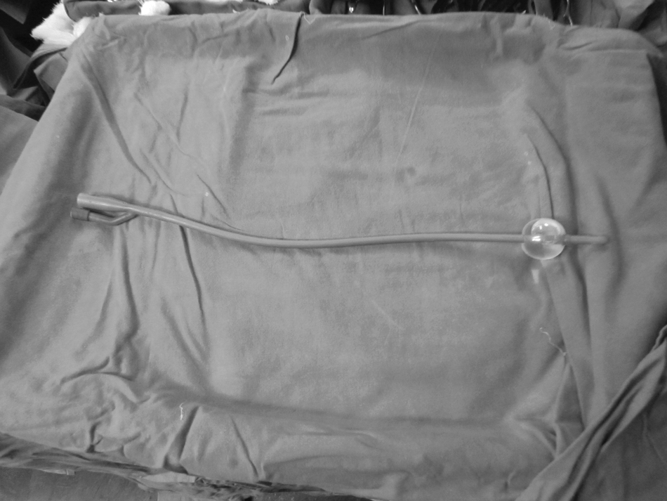
Foley catheter bulb inflated with saline.
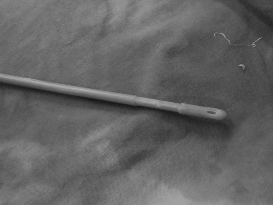
Foley catheter with denuded bulb after deflation.
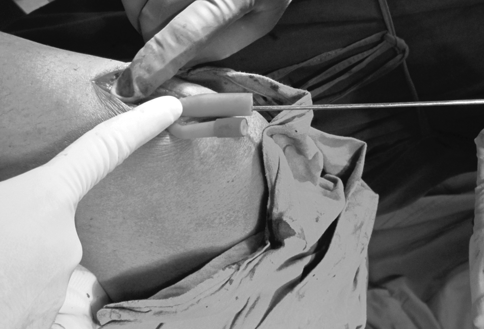
Modified Foley catheter introduced into the tibial medullary cavity with the help of a guide wire.
The system was ready now for irrigation. We used physiologic saline (generally 1-2 L) without antibiotic agents for irrigation. Through the smaller lumen of the catheter that was open distally, copious irrigation is possible, delivering the fluid to the area required; at the same time, the suction device was connected to the proximal tip of the Foley catheter using a sterile adaptor (Fig. 4). The modified Foley catheter was thus used as a suction-irrigation apparatus.
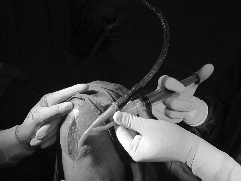
Foley catheter as a suction-irrigation device.
Suction-irrigation was continued until the fluid was clear and devoid of any debris. The lumen of the Foley catheter was reasonably large so as to prevent frequent blockage from debris. During lavage, the distal locking hole of the intramedullary nail after screw removal was physically sealed by external pressure to keep the medullary canal closed for the effective suction.
Once the aspirated fluid appeared clear, it was sent for Gram staining and culture and sensitivity tests. The end point of the lavage was determined when the numerical grade of the semi-quantitative Gram stain was reported one plus by the microbiologist in the operating theater. The semi-quantitative Gram stain method was based on the number of bacteria per high power (x1,000) oil immersion field. Zero (0) = no bacteria in the field, one plus = fewer than one bacterium per field, two plus = one to five bacteria per field, three plus = six to 30 bacteria per field, 4 plus = more than 30 bacteria per field [4]. This procedure took an extra eight to 10 minutes each time it was performed. The test had to be performed two times in three patients to ensure that the numerical grading of the Gram stain was one plus.
In the event of any blockade, the catheter can be removed, the guide wire can be used to clear the intra-luminal debris, and the same catheter can be inserted into the medullary cavity to complete the procedure.
Patients received IV antibiotic agents post-operatively for 10 days and continued receiving oral antibiotic agents for a total of 6 weeks post-operatively. We considered the patients to be healed if there were no symptoms for 12 months of follow-up and the patients did not require antibiotic agents 6 weeks after the procedure.
Results
In all nine patients, the semi-quantitative Gram test was one plus, suggesting the bacterial population was less than one per oil immersion field. The culture reports in all nine patients were negative post-operatively. All nine patients experienced primary healing. We followed the patients radiologically for 3 months, 6 months, and 12 months, and no patient showed any radiologic features of infection. There was no evidence of recurrence in any patient, even after 12 months of follow-up.
Discussion
There have been methods to bring about thorough washouts in infected cavities such as using a double-lumen catheter prepared on the operating table, using the introduction of an epidural catheter into a blood giving set with the help of appropriate connecters. This method, however, is used essentially when the surgeon believes that a closed-suction irrigation system is to be maintained for 3 weeks, wherein an appropriate antibiotic solution is used to irrigate for 30 minutes followed by suction for 3 hours and 30 minutes. The possibility of blockage in the system is always present, necessitating the use of streptokinase. Maintenance of such a closed irrigation and suction system for 3 weeks will need close monitoring in specialized centers to avoid secondary (iatrogenic) infection.
Although the system we describe can be used as an indwelling system, we have not used it primarily for that purpose. This method is effective for clearing infective foci after reaming of the medullary canal. The reamed medullary canal needs a thorough irrigation and suction, which can be obtained effectively on the operating table and even confirmed bacteriologically intra-operatively by sending the aspirated fluid for Gram stain and culture sensitivity testing. The appearance of the aspirated fluid also aids the surgeon.
The concept of irrigation in the management of chronic osteomyelitis dates back to 1917 [5]. Chronic osteomyelitis involving the entire medullary canal has always been a surgical challenge for orthopedic surgeons. The importance of surgical debridement cannot be over-emphasized; some surgeons have even tried introducing an arthroscope into the medullary cavity as a method to confirm the adequacy of the debridement.
Although there are lot of methods described to irrigate the medullary canal with antibiotic agents, followed by suction using double-lumen suction irrigation, there is a paucity of literature that highlights the importance and techniques of performing mechanical fluid washout of the medullary canal. This is the primary pre-requisite for the irrigation-suction method using antibiotic agents to act and provide significant therapeutic benefit.
The double-lumen technique involves insertion of an epidural catheter or a central IV catheter into a suction catheter using adaptors. The lumen of the internal catheter is considerably small and will be suitable essentially for the administration of an antibiotic solution over time (1.5 hours) so that the same can be suctioned over 3.5 hours as a part of a cycle of 4 hours, which can be repeated and maintained up to 3 weeks to chemically sterilize the medullary canal. The same system, because of the size of the catheter, is not suitable for mechanical lavage, irrigation, and suction.
The other method used for mechanical clearing of the medullary canal is the RIA device that involves simultaneous reaming, irrigation, and aspiration. Although it serves the purpose, the RIA is not readily available and is a non-reusable, expensive device.
The method we are using consists of universally available, sterile, inexpensive Foley catheters (less than £3/RS170/$4). These are available in different sizes for different sized medullary canals after preliminary reaming for mechanical medullary debridement.
The Foley catheter without its distal bulb is a freely available double-lumen catheter of sufficient diameter for both irrigation and suction. The chances of blockage of the outer, larger diameter tube by debris are very low. Any blockage encountered can be addressed adequately by the withdrawal of the Foley catheter and removal of the blocking debris using a guide wire.
We have not encountered any incarceration or breakage of the Foley catheter in the medullary canals. Such a possibility can be addressed using a Foley catheter with a radio-opaque line on its surface to identify the location of the broken catheter piece for withdrawal.
Limitations
This series is very small and without any control study to prove efficacy of the method that we are trying to describe. The purpose of describing this method is the non-availability of the commonly accepted method (RIA) in most of the centers in developing countries. We believe that this is an important technical tip and improvisation in achieving complete clearance of infective foci that orthopedic surgeons commonly encounter. The alternatives we have are expensive and not readily available (Table 1).
We have been using this method successfully for irrigation and suction of the infected medullary canal. We have also used the same method for irrigation and suction of closed compartments such as an infected knee joint when the patient is not fit for open arthrotomy, such as septic arthritis in patients with psoriasis with skin lesions around the knee joint.
Conclusion
We have described an effective, inexpensive, and readily available method to aid in the debridement of an infected medullary cavity.
Footnotes
Acknowledgment
We would like to thank Dr. Varghese Joe and Sherin Babu for helping us in the formatting of this article.
Author Disclosure Statement
No competing financial interests exist.