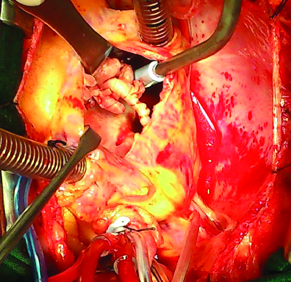
A 53 year-old male with a past history of two pacemaker implantations was referred to our institution because of high (up to 39°C) intermittent fever with 2 y of duration. Past transthoracic and transesophageal (TEE) echoes were reported as normal. A new TOE revealed a 2 cm x 2 cm mass originating from the tricuspid valve (TV) attached to the pacing wire. At the operative field, the septal leaflet of the TV was penetrated from the pacing wire forming a concrete mass (Fig. 1). The pacing wire was covered by multiple large colonies, of different time formation, occupying most of the right atrium (Fig. 2). The TV was replaced with a 27 mm Sorin Pericarbon bioprosthetic valve and the pacing wire was totally removed along with the battery. Cultures revealed Staphyloccocus epidermidis, which was sensitive to daptomycin, and which was continued for 4 wks at 850 mg intravenously daily. Seven mo later, the patient remains asymptomatic.
The septal leaflet of the tricuspid valve was penetrated from the pacing wire, forming a concrete mass occupying most of the right atrium. Color image is available at www.liebertpub.com/sur
Multiple large colonies, of different time formation, covering most of the pacing wire. Color image is available at www.liebertpub.com/sur