
Abstract
Background:
Human cystic echinococcosis (CE), most commonly caused by echinococcosis granulosis, is the most common presentation of hydatid disease of the liver and is considered endemic in the Middle East region.
Patients and Methods:
This study is a retrospective single-center analysis of all patients with hepatic hydatid disease presenting for surgical management from 2001 to 2019.
Results:
From 2001 to 2019, 100 patients (54 males, 46 females) were diagnosed with hydatid disease of the liver with a mean age of 45 years (range, 19–82). The most common presenting symptom was right upper quadrant abdominal pain followed by incidental finding of cyst on imaging. Thirteen patients (13%) presented with signs and symptoms of obstructive jaundice. Of the 100 patients, 39 underwent laparoscopic surgery and 61 underwent open surgery. The most common complications were as follows: 16 bile leaks (16%), 14 intra-abdominal fluid collections (14%), 8 wound infections (8%), and 3 patients had biliary strictures (3%). Of the 100, 8 patients(8%) had recurrence of their hepatic hydatid disease.
Conclusions:
Hydatid disease of the liver is not a common disease, and its management can include medical, surgical, and interventional radiology. The decision depends on the size and complexity of the cyst and its location. Bile leak is a common complication and should be managed conservatively or through intervention by radiology or endoscopic retrograde cholangiopancreatography (ERCP).
Human cystic echinococcosis (CE), most commonly caused by echinococcosis granulosis, is the most common presentation of hydatid disease, and it accounts for more than 95% of the estimated two to three million cases worldwide [1]. Among the main regions in which CE is endemic is the Mediterranean region. Moreover, it has a cosmopolitan distribution and is also endemic in areas such as southern Brazil, central Asia, western China, and East Africa, where it represents a major public health problem [2]. The clinical presentation of CE depends on the organ involved, the location of the cyst within it, the relation of the cyst to the surrounding structures, and finally, the integrity of the cyst wall [2]. Unless complications occur, CE is commonly asymptomatic. Most cases have one cyst located in one organ, with the liver, specifically the right lobe, being the most common organ affected. The next most frequently affected organ is the lung, which accounts for approximately 20% of the cases [2]. The presentation of symptomatic CE patients with liver cysts is commonly upper abdominal discomfort and poor appetite. Jaundice may result when the bile ducts are compressed. Abdominal distension, hepatomegaly, or a tumor-like mass may be found on palpation [3].
The World Health Organization Informal Working Group on Echinococcosis (WHO-IWGE) classification sets the staging of hepatic hydatid cyst based on its ultrasound aspect. The therapeutic approach is based in consonance with the staging [4]. Several complications are seen in liver hydatid disease. Examples include biliary tree obstruction accompanied by superimposed infection resulting in cholangitis, abscesses, and intra-peritoneal leakage causing peritonitis can occur. In extreme cases, leakage may result in anaphylactic shock potentially leading to death [5]. However, the most frequent complication is the development of a cysto-biliary fistula that in turn can be either frank or occult [6]. Diagnosing a frank fistula clinically and radiographically is usually easy, whereas diagnosing an occult fistula is difficult. Failure to diagnose a frank fistula preoperatively can imply a complicated postoperative course [6]. Hence, to predict its presence pre-operatively, different scoring systems were used [6,7]. Finally, a gastric ulcer can also occur as rare complication of hepatic hydatid disease [8].
In this study, we present our experience in the management of hydatid disease of the liver from 2001–2019, with emphasis on symptoms, location of cyst, complications, management and surgical treatment, and hospital stay.
Patients and Methods
This study is a retrospective single-center analysis of all patients with hepatic hydatid disease presenting for surgical management from 2001 to 2019. In total, 100 patients were diagnosed with hydatid disease of the liver. The Institutional Review Board (IRB) committee approved the study protocol and data were retrieved from the medical records of the American University of Beirut Medical Center (AUBMC).
Data were collected using an approved worksheet designed for this study. The patients were either diagnosed primarily at our institution or referred to us (being a tertiary referral center). The age, gender, weight, duration of hospital stay, clinical presentation, computed tomography scan findings, post-operative management, morbidity and mortality, and other parameters were reported.
Results
Demographics and clinical presentation
Of the 100 patients diagnosed with hydatid disease of the liver, 54 (54%) were males and 46 (46%) were females. The median age was 45 years (range, 19–82) and body mass index (BMI) was 26.2 (range, 23.9–26.8). The most common presenting symptom was abdominal pain followed by incidental finding, and 13 (13%) patients presented with signs and symptoms of obstructive jaundice. Of the 13 patients with jaundice, 8 (8%) patients had severe cholangitis with fever, and 11 (11%) patients underwent endoscopic retrograde cholangiopancreatography (ERCP). Six (6%) patients presented with ruptured cyst and were operated on an emergency basis (Table 1).
Patient Demographics and Clinical Presentation
IQR = interquartile range; ERCT = endoscopic retrograde cholangiopancreatography; BMI = body mass index; Hb = hemoglobin; Hct = hematocrit; WBC = white blood cells; AST = aspartate aminotransferase; ALT = alanine aminotransferase; GGT = γ-glutamyl transferase; INR = international normalized ratio; CT = computed tomography.
Radiologic imaging and biochemical profile
Pre-operative radiologic assessments using computed tomography (CT) scan was performed for all patients. The median number of cysts in the liver was 2 cysts (range, 1–8), with the smallest measuring 1 × 1 cm and the largest reaching 15 × 11 cm (Fig. 1).
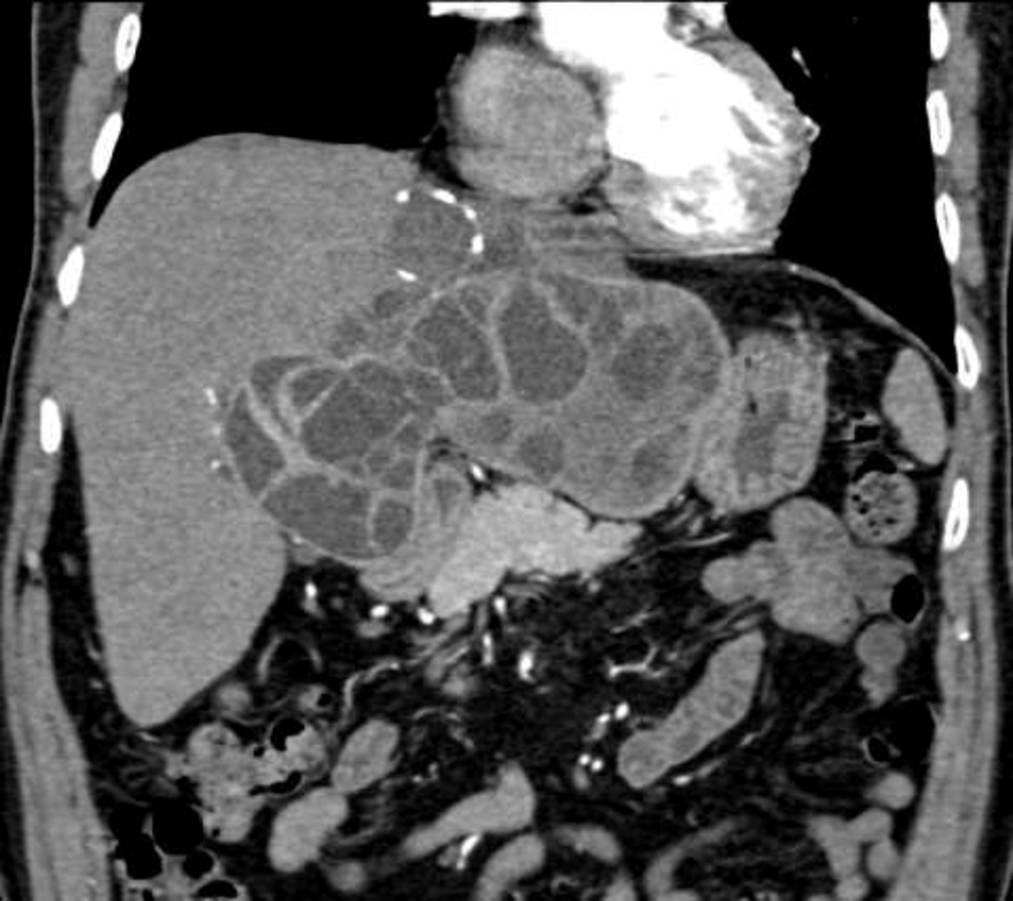
Complex hydatid disease of the liver, cyst involving the left lobe of the liver measuring 11 × 15 cm.
The biochemical profile included a median (range) white blood count of 8,000/mm2 (range, 3,000–25,700); hemoglobin, 13.3 g/dL (7 · 5–16.5); eosinophils 8% (range, 1%–16); aspartate aminotransferase level, 40 U/L (range, 8–691); total bilirubin, 0.5 mg/dL (range, 0.1–21.5); γ-glutamyl transferase, 45 U/L (range, 8–966); alkaline phosphatase, 77 U/L (range, 25–480); creatinine, 0.9 mg/dL (range, 0.4–2.2); and international normalized ratio, 1 (range, 0.9–1.2) (Table 1). Of the 80 patients, 50 (50%) had serology hydatid test done (indirect hemagglutination [IHA]), of which 40 (40%) were positive (Table 1). Thirty (30%) patients were taking albendazole pre-operatively at a dose of 400 mg twice daily for two weeks followed by another treatment for six more weeks.
Surgical procedures
Of the 100 patients, 39 (39%) underwent laparoscopic surgery and 61 (61%) underwent open surgery. The principles of surgery for hydatid disease of the liver are the same for laparoscopy and open surgery, and the only difference is the approach (Table 2).
Patient Outcome
IQR = interquartile range; SICU = surgical intensive care unit.
Surgical intervention involves the following steps.
Coverage of the cyst and surrounding area with cetrimide soaked gauzes. This is an antiseptic scolicidal solution made of a mixture of three quaternary ammonium compounds: tetradonium bromide, cetrimonium bromide, and laurtrimonium bromide.
Aspiration of the cyst fluid, followed by injection of cetrimide solution into the cyst cavity to kill the daughter cysts. It should remain in the cyst for five to 10 minutes before it is aspirated.
Complete evacuation of the cyst without spillage with removal of all membranes and daughter cysts (Gombo suction system is used most of the time).
Complete unroofing of the cyst.
Identification of any biliary fistula, and suturing it when found. Irrigation with normal saline and aspiration of all fluid.
Placement of intrabdominal drain.
Mortality and morbidity and disease recurrence
All patients were alive at 30 days post-surgery. The median hospital stay was five days (range, 2–29). Of the six patients who had ruptured cysts, two (2%) developed anaphylactic shock secondary to spillage of cyst material into circulation, and both patients recovered well after surgery.
The most common complications were as follows: 16 (16%) bile leaks, 14 (14%) intra-abdominal fluid collections, 8 (8%) wound infections, and 3 (3%) patients had biliary strictures. Management of bile leaks was with CT or ultrasound-guided drainage with or without ERCP. Intra-abdominal fluid collections were managed with simple drainage, and biliary stricture were managed with balloon and stenting.
Of the 100 patients, six (6%) patients had recurrence of their hepatic hydatid disease. Four (4%) of them were re-operated and two (2%) were treated with interventional radiology using the percutaneous aspiration-injection-re-aspiration (PAIR) technique plus a course of albendazole.
Discussion
Echinococcosis granulosis infection of the liver usually shows no symptoms. Most patients have a single cystic lesion located in a single organ. The liver, particularly its right lobe, and the lungs are the most frequent organs affected [2].
The presentation of symptomatic CE patients with liver cysts is commonly upper abdominal discomfort and poor appetite. Compression of the bile ducts may lead to jaundice. Examples of complications associated with hepatic hydatid disease are biliary tree obstruction accompanied by superimposed infection resulting in cholangitis, abscesses, and intra-peritoneal leakage causing peritonitis [5].
Notably, the development of a cysto-biliary fistula is most frequently seen. A fistula can be frank or occult (Figs. 2 and 3) [6]. Diagnosing a frank fistula clinically and radiographically is usually easy, whereas diagnosing an occult fistula is difficult. If a frank fistula is not identified prior to surgery, the patient's postoperative course would commonly be complicated [6]. For this reason, several scoring systems have been used to aid in its detection pre-operatively [6,7]. Gastric ulcer can also occur as rare complication of hepatic hydatid disease [8].

Endoscopic retrograde cholangiopancreatography (ERCP) showing bile leak at the site of the intra-abdominal drain in the right lobe of the liver.

Suturing of a bile leak in the cyst cavity.
Suspicion of hydatid cyst should be present in endemic areas after careful history taking and physical examination are performed [3]. The management of hydatid disease of the liver or other organs is managed by several specialties in our institution, which includes surgery, infectious disease, and gastroenterology. Some patients are only medically treated, whereas others are referred to surgery or interventional radiology. In our series, some patients had multiple cysts that explains the wide range (1–15 cm) of cyst sizes in this surgical series.
Ultrasound has a sensitivity of 90%–95% and thus, offers a fast and accurate approach of detecting liver lesions. It is common for hydatid cysts to have a simple fluid-filled appearance. Based on the variations in ultrasound appearance and characteristics, the WHO has classified the different cyst types and each type has a specific treatment regimen [5].
Computed tomography scan and magnetic resonance imaging offer a better sensitive and specific approach to detecting and characterizing hydatid disease; CT scan findings are very specific to hydatid disease, which includes the presence of septation, membranes, and sometimes calcifications. However, these modalities are mostly referred to in cases of extra-hepatic involvement. Therefore, they are not always utilized in the diagnosis albeit a CT scan comes into play in cases of cystic rupture because it points to the exact location and type of rupture (Fig. 1) [5].
Additionally, a variety of serologic tests have been established to aid in the diagnosis of hydatid disease. Initial screening includes indirect hemagglutination (IHA) and enzyme-linked immunosorbent assay (ELISA) for immunoglobulin (Ig) G, IgM, or IgE antibodies. Indirect hemagglutination was sensitive in approximately 80% of liver hydatid disease. Next, if necessary, confirmatory tests such as arc-5 immunoelectrophoresis and immunoblotting using specific antigens are performed [5].
Percutaneous aspiration is another method that is not required in most cases. However, it may be done prior to surgery to confirm the diagnosis by directly visualizing the protoscolices under a microscope. To avoid anaphylaxis from fluid leakage, the patient can be pre-treated with oral albendazole for four days prior to the procedure. After the aspiration, the patient can then start oral albendazole for a month. In fact, most of the protocols initiate PAIR as a diagnostic and therapeutic intervention once the cyst has been punctured [5]. WHO-IWGE has staged the hepatic hydatid cyst based on its ultrasound characteristics and accordingly outlined the appropriate therapeutic approach [4].
Medical management
Cyst disappearance on imaging is achieved by anti-helminthic chemotherapy, which has a 30% cure rate. Albendazole is the drug of choice for liver hydatid disease. It is given initially as a short-term therapy pending any surgical or percutaneous intervention. Prior to surgery, treatment with albendazole has resulted in a higher rate of non-viable cysts at the time of surgery. In inoperable cases such as having multiple cysts or peritoneal spread, medical therapy is indicated [5]. The optimal duration of treatment with albendazole is uncertain, however, treatment should be initiated at least four days prior to surgery. The WHO suggests a duration of four to 30 days prior to surgery and should be maintained for at least one month (in case of albendazole) or three months (in case of mebendazole) after surgery. It can be given pre-operatively or post-operatively. Patients who have seen an infectious disease specialist before visiting a surgeon would have usually begun medical treatment earlier on. If the patient is seen directly by a surgeon, the albendazole would be most likely administered post-operatively. The protocol for pre-operative treatment at our institution is two weeks pre-operatively followed by six weeks post-operatively.
Watch and wait approach
Dormant uncomplicated cysts are monitored for changes using ultrasound. This approach is mainly dependent on the cyst stage, and the only candidates are CE4 and CE5 cysts. Additionally, administration of treatment is not required [5].
Percutaneous drainage
In uncomplicated single compartment cysts (CE1 and CE3a), smaller than 5 cm in size, percutaneous treatment may be an option instead of surgery [5]. The procedure, referred to as PAIR, is applicable to CE1, CE2, and CE3 cysts [4]. It can be used in cases of a cyst with daughter vesicles, detached proligere membrane, multiple cysts if accessible to puncture, superinfected cyst, post-surgical relapse, patients who refuse surgery or who cannot undergo surgery, lack of response to medical therapy, and pregnancy. In cases in which the patient was non-cooperative or when cysts cannot be punctured, are inactive or calcified, or communicating with biliary tree, PAIR is contraindicated [4]. A variant, PAIRD (D = drainage), is when a catheter is inserted in the cyst at the end of the procedure. The cavity is then irrigated with saline solution and drained for 24 hours [4]. Surgical or radiologic intervention is required if the size increases on treatment (more than 5 cm).
If the PAIR technique is not recommended in cases in which the cyst communicates with the biliary tree or the cyst has multiple vesicles within or has contents that cannot be suctioned, the percutaneous evacuation/modified catheterization technique (PEVAC/MoCat) can alternatively be used. In this technique, a catheter is inserted into the cyst and successive injections and aspirations of an isotonic solution are done to evacuate its solid contents (i.e., daughter vesicles and endocysts). The target drainage is less than 10–15 mL over 24 hours [4].
Surgical management
The treatment of hepatic hydatid cysts requires surgical procedures that do not include peri-cyst resection (cystectomy) or that include pericyst resection [4]. The latter includes partial hepatectomy, peri-cystectomy, and peri-cystoresection. Additionally, the remaining cavity is addressed through several procedures that include external drainage, bipolar drainage of the cavity and the main bile duct padding, omental plombage, and peri-cystobiliary drainage. Moreover, drainage of the cavity may be done by anastomosis with stomach or jejunum [4].
Hypertonic saline solution, ethyl alcohol, hydrogen peroxide, or albendazole are options used to inactivate the parasites prior to opening the cyst cavity. Furthermore, isolating the cyst from the peritoneal cavity can be attained by enfolding the nearby areas with dressings drenched in anti-helminitic solutions. Another way is to apply adherent cones to the cyst through the icing technique or suction [4].
Treatment of liver hydatid cyst in complicated cases is done through open surgery, which is an accepted procedure worldwide [9]. Surgical removal of small cysts (<6 cm) located peripherally and anteriorly is done laparoscopically [4]. Laparoscopic surgery for hydatid disease is minimally invasive and has several advantages such as shorter operative time, fewer incisional complications, shortened hospital stay, and good cosmetic results.
A recent systematic review and meta-analysis [9], has shown that for four major outcomes, no advantages exist between open surgery and laparoscopic surgeries. The particular outcomes were complications, mortality, cure rate, and recurrences. No statistically significant dissimilarities were shown between the outcomes of the two procedures. Conversely, there may be misleading successful results regarding the laparoscopic approach because most of the published literature reports were either case reports or series [9].
Commonly, the indications for laparoscopic surgery are simple anterior cysts and hepatic hydatid cysts that are uncomplicated. Deep cysts and cysts located in the segment 1 or 7 are not indicated for laparoscopic surgery [9].
Some disadvantages of laparoscopic surgery include the risk of anaphylactic shock, peritoneal seeding, and even death. An additional disadvantage is the probability of its failure. In such cases, the procedure should be converted into open surgery. Other instances in which a procedure is converted to open surgery include failed and recurrent cases of medical and PAIR approaches. Other cases in which converting to open procedure is considered include cystic rupture, life-threatening bleeding, and residual daughter cysts that are irremovable. If the cystic locations are adjacent to the inferior vena cava, and there is intra-operative spillage of cyst contents, open surgery may then be necessary. The conversion rate in our experience is 3.3% compared with 1.7% reported in the literature [9]. The high cost of this alternative makes it not cost-effective. However, laparoscopic surgery has the advantages of less time and hospital stay than open surgery [9]. Not all surgery can be performed laparoscopically; multiple, complex, recurrent, and complex hydatid cysts with biliary fistulas are treated in an open technique, whereas simple, or large multiple hydatid cysts can be treated in a minimally invasive surgery.
Redo-hydatid disease of the liver is challenging, and in some cases, it is contraindicated. In such cases, we elect to treat the patient medically or refer them to interventional radiology.
Endoscopic retrograde cholangiopancreatography for hepatic hydatid disease complications
The role of ERCP with sphincterotomy was studied in cases of intra-biliary rupture of hepatic hydatid disease where 2,860 patients, average age of 41 years, received therapeutic ERCP. Of those patients, 151 (5.3%) had the diagnosis of hepatic hydatid disease. Intra-biliary disruption of hepatic hydatid occurred in 112 of the patients. Thirty-nine patients were admitted for sphincterotomy for flow reversal after surgery of hydatid cyst [10].
Surgery has traditionally been used for treatment of biliary fistulas where hydatid cysts have failed to close voluntarily. It was shown, however, that ERCP can be used in cases of cysto-biliary fistula or in cases of hydatid membrane and/or daughter cysts causing biliary obstruction. Sphincterotomy can empty out hydatid material present in bile ducts in cases of major rupture. A biliary occlusion balloon and Dormia basket can be used to clean out the common and main bile ducts (Fig. 2). Biliary flow is facilitated by sphincterotomy through the decrease in duodenobiliary pressure gradient. Eighty-one percent had an overall rate of fistula closure within 10 to 20 days, and success rate of 70% to 100%.
It was thus found that ERCP with sphincterotomy offers exceptional instant and short-term post-operative results. Consequently, a reasonable rate of morbidity with no mortality was also achieved. It was therefore suggested as an alternative in cases in which the surgical risk is high and given adequate clinical benefit [10].
Hydatid disease in Lebanon
In a review of the Lebanese contribution to hydatid disease done by Araj et al. [11] in 2014, the most common presentation site was the hepatic (primarily in the right lobe). The second most common presentation site was pulmonary. Other sites of presentations were also reported including vertebral, brain, cardiac, pancreas, spleen, female pelvis, eye orbit, kidneys, peritoneum and mesocolon, and thigh muscles. The highest rates occurring was in females between ages 20 and 60, in Mohafazat Beirut and Mount Lebanon, and in dog owners. No differences were observed among social classes [11].
Conclusions
Human CE, most commonly caused by echinococcosis granulosis, is the most common presentation of hydatid disease, and is considered endemic in the Middle East region. In the present study, the most common presenting symptom was abdominal pain followed by incidental finding. Patients were treated with albendazole prior to surgery. Most patients underwent open surgery by covering the cyst with anti-helmenthic solution, injecting anti-helmenthic solution to inactivate daughter cysts, and surgically evacuating and un-roofing the cyst. The most common complication post-operatively was bile leak, followed by intra-abdominal fluid collection, wound infection, and biliary strictures. Bile leaks were managed with CT or ultrasound-guided drainage with or without ERCP, intra-abdominal fluid collections were managed with simple drainage, and biliary stricture were managed with balloon and stenting.
Footnotes
Funding Information
This research did not receive any specific grant from funding agencies in the public, commercial, or not-for-profit sector.
Author Disclosure Statement
No competing financial interests exist. No benefits in any form have been received or will be received from a commercial party related directly or indirectly to the subject of this article. This study was approved by the Ethics Committee and after the approval of the Institutional Review Board (IRB) at our institution.