




Abstract
Background:
Cystic echinococcosis (CE) is caused by Echinococcus granulosus sensu lato. In Central Iran, no molecular information is available on CE in humans. Therefore, in this study, we identified the genotyping of hydatid cysts obtained from patients with CE in central Iran using mitochondrial cytochrome c oxidase subunit I (cox1) gene.
Patients and Methods:
Hydatid cysts were obtained from 19 patients referred to Shahid Sadoughi, Mojibian, and Mortaz Hospitals, Yazd, Iran from 2018 to 2020. Informed consent was obtained from all included patients. After DNA extraction, amplification was done using cox1 gene. Phylogenetic analysis was performed using MEGA7.
Results:
Of the 19 patients, 11 (57.9%) were male and eight (42.1%) were female. The mean age of the patients was 35.645 ± 2.55 years old. Regarding cyst location, of eight isolates from lung, six and two belonged to G1 and G6, respectively; and all liver cysts were G1 genotype. The spleen and neck cysts had G1 and G6 genotypes, respectively (p > 0.05). All cysts with a diameter in the range of 5–10 cm (n = 9) and large cysts (>10 cm; n = 5) were identified as G1 (p = 0.002). The maximum likelihood tree topology demonstrated the maximum similarity of G1 among Iran and worldwide (99%–100% likelihood).
Conclusions:
Based on our results, it seems that the sheep–dog cycle in the infection of humans by Echinococcus granulosus in this study area has the most important role compared with the other cycles such as the camel–dog one.
Cystic echinococcosis (CE) is a zoonotic infection caused by Echinococcus granulosus sensu lato. Carnivores, particularly dogs, act as the definitive hosts, whereas livestock are considered as intermediate hosts [1]. Humans can be infected through the consumption of food contaminated with eggs, transformed to hydatid cyst [2]. In endemic regions, incidence rates exceed 50 per 100,000 person-years and global prevalence estimates are 50 million [3]. In Iran, the cost of CE is estimated to be $1,027 and $851 for hepatic and pulmonary CE, respectively. This issue is $1,911 and $2,458 for hepatic and pulmonary CE in a private hospital [4]. Cystic echinococcosis as a neglected tropical disease (NTDs) and neglected zoonotic disease (NZDs) [5,6] is worldwide [7]. Iran is considered a hyperendemic area for CE with the rate of 1.1 to 18.3 per 100,000 individuals [8]. Cystic echinococcosis is an important public health problem in Iran because of effect on critical organs such as the liver (70%), lung (12%) [9,10], and other organs (e.g., spleen, brain, muscle, etc.) [11].
Early diagnosis of CE is important to prevent complex clinical aspects [12] using imaging techniques [13] and serologic tests [14]. Knowledge about the prevalence of Echinococcus granulosus s.l. genotypes for the development of control or eradication strategies is important [15]. Echinococcus granulosus s.l. has four groups [16,17] including Echinococcus granulosus sensu stricto (G1–G3), Echinococcus equinus (G4), Echinococcus ortleppi (G5), and Echinococcus canadensis (G6/7, G8, G10). To design proper planning to prevent parasite transmission [18], several studies have been conducted to identify genotypes of Echinococcus granulosus s.l. in different parts of Iran in humans [19–22]. There are no studies available about the genotyping of CE in humans in Central Iran, therefore in the present study, along with the consideration of some clinical features of patients with CE undergoing surgery at Yazd Hospital in Central Iran, parasite genotypes were also studied by molecular characterization cytochrome oxidase c subunit 1 (cox1) gene.
Patients and Methods
Ethical statement
All experiments and study protocols were reviewed and ethically approved by the Ethics Committee of Shahid Sadoughi University of Medical Sciences, Yazd, Iran (approval ID: IR.SSU.SPH.REC.1398.068) and informed consent was obtained from all participants. If the participant was a minor child, their parents completed and signed the related forms.
Sample preparation
A total of 19 cysts were collected from patients with CE after diagnosis based on abdominal ultrasound, computed tomography, radiography, and clinical manifestations, through surgery in Mortaz, and Mojibian Hospitals, from 2018 to 2020. The cysts were encoded from 101 to 119. All samples were transported to the Molecular Laboratory of Research Center for Food Hygiene and Safety, Shahid Sadoughi University of Medical Sciences, Yazd, Iran. A questionnaire was developed to record the demographic and diagnostic data from each patient, including occupation, gender, age, residence, location, size of the cyst, using drug before surgery, clinical manifestations, history of dog ownership or contact, history of the disease in family members, and drinking water sources.
Gross examination and histopathology
The mass of each cyst was measured and then each specimen was fixed in 10% neutral buffered formalin. Then samples were cut into 5 mcm sections, stained with hematoxylin and eosin (H&E), and were observed by two pathologists for conventional histopathologic examinations.
DNA extraction
The genomic DNA was extracted from germinal layers with a commercial kit (GeneAll, South Korea) according to the manufacturer's instructions. The quantity of the extracted DNA was analyzed using a NanoDrop (Thermo Fisher Scientific, Waltham, MA) and stored at −20°C until subsequent analysis.
Molecular analysis
Genotyping was identified based on the study by Hajimohammadi et al. [23]. The sequences respecting to the mentioned isolates were alignment using the T-COFFEE software. Phylogenetic analysis was applied using the maximum likelihood (ML) method with MEGA7 based on the Tamura-Nei model with 1,000 bootstrap replicates [24].
Statistical analysis
The obtained data were analyzed using SPSS Statistics, version 20 (IBM Corp, Armonk, NY). The relation between genotypes and clinical/demographic data were analyzed using χ2 and Fisher tests. A p value <0.05 was considered as statistically significant.
Results
Patient clinical and demographic data
We reviewed the related demographics data from patients with CE. Of 19 patients entered in this study, 11 (57.9%) were male and eight (42.1%) were female. The mean age of the patient was 35.64 ± 2.55 years old (range, 8–75). The infection rate in the age group is shown in Figure 1a. Most patients were housewives and students (n = 12; 63.1%) followed by workers (n = 2), farmers (n = 2), salaried employees (n = 2), and the least affected groups self-employed (n = 1). The results showed that liver cysts were common followed by lung cysts. The unusually located cysts such as spleen and neck were also diagnosed. The percentage of infected organs is given in Figure 1b.

The categorizes of the samples used in this study. (
Abdominal pain was the most common clinical symptom (52.6%), and in seven patients (36.8%), complaints included chest pain and shortness of breath. Other signs and symptoms were swelling of the back of the neck (5.2%) and shoulder pain (5.2%). As for size, there were nine (47.4%) medium size (5–10 cm), four (21.1%) small (<5 cm), and five (26.3%) large (>10 cm). No information was available for the size of isolate 117.
The results of the study showed that the source of water for most patients is tap water (n = 18; 94.7%) and only one case of well water consumption was reported. Also, more than 78% of patients had not been in contact with a dog, and of 19 infected patients, four (21%) had a history of dog ownership. Computed tomography (CT) scans and radiography of patients are shown in Figure 2.
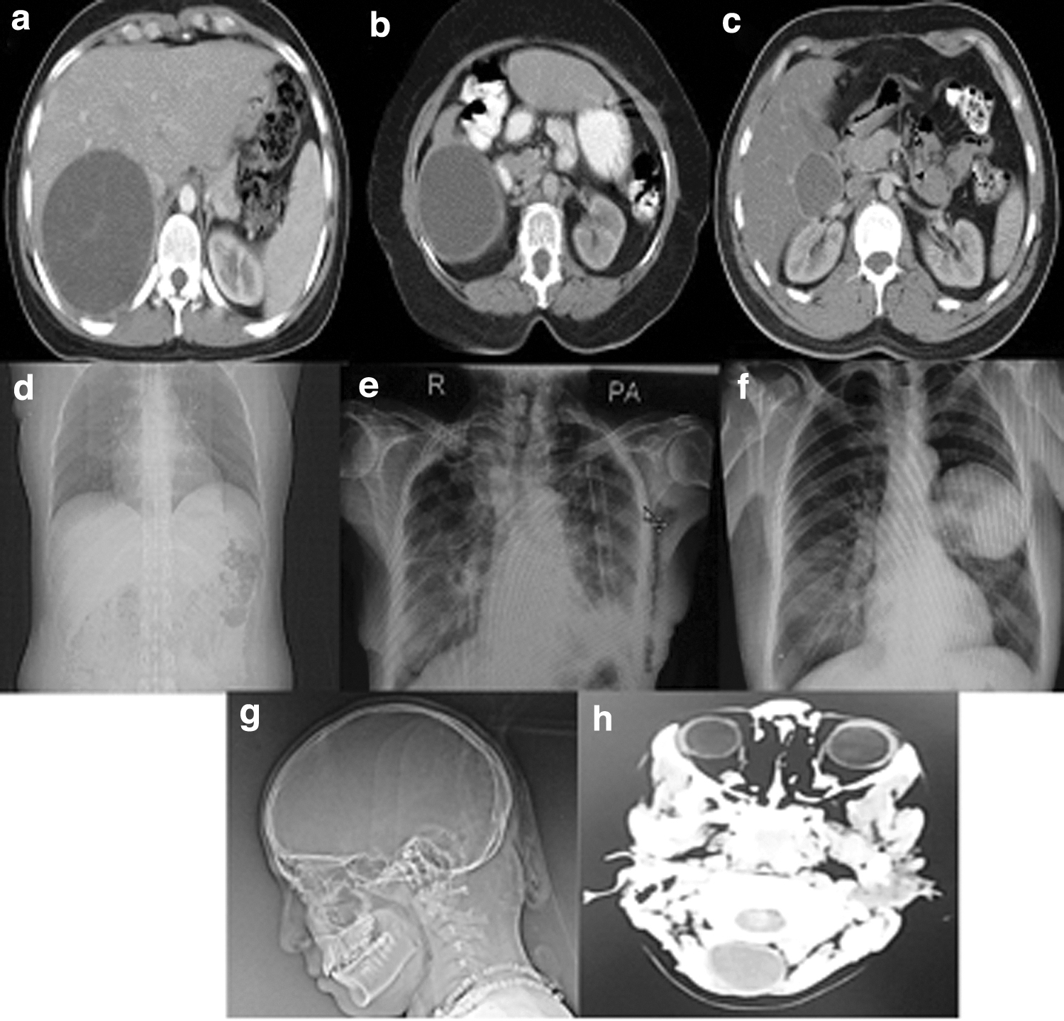
Radiologic appearance of hydatid cysts. (
Histopathology
The sections showed cyst wall composed of acellular lamina lucida with fibrous connective tissue that in some cases there was severely infiltrated by inflammatory cells containing lymph plasma cells, eosinophil, neutrophil, and multinucleated giant cell (foreign body).
In the spleen specimen, fibrous cyst wall with absence epithelial lining favor secondary (false) cyst was observed. In isolate 117, fibrous connective tissue with areas of necrosis and calcifications liver parenchyma was present. Among some necrotic materials, protoscoleces and hooklets of Echinococcus granulosus were identified. Overall, the results of the pathologic examination confirmed cysts to be a hydatid cyst. Some sections of specimens are shown in Figure 3.

The histopathologic section of hydatid cyst. (
Molecular analysis
The polymerase chain reaction (PCR) amplification was performed successfully on 19 isolates for cox1 with the amplicons of approximately 450 bp (Supplementary Figure S1). The mentioned sequences of these isolates were deposited in the GenBank, National Center for Biotechnology Information under the accession numbers that are shown in Table 1.
Sequences Used in Phylogenetic Tree Analysis
Overall, the analysis of cox1 gene sequences revealed that 84.2% of isolates (n = 16) had G1 genotype in which 11 cases were male and five were female. However, three of 19 cysts (15.8%) belonged to Echinococcus canadensis (G6 genotype), all of which were isolated from female patients. There was a statistically significant relation between gender and genotypes (p = 0.027).
With respect to the site of the cysts, of eight isolates of the lung, six and two belonged to G1 and G6, respectively, and all of the isolates of the liver were G1 genotype. Two cysts from patients with spleen and neck involvement had G1 and G6 genotypes, respectively. There was no statistically significant relation between genotypes and involved organ (p > 0.05).
The results showed that there was a statistically significant relation between genotype and the size of the cysts (p = 0.002). All cysts with a diameter in the range of five to 10 cm (n = 9) and large cysts (n = 5) had G1 genotype. On the other hand, all isolates with G6 genotype were small size.
Other clinical and demographic data including occupation, water source, clinical manifestation, history of dog ownership, and age group showed no statistically significant relation with G1/G6 genotype (p > 0.05). The maximum likelihood tree topology of the mentioned gene demonstrated the maximum similarity of G1 in Iran with those worldwide (99%–100% likelihood) (Fig. 4).

Molecular phylogenetic analysis of Echinococcus granulosus isolates obtained from patients with cystic echinococcosis. The maximum likelihood method was used in this analysis based on Tamura-Nei model using MEGA7.
Discussion
Cystic echinococcosis is caused by the larval stage of Echinococcus granulosus s.l for which livestock such as sheep, cattle, goat, and camel act as an intermediate host. The first involved organ in hydatid disease is the liver, but it can also spread to other organs via the hematogenous route [25]. Therefore, CE can be seen in the lung, kidney, spleen, and brain as well as the heart [26]. On the basis of our results, most sites of echinococcosis were in the liver and a small portion of the total cases are in rare sites including the spleen and neck. Also, the results of different animal studies presented a vast distribution of the cyst in organs and reported the highest involvement in liver and lung, respectively [27,28].
Our results showed that 78% of patients had no contact with dogs, whereas dogs are known as definitive hosts for Echinococcus granulosus s.l. This indicates that in some cases of infection, indirect modes of transmission including contaminated plants with feces from dogs are responsible for the disease. Several studies have shown that transmission of this parasite via contaminated water is possible [29]. However, in the current study, approximately 94% of patients had access to tap water and less than 10% of patients used well water. Therefore, given that this parasite can cause disease in different modes, different measures including dog deworming, health education, and preparation of tap water can be effective in reducing Echinococcus granulosus s.l. transmission to humans.
In this study, the predominance of G1 genotype of Echinococcus granulosus s.s. identified in the study area, which is consistent with its global results [30,31]. Consistent with our study, in Iran, many types of research demonstrated that G1 is the most frequent genotype from CE isolates and is considered as the main causative agent of the disease [16]. These results may be because of the higher number of sheep as intermediate host. Also, Fasihi et al. [32] showed that in human and livestock hosts, G1 is the most dominant. In a study performed by Ahmadi et al. [33] on human, sheep, and camel samples indicated that G1 is dominant genotype in human by targeting ITS1 region of rDNA. Also, global studies in neighboring countries that share strong cultural and socioeconomic ties have reported that the most common genotypes of human hydatid cyst isolates are G1 or G3 [25,34]. Similarly, in Algeria, Moussa et al. [35] investigated the genotypic diversity of 46 Echinococcus granulosus s.l. samples and reported G1 as a dominant genotype. A study supports that G1–G3 is considered as a dominant genotype [36] and in our recent comparative study conducted on livestock, the G1–G3 genotype had the most frequency in samples and the results of this study confirmed that the G1 genotype is circulating between humans and livestock [37].
Along with our study, G6 was also identified from human isolates. In most human reports from various countries with similar weather including Egypt [38] and Sudan [39], G6 was identified in human cases as well as our study and some reports around the world indicated the G6 genotype in humans [40,41]. However, as mentioned previously, several studies indicated the G1 genotype as the only genotype found in humans. The study conducted by Fasihi et al. [32] using restriction fragment length polymorphism (ITS1-RFLP) method, G1 (30 cases) and G6 (3 cases) genotypes were identified from different provinces of Iran, which is similar to our results. In a systematic review study was conducted by Manterola et al. [36], 8,643 liver and lung samples from 48 different countries were studied. The majority of hosts were cattle, sheep, and human. The results of this report indicated G1, complex G1–G3, and G6 as the most frequent genotypes, which is in agreement with our results [36]. However, further studies are required including larger number of human isolates to improve knowledge.
Cystic echinococcosis transmission in humans can be influenced by a set of behavioral and socioeconomic factors such as farming activities, including traditional sheep raising and agricultural practice, contact with dogs, geophagy, outdoor activities, or contaminated matrices that may facilitate acquiring a high egg concentration of Echinococcus granulosus s.l. [42,43]. Indeed, the evidence for the comparative significance of food and water to CE transmission need further study. Free-roaming dogs could contaminate surface water as well as farm produce intended for human consumption. Therefore, eating contaminated food and raw vegetables can be one of the major risk factors of human CE in Iran [42]. However, little information is available regarding the presence of Echinococcus granulosus eggs in water, and more studies are required in this region. In the northern province of Mazandaran, two of 989 water samples (0.2%) were found contaminated with taeniid eggs by using conventional parasitological techniques [43].
Conclusions
The current study confirmed the presence of G1 and G6 genotypes of Echinococcus granulosus s.l. from human isolates in Yazd Province, Iran. To our knowledge, this is the first molecular identification of echinococcosis in Central Iran. Based on our results, it seems that the sheep–dog cycle in the infection of humans by Echinococcus granulosus s.l. in this study area has the most important role in comparison with the camel–dog cycle.
Footnotes
Acknowledgments
We thank all technical staff from Research Center for Food Hygiene and Safety, Shahid Sadoughi University of Medical Sciences, Yazd, Iran to finalize this project (Project ID 6739).
Authors' Contributions
All authors have read and approved the manuscript.
Funding Information
This research was financially supported by Iran National Science Foundation (grant no. 97018792).
Author Disclosure Statement
The authors declare that they have no competing interests.

References
Supplementary Material
Please find the following supplemental material available below.
For Open Access articles published under a Creative Commons License, all supplemental material carries the same license as the article it is associated with.
For non-Open Access articles published, all supplemental material carries a non-exclusive license, and permission requests for re-use of supplemental material or any part of supplemental material shall be sent directly to the copyright owner as specified in the copyright notice associated with the article.
