




Abstract

To the Editor:
Pyogenic spondylitis is an infrequent infectious disease of the vertebral body, intervertebral disc, or paraspinal tissues, the reported incidence of pyogenic spondylitis approximates between 0.4 and 2.0 cases per 100,000 [1]. The most common microbial species that cause pyogenic spondylitis are Staphylococcus aureus, Streptococcus, Escherichia coli, Klebsiella pneumoniae, and Enterococcus [2]. Nocardia is widely distributed in nature and can cause purulent infections (especially in immunocompromised individuals); the commonly affected organs are lung, brain, and skin. Here, we report a rare case of pyogenic spondylitis caused by Nocardia brasiliensis. The pathogen was identified by metagenomics next-generation sequencing (mNGS).
A 67-year-old male presented to the hospital with back pain for more than two months and fever for seven days. He had a history of diabetes mellitus for three years. Results of laboratory studies showed erythrocyte sedimentation rate was 120 mm/hr, C reactive protein was 124.87 mg/L, and procalcitonin was 1.57ng/mL. Magnetic resonance imaging (MRI) indicated bone destruction of T9–T10 vertebral bodies, narrowing of intervertebral space, abnormal shape of intervertebral disc, swelling of paravertebral tissue, and paravertebral abscess (Fig.1A and 1B). Pyogenic spondylitis was considered.
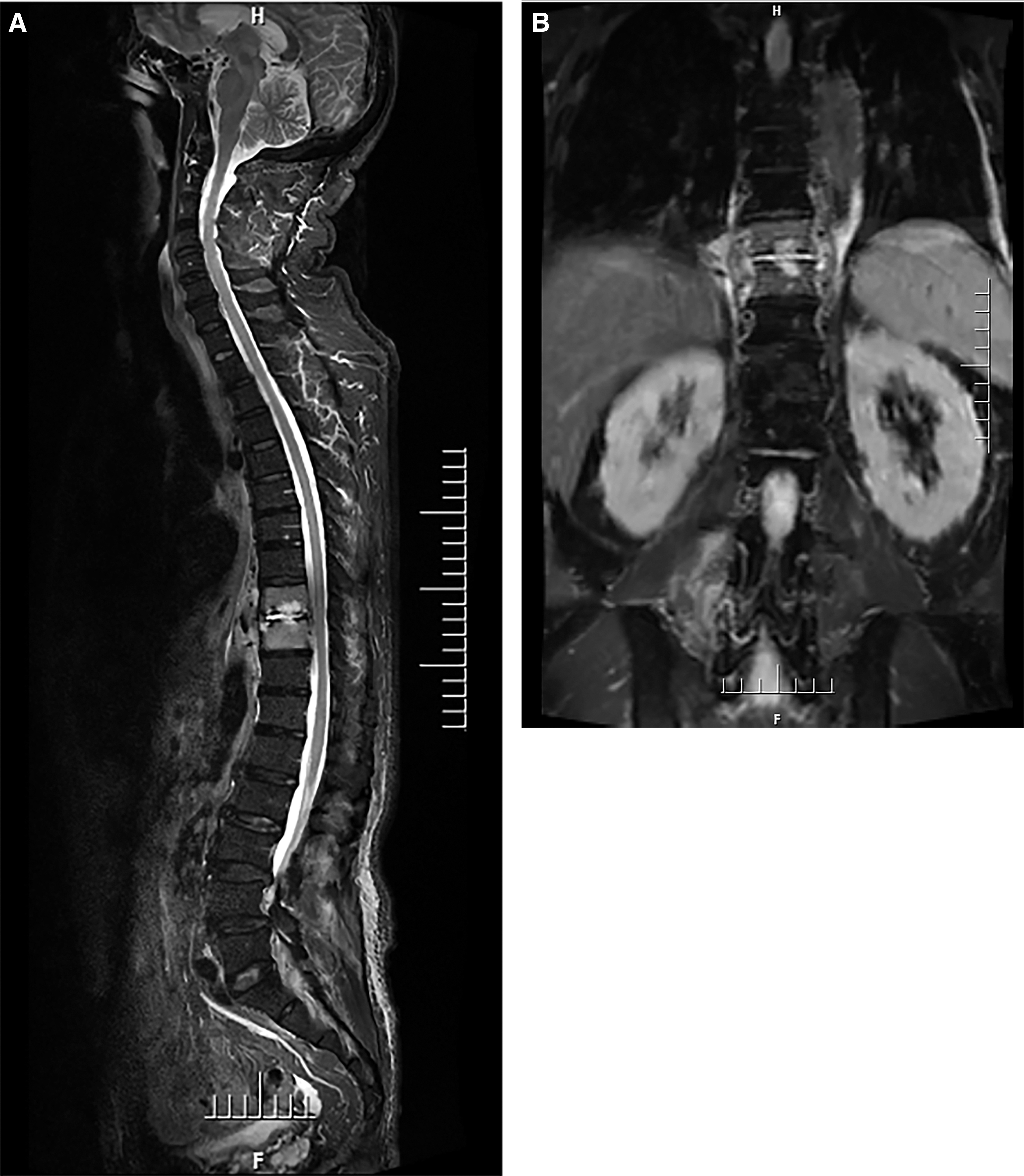
(
The patient underwent surgery on the basis of antibiotic treatment (piperacillin/sulbactam). Histopathologic examination revealed hyperplasia of fibrous connective tissue with neutrophil infiltration and sequestrum formation. Blood and tissue cultures were negative. After two weeks of antimicrobial therapy, his inflammatory markers and back pain did not improve. Repeat MRI showed paravertebral abscess formation again. Abscess puncture and drainage were performed. The pus was sent for culture and mNGS detection, which detected Nocardia brasiliensis; no other pathogenic bacteria were detected and pus culture was still negative. Then his antibiotic regimen was adjusted to trimethoprim-sulfamethoxazole combined with amikacin. The medication was prescribed for at least eight weeks. The patient's pain and inflammatory markers improved from the second treatment. Currently, there has been no evidence of recurrence; the patient undergoes regular clinical and radiologic follow-up.
Pyogenic spondylitis requires long-term antibiotic treatment and identification of the etiologic micro-organism is essential. Blood and tissue cultures require a long time and results may be negative. Compared with traditional methods, mNGS has obvious advantages in pathogen detection. Not only are the sensitivity and timeliness better than traditional culture methods, but also many types of pathogens can be detected at one time, especially for some pathogens that are difficult to detect by conventional methods. This case indicates that mNGS may be an effective method for acquisition of pathogenic bacteria of cultured-negative pyogenic spondylitis, especially for some rare pathogens.