
Abstract

To the Editor:
C
A 46-year-old female presented to the hospital with abdominal pain, nausea, and vomiting of 12 days' duration. She also reported that she had eaten raw fish 10 years ago. The results of liver function tests were abnormal, aspartate aminotransferase was 236.9 IU/L, alanine aminotransferase was 364.4 IU/L, alkaline phosphatase was 550.7 IU/L, γ-glutamyltransferase was 391.5 IU/L, total bilirubin was 52.5 mcmol/L, and conjugated bilirubin was 36.6 mcmol/L. The eosinophil count was also elevated (1,600 per cubic millimeter). Magnetic resonance imaging showed middle and lower choledocholithiasis, with low biliary obstruction. She underwent surgery, leaf-shaped worms were found in the bile ducts, pathological sections were identified as Clonorchis sinensis (Fig. 1). The patient was treated with praziquantel and recovered.
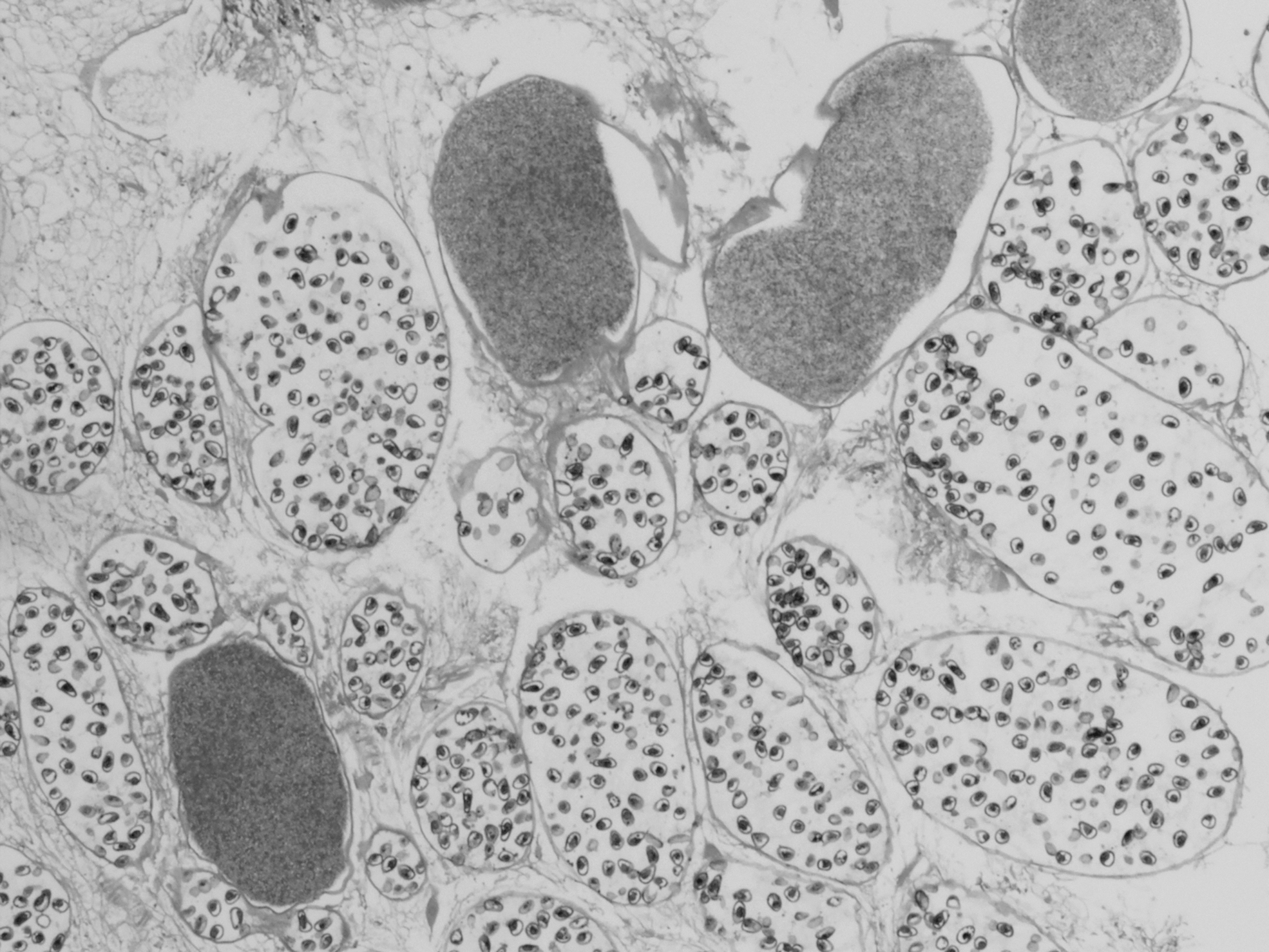
Pathologic examination of Clonorchis sinensis (hematoxylin and eosin staining, × 100).
Clonorchis sinensis may persist in the bile ducts for decades. Chronic infection results in various complications, mainly cholecystitis, cholangitis, cholelithiasis, and prolonged infestation is associated with the development of cholangiocarcinoma [2]. Symptoms caused by clonorchiasis are related to worm burden. Patients with low infection intensity are often asymptomatic or show mild symptoms, whereas patients with high infection intensity often show unspecific symptoms [2], this can easily lead to missed diagnosis. Clinicians should be alert to Clonorchis sinensis infection for patients in areas where Clonorchis sinensis is endemic or who have the habit of eating raw freshwater fish. Diagnostic methods mainly include traditional approaches (detecting the eggs in fecal samples, parasite biopsy), immunologic approaches (enzyme-linked immunosorbent assay), and molecular approaches (polymerase chain reaction, loop-mediated isothermal amplification). Praziquantel and albendazole are effective drugs for the treatment of clonorchiasis.