
Abstract

To the Editor:
A 35
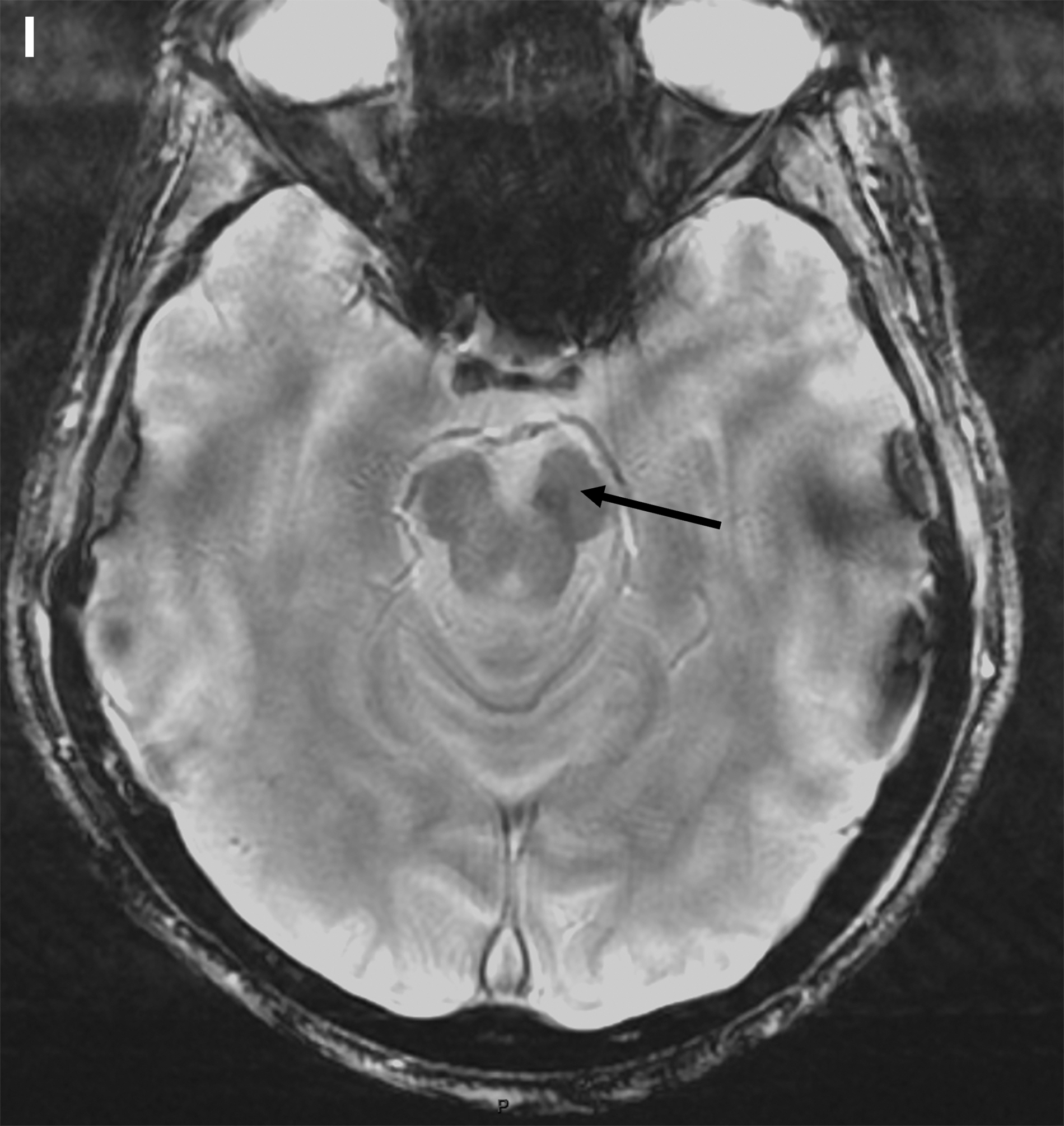
Magnetic resonance imaging (MRI): multiplanar reformat (MPR) image from contrast-enhanced three-dimensional T1 (CE-3DT1) acquisition, in the axial plane, at the level of the thalamus shows brain abscess, presenting as rim-enhancing rounded lesions. The right medial thalamus (
The course of illness was characterized by the regression of the left deficit (5/5 to 3/5) and a gradual decrease in abscesses size until their almost complete resolution at three months (Fig. 1D–1If). Among HACEK-IE, Hemophilus spp. IE are the most frequent. In our cohort of 424 patietns with IE, two (0.4%) were HP-IE. Typically, HP-IE presentation includes large vegetations, native mitral valve involvement, and embolic phenomena, leading to a severe disease. The HP-IEs are subacute in progress and are not associated with cardiac failure. Only half of the patients need valve surgery. 1 In contrast to the above considerations, our case shows that IE can develop rapidly and can be complicated by cardiac failure even in a young patient without comorbidities.
The diagnosis of HP-IE remains a challenge because of the slow growing characteristics of the organism. In our cohort of 424 patients with IE, two (0.4%) were HP-IE, and one presented with brain abscess (BA) (0.2%). To note, Darras-Jolly et al. 1 in a study of 26 HP-IE did not report BAs. The thalamus and the brainstem are unusual locations for BAs: 1.8% to 3%2,3 and 3%, respectively. 2 Thalamic abscesses may be revealed by hemiparesis, present in 44%. Subcortical aphasia has only been reported in a few cases. 4 Common symptoms of brainstem abscess are unilateral face or extremity numbness or weakness (55%), headache (50%), diplopia (38%), vomiting (38%), and ataxia (30%). Our patient had isolated hemiparesis, most likely related to the thalamic abscess. The brainstem abscesses were small in size, which would explain the absence of brainstem syndromes.
Hemophilus spp. represent 2% of 7,340 of 9,699 BAs with positive cultures in the review by Brouwer et al. 3 Hemophilus parainfluenzae is not listed in the series by Nathoo et al. 2 To the best of our knowledge, only one prior case of HP thalamic BA has been reported in a non-IE patient. 5
In IE, the differential diagnosis between BA and acute ischemia may be difficult. Contrast enhancement, not observed at the acute phase of the ischemia, strongly indicated a BA. The susceptibility ring, related to hemorrhage in the abscess wall, helps in differentiating BAs from necrotic malignant tumors; it is not observed in brain infarcts. Combined with apparent diffusion coefficient (ADC), it can help to differentiate BAs.
Our case highlights the effectiveness of antibiotic treatment alone, in a patient with HP-IE with multiple and small abscesses (≤2.5 cm). The duration of the treatment varies from six to eight weeks or more, and depends on the resolution of the lesions on the follow-up imaging.