
Abstract
Abstract
Tissue engineering is a promising approach, not only for cartilage, but also for osteochondral repair. Recent studies have demonstrated that scaffold-free cartilaginous tissue can be engineered using the alginate-recovered-chondrocyte (ARC) method. This method has also been applied to form osteochondral tissue using bovine articular chondrocytes and coralline hydroxyapatite (HA). The purpose of this study was to test whether osteochondral tissue, fabricated in vitro using the ARC method combined with a block of HA, would undergo maturation in vivo using a subcutaneous model in immunodeficient mice. Articular chondrocytes were isolated from the cartilage of New Zealand white rabbits and cultured in alginate beads. The cells with their associated matrix were recovered by dissolving the alginate beads with a sodium citrate buffer, resuspended in media and seeded onto a porous HA block. After 4 weeks of culture, some samples were analyzed, and others were implanted into subcutaneous pockets in nude mice. The analysis involved removing the cartilage portion of the de novo–formed ARC-HA graft and performing biochemical and histological examinations. Some samples were subjected to nondecalcified histology. Histological and immunohistochemical analyses of cartilaginous tissue were performed at 0, 2, 4, and 8 weeks after implantation. Biochemical characteristics were examined at 0, 4, and 8 weeks. The size and shape of the implanted ARC osteochondral tissue changed with time. The histological and immunohistochemical examination of the tissue revealed that it contained a cartilage-like matrix that stained strongly with Toluidine blue and for collagen type II. The proteoglycan (PG) content had increased significantly at 4 weeks from baseline. However, by 8 weeks, the PG content had decreased from 4 weeks. The results presented here represent a possible approach to form a tissue-engineered osteochondral implant. Further studies are needed to improve biomechanical properties of the osteochondral implant to be suitable for surgical transplantation.
Introduction
However, in the twenty-first century, articular osteochondral repair remains a clinical challenge.8–10 The treatment of osteochondral defects must integrate the repair of bone and cartilage, which have different structures and physiological functions, to restore weight-bearing and functional joint movement. 11 These requirements could potentially be met using a tissue-engineered osteochondral (bone–cartilage) composite of predefined size and shape generated in vitro using autologous cells. The advantages of such a graft would be to minimize donor site morbidity using cell-expansion techniques, to eliminate complications related to the use of allografts and mechanical devices, and to provide mechanical stability from the time of implantation. 12 Because a bone-to-bone interface integrates better and more quickly than a cartilage-to-cartilage interface, the bone region of the engineered osteochondral composite may help to anchor the graft within the defect. 13
To achieve these advantages, and to generate a bone–cartilage interface along the entire length of a construct, a biphasic scaffold may be required. 14 Biphasic scaffolds, which were designed to guide the growth of two different tissues to satisfy different mechanical and biological requirements, have been investigated for osteochondral regeneration.10,15 Synthetic hydroxyapatite (HA), which is a popular biomaterial in the orthopedic field, could be a candidate for the subchondral layer of such biphasic scaffolds. The use of ceramics (beta-tricalcium phosphate or synthetic HA) in three-dimensional bone-like tissue-generation applications is based upon their structural similarity to the mineral phase of bone. 16 HA has properties, such as pore size, continuity between pores, and shape (i.e., convex or concave) that can be controlled. In addition, the biological qualities (biocompatibility, osteoconductivity, and, under specific conditions, osteoinductivity) of HA ceramics are well established.16,17 Synthetic HA has been used as a bone-void filler or a spacer. 18 Thus, the use of HA is practical and may be clinically useful in biological resurfacing.
Previous studies have reported that chondrocytes, cultured in combination with HA, formed a mineralized cartilaginous tissue in vivo. 19 These studies, however, focused on the potential that chondrocytes loaded onto a HA graft would stimulate endochondral ossification in a bone defect. In this experiment, the use of such a composite graft was evaluated for the repair of large articular cartilage defects with involvement of the subchondral bone.
Van Susante et al. 20 attempted to restore 10-mm cartilage defects in goat femurs with chondrocytes suspended in fibrin glue on top of HA cylinders. Because of inadequate fixation of the implant, fibrocartilaginous tissue resulted. However, the study put forth the idea that using porous HA and chondrocytes as a biphasic osteochondral composite graft may be used for osteochondral repair. When a biphasic implant for an osteochondral graft is designed, the most difficult, but important, goal is to make an adequate cartilage layer for implantation; this is because cartilage is more limited than bone tissue in its ability to self-repair.
Most attempts to tissue engineer cartilage have involved the seeding of cultured cells into biological or synthetic scaffolds.21,22 Although the use of a scaffold for articular cartilage repair has been a popular approach, stable long-term results have not yet been achieved. To repair cartilage defects and obtain hyaline-like repair cartilage, a scaffold-free method to form a cartilaginous tissue may eliminate the difficult issues of degradation properties and biocompatibility of scaffold materials. Waldman et al. introduced a method to create a biphasic scaffold-free cartilage implant using porous calcium polyphosphate substrates.23–25 It has recently been demonstrated that scaffold-free, three-dimensional, mechanically functional cartilaginous tissue can be engineered using mature bovine and human articular chondrocytes using the alginate-recovered-chondrocyte (ARC) method. 26 This two-step culture approach makes possible the in vitro formation of a cartilaginous-like tissue by mature adult chondrocytes without the aid of a synthetic scaffold. We hypothesized that the preculture of chondrocytes in alginate beads would accelerate the formation of a cartilaginous tissue in a biphasic implant. We also hypothesized that the use of a biomaterial with a large pore size might enhance the integration of the implant with the subchondral bone by allowing the infiltration of mesenchymal stem cells to form the bone portion. The ARC method has been used to form osteochondral tissues using bovine articular chondrocytes and HA in vitro. 27 In that study, the similarity of the cartilaginous ARC tissues formed on an HA block to cartilage itself suggested that HA can serve as a biologically compatible support for chondrocytes in osteochondral tissue engineering. However, a study examining the in vivo maturation of the composite graft remains to be done.
The purpose of this study was to test whether osteochondral tissue, fabricated in vitro using the ARC method and an HA block, could undergo maturation in vivo using a subcutaneous model in immunodeficient mice.
Materials and Methods
Harvest of rabbit chondrocytes
New Zealand white rabbits (n = 6) weighing approximately 1.5 to 2.0 kg were used (Institutional Animal Care and Use Committee approval #04–042). After general anesthesia with an intramuscular administration of ketamine (40 mg/kg), xylazine (5 mg/kg), and acepromazine maleate (1 mg/kg), the rabbits were euthanized using an intravenous injection of a supersaturated pentobarbital solution (90 mg/kg). Using sterile technique, full-thickness articular cartilage tissue was harvested from the knee and shoulder joints. The cells were isolated from the cartilage tissue using sequential enzymatic digestion at 37°C in a humidified atmosphere of 5% carbon dioxide (CO2) using 0.2% pronase (Calbiochem, La Jolla, CA) for 1.5 h followed by 0.025% collagenase-P (Boehringer Mannheim, Indianapolis, IN) overnight at 37°C. After digestion, the cells were washed, filtered through a 70 μm mesh (Becton Dickinson, Lincoln Park, NJ) to obtain a single cell suspension, and counted. Freshly isolated cells were resuspended in 1.2% low-viscosity alginate solution (Keltone LV, ISP Alginate, Inc., San Diego, CA) at 4 million cells/mL. Alginate beads were formed by dispensing the alginate–cell suspension drop-wise into a 102 mM calcium chloride solution using a 22-gauge needle attached to a syringe pushed by a syringe pump at a constant flow rate. After 10 min, the newly formed beads (containing ∼40,000 cells/bead) were washed with a sterile 0.9% saline solution followed by washes with Dulbecco's modified Eagle medium (DMEM)/F12. The seeding density was selected to obtain the maximum matrix without a significant increase of cell number (data not shown). The beads were cultured in complete DMEM/F12 containing 20% fetal bovine serum (FBS), 25 μg/mL of ascorbate and 10 μg/mL of gentamicin in separate 100 mm suspension culture dishes. The cultures were maintained with daily media changes for 14 to 21 days.
Formation of osteochondral constructs
HA scaffolds (300 μm pore size, 70% porosity, 5 mm diameter × 2 mm thickness; CELLYARD, Pentax, Japan) were placed in individual wells of a 96-well cell culture plate (Corning, Corning, NY) containing DMEM/F12. After 30 min, the media were removed, and 50 μL of a solution of sodium hyaluronate (ARTZ, 10 mg/mL, average molecular weight 600,000–1200,000 Dalton, Seikagaku Corp., Tokyo, Japan) was added to the wells containing the HA scaffold. The HA scaffold was allowed to incubate for 1 h in this solution to facilitate the infiltration of the sodium hyaluronate into the HA scaffold (Fig. 1A). After this procedure, the bottom two-thirds of the pores of the HA scaffold were filled with the high-viscosity sodium hyaluronate solution. While this process was ongoing, the alginate beads were collected and dissolved in a buffer containing 55 mM sodium citrate, 0.15 M sodium chloride, pH 6.8, for 20 min at 4°C. The resulting suspension of chondrocytes with their cell-associated matrix (CM) was centrifuged, and the pellet was washed and resuspended in 5 mL of complete medium containing 20% FBS. The recovered cells with their CM were layered on the HA scaffolds incubating in sodium hyaluronate (Fig. 1B). Because of the viscosity of the sodium hyaluronate that had filled the bottom two-thirds of the HA scaffold, the seeded cells were not able to sediment into that part of the HA scaffold. The cells with their CM infiltrated only the top one-third of the HA scaffold that was not filled with sodium hyaluronate. The remaining cells with their CM formed a visible layer on the surface of the HA (Fig. 1C). Because the chondrocytes with their CM formed a cohesive tissue within 2 days, the sodium hyaluronate solution was replaced with complete medium supplemented with 20% FBS 2 days later. After this point in time, most of the chondrocytes remained on the top one-third of the HA scaffold without migrating into the bottom two-thirds. A preliminary experiment had indicated that cells seeded directly onto HA scaffolds, without the addition of sodium hyaluronate, did not remain on the top of the scaffold and thus did not form a desired layered structure (data not shown). The implants were cultured with daily changes of complete medium with 20% FBS in a 96-well cell culture plate (Corning) at 37°C, 5% CO2 for 2 to 3 weeks. At the end of the in vitro culture period, a majority of the sodium hyaluronate solution that had filled the pores in the bottom two-thirds of the HA scaffold had diffused out and been replaced by culture medium (Fig. 1D).

The method used to engineer a biphasic osteochondral implant. Hydroxyapatite (HA) scaffolds (300 μm pore size, 70% porosity, 5 mm diameter × 2 mm thickness) were placed in individual wells of a 96-well cell culture plate containing Dulbecco's modified Eagle medium/F12. After 30 min, the medium was removed, and 50 μL of a solution of sodium hyaluronate (1%) was added to fill the bottom two-thirds of the HA scaffold. One hour after adding sodium hyaluronate to the wells, the bottom two-thirds of the HA scaffold pores were filled with the high-viscosity sodium hyaluronate solution (
Nude mouse implantation model
Athymic mice (nu/nμ) (3–4 weeks of age, Harlan, Indianapolis, IN) were allowed to acclimate to the experimental condition for 1 to 3 weeks before the surgical procedure. All animal procedures were performed in accordance with the guidelines of the Institutional Animal Care and Use Committee at the authors' institution.
The surgical procedure was performed under general anesthesia induced using isoflurane (4–5%; Pittmann and Moore, Mundelein, IL) in oxygen administered in an anesthesia chamber. Once sedated, each animal was removed from the chamber, and isoflurane was administered using a nose cone to maintain anesthesia (1.5–2.5%). The surgical site was scrubbed with a povidone iodine-based surgical scrub (Scrub Care, Allegiance Healthcare Corporation, McGaw Park, IL) and rinsed with alcohol. Using sterile technique and equipment, four 1-cm skin incisions were made bilaterally, and subcutaneous pockets were created on the dorsa. Each animal received three to four osteochondral constructs. The incisions were closed using a surgical stapler (3M, St. Paul, MN). After surgery, the mice were allowed to recover and move freely without immobilization. At 2, 4, and 8 weeks after implantation, the mice were sacrificed using an intravenous injection of sodium pentobarbital. Half of the collected tissues were used for histological and immunohistochemical studies and cellular origin methodology, and the other half were used for biochemical studies.
Macroscopic observations
Two mice were sacrificed at 2, 4, and 8 weeks after surgery, and the implanted constructs were collected. Constructs were analyzed at each time point. The surfaces of the specimens were inspected for color, integrity, contour, and smoothness.
Histology
Harvested construct specimens were immediately fixed with 10% neutral buffered formalin for at least 24 h at room temperature. After rinsing in running tap water, samples were dehydrated using graded alcohols (70%, 80%, 90%, 95%, and 100%). The specimens were defatted in xylene for 6 to 12 h twice, again dehydrated with 100% alcohol, and infiltrated and polymerized in methyl methacrylate (Technovit 9100 New, Heraeus Kulzer Gmbh, Wehrheim, Germany).
For histological observations, hematoxylin-eosin (H-E) and toluidine-blue (T-B) staining were obtained for each specimen. H-E was used to identify the distribution of cells within the constructs. T-B was used to identify glycosaminoglycans (GAGs) in the construct extracellular matrix (ECM).
Immunohistochemistry
The expression of type I and type II collagen in the tissue was analyzed immunohistochemically. After fixation in 0.4% paraformaldehyde, the specimens were serially sectioned into 10-μm-thick sagittal sections using a cryostat, followed by air drying and incubation for 3 min in phosphate buffered saline (PBS). To abolish endogenous peroxidase, the sections were first incubated with 0.3% hydrogen peroxide (H2O2) in methanol for 10 min. After three washes in PBS and 0.2% TWEEN 20 (Sigma-Aldrich, St. Louis, MO), pH 7.4, the sections were enzymatically digested using testicular hyaluronidase type VIII (Sigma-Aldrich; 0.2% in PBS, pH 7.4) for 30 min at 37°C. These sections were then incubated in a 1:400 dilution of mouse monoclonal anti-rabbit type I collagen (Sigma-Aldrich) or mouse monoclonal anti-rabbit type II collagen (Developmental Studies Hybridoma Bank, Iowa City, IA) antibodies overnight at 4°C. Immunoreactivity was detected by incubating sections with biotinylated rabbit anti mouse antibody, followed by streptavidin-horseradish peroxidase (SAB-PO kit; Nichirei, Tokyo, Japan). The signal was developed as a brown reaction product using peroxidase substrate 3, 3′-diaminobenzidine, and H2O2. The sections were counterstained with Harris hematoxylin, dehydrated, cleared, and mounted.
Biochemical characterization of the cartilaginous tissue formed in vivo
All cartilaginous ARC tissues were separated from the HA block, and wet weights were obtained. The specimens were digested overnight at 55°C with papain at 20 μg/mL in 0.1 M sodium acetate, 50 mM sodium ethylenediaminetetraacetic acid, and 5 mM L-cysteine hydrochloride at pH 5.53, after which the specimens were boiled for 10 min to deactivate the papain and stored at −20°C until analyzed. 28 After papain digestion, the DNA content of the cells in the cartilaginous ARC tissues was measured using the bisbenzimidazole fluorescent dye method (Hoechst 33258: Polysciences, Inc., Warrington, PA). 28 The content of sulfated proteoglycans (PGs) in the papain digest was determined using a modified dimethylmethylene blue (Polysciences, Inc.) dye-binding method. 29
Statistical analysis
The data are expressed as means ± standard deviations. The data were subjected to one-way analysis of variance using the between-subject factors for the different time points. The post hoc analyses were performed using the Fisher protected least significant difference test; significant differences are defined at p < 0.05.
Results
Wound healing was uneventful, and all animals could be followed as planned. No bacteriological contamination was detected in implanted tissues.
Macroscopic observations of the tissue-engineered osteochondral construct (ARC tissues on HA blocks)
Macroscopic observations were made after removal of the granulation tissue that surrounded the implanted constructs. A gross examination of the implant showed that there were no abrasions of the cartilaginous ARC tissues and that the ARC tissues appeared to be well integrated with the HA block interface.
The size and shape of the implanted ARC osteochondral tissue changed with time. At implantation, the surface of the implanted specimens was rough and fragile. A significant change in the shape of the retrieved constructs was noticed between 2 and 4 weeks post-implantation. After the 2-week time point, the overall size gradually reduced, and the surface of tissue became more stiff and opaque.
Histology
H-E staining
The ARC tissue was formed on the surface and the top one-third of HA block at implantation. Chondrocytes from the ARC tissue did not significantly migrate into the deep portion of the HA scaffold throughout the study. The cells that did grow into the HA pores showed vascular cell shapes or a fibrous shape, unlike chondrocytes. Those cells were assumed to originate from the nude mouse host tissue. A uniform distribution of chondrocytes was observed in the cartilaginous tissues at the 4-week time point (Fig. 2A). At the 8-week time point, the chondrocytes were well distributed in the interface area, whereas the cell density in the surface area had decreased from the 4-week time point (Fig. 2B).
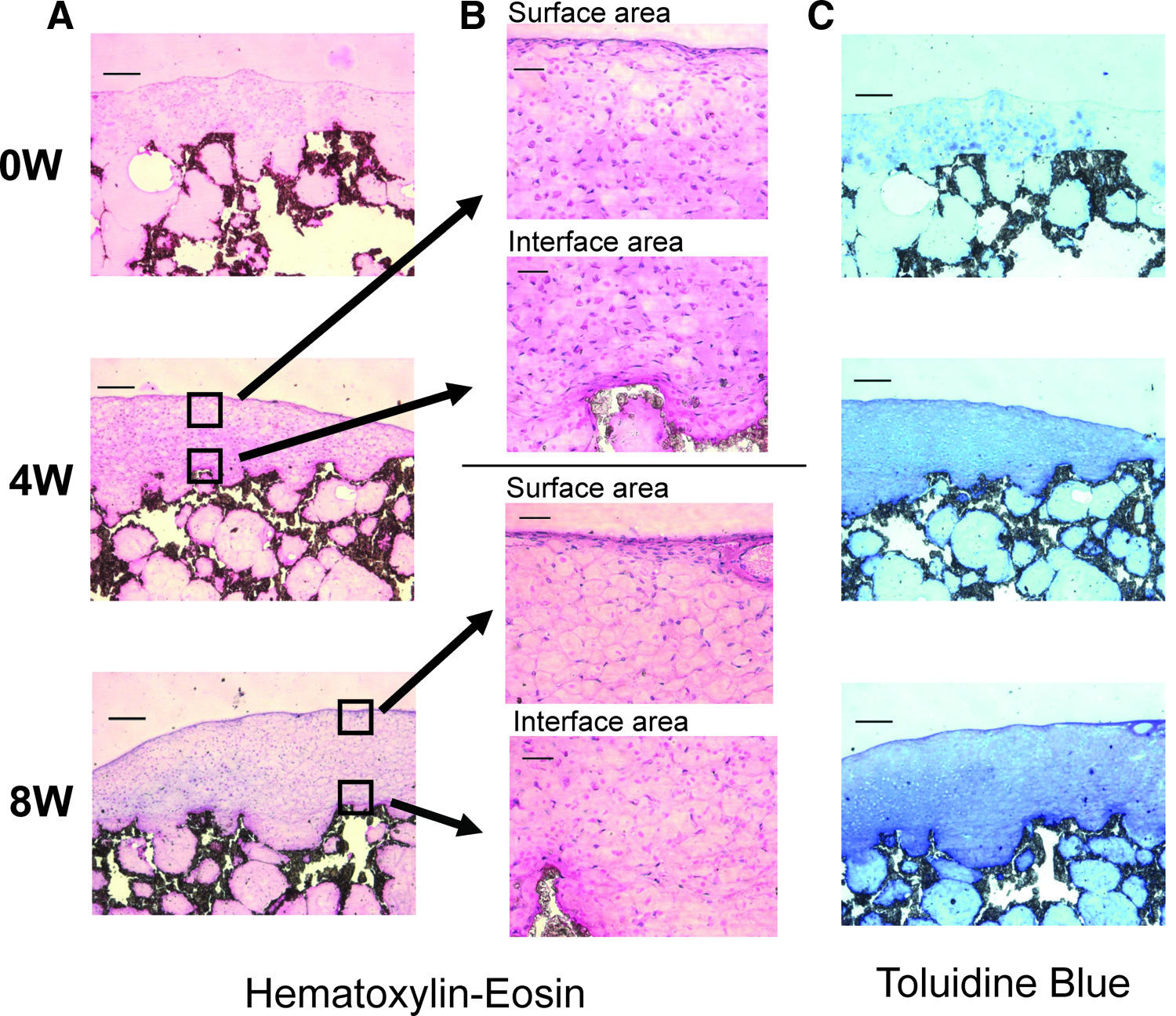
The histological appearance of osteochondral composite implants before (0-week time point) and after in vivo maturation (4- and 8-week time points) in a subcutaneous model in immunodeficient mice. (
T-B staining
Histological examination of the ARC tissues revealed that they contained a cartilage-like matrix that stained strongly with T-B, which reflects the GAG content. The distribution of T-B positive-staining areas showed a time-dependent difference between the cartilaginous constructs. Before implantation (the 0-week time point), the accumulation of GAGs was restricted to scattered areas, but after 2 weeks in vivo, a more-continuous ECM containing GAGs was observed. At the 4-week time point, the specimens showed massive areas of T-B–positive staining from the surface to the deep zone, although the T-B–positive–stained areas seemed to shift to the middle and deep zones at the 8-week time point (Fig. 2C).
Immunohistochemistry
At the 2- and 8-week time points, immunostaining of ARC tissues with a monoclonal antibody against type II collagen revealed the expression of type II collagen, particularly in the territorial area around the chondrocytes.
A time-dependent coalescence of matrices formed by the individual chondrocytes was evident. The amount and coalescence of ECM positively stained for type II collagen were higher at the 8-week time point than the 2-week time point (Fig. 3). Positive immunostaining for type I collagen was observed only on the surface of the retrieved ARC tissues (data not shown).
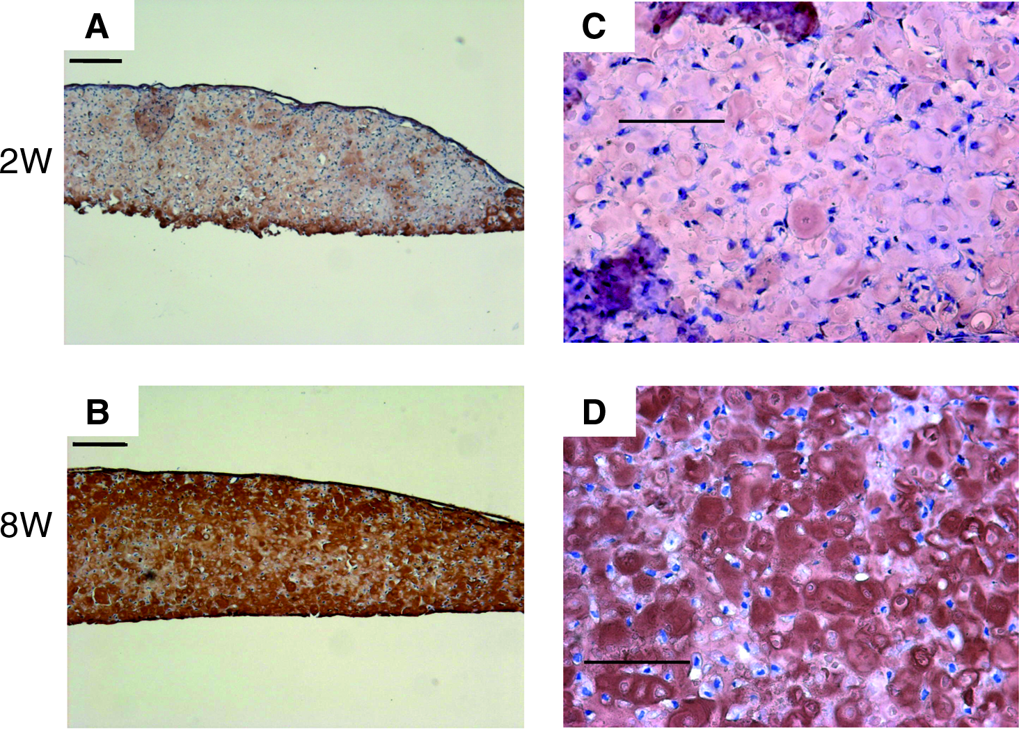
An immunohistochemical analysis of osteochondral composite implants after in vivo maturation (2- and 8-week time points) in a subcutaneous model in immunodeficient mice, using an antibody directed against type II collagen, shows a uniform and intense staining of the matrix that is time-dependent. (
Biochemical analyses
At the 8-week time point, the wet weights of the ARC tissues formed on the osteochondral constructs had increased significantly from the 0- and 4-week time points (0 weeks, 7.50 ± 1.25; 4 weeks, 10.0 ± 0.75; 8 weeks, 14.63 ± 2.28 mg; 8 weeks, p < 0.01 vs 0- weeks, p < 0.05 vs 4 weeks) (Fig. 4A). The DNA content of the ARC tissues had decreased significantly at the 4-week time point from implantation (0 weeks, 2.73 ± 0.3; 4 weeks, 1.06 ± 0.06 μg, p < 0.05). However, at the 8-week time point, the DNA content had increased significantly from the 4-week time point (8 weeks, 2.69 ± 1.37 μg, p < 0.05). The PG content, normalized to the DNA content, had increased significantly at the 4- and 8- week time points from implantation (0 weeks, 3.62 ± 0.6; 4 weeks, 23.3 ± 6.24; p < 0.01 vs 0 weeks, 8 weeks, 13.1 ± 0.72, p < 0.05 vs 0 weeks and 4 weeks, μg/μg DNA). However, at the 8-week time point, the PG content had decreased significantly from the 4-week time point (p < 0.05) (Fig. 4B).

The wet weight and proteoglycan (PG) content of cartilaginous alginate-recovered-chondrocyte (ARC) tissues before (0-week time point) and after in vivo maturation (4- and 8-week time points) in a subcutaneous model in immunodeficient mice. At implantation and after 4 and 8 weeks, osteochondral grafts were removed from the nude mice, and the ARC tissues on the scaffold were excised. (
Discussion
This study demonstrated that, using the ARC method, rabbit chondrocytes can be cultured in vitro on porous HA blocks to generate tissue-engineered osteochondral constructs with a well defined cartilage–bone-like interface. When placed in subcutaneous pockets in the nude mouse for up to 8 weeks, the ARC layer of the graft exhibited cartilage maturation. The newly developed methodology was established to obtain the attachment of tissue-engineered cartilage, formed using the ARC method, to the HA scaffold with a limited penetration of chondrocytes into the pores of the HA scaffold.
The ARC method used in this study consisted of culturing chondrocytes in alginate beads under conditions that maintain the chondrocytic phenotype and modulate the formation of a CM rich in aggrecan molecules. 26 After 2 weeks, the alginate gel was solubilized, and the cells with their CM were recovered and cultured on an HA block. The initial use of sodium hyaluronate to limit the migration of seeded chondrocytes with cell-associated matrix successfully localized chondrocytes on the surface and top one-third of HA blocks. After in vitro culture for another 2 weeks, the integration of the ARC tissue with the HA interface in the composite construct was already histologically observable. To the best of our knowledge, the present study is the first to report the in vivo maturation of a biphasic osteochondral implant consisting of a type of scaffold-free cartilaginous tissue and an HA block.
The histological results of this study indicated that the porous structure of the HA block serves as subchondral bone substrate and plays an important role in forming the biphasic structure. After 2 to 3 weeks in in vitro culture, the pores in the HA block (bottom two-thirds) became open because a majority of the sodium hyaluronate had diffused out. The chondrocytes seeded onto the HA scaffold did not migrate into the bottom two-thirds at this time because these cells had already formed a cohesive tissue. The fact that the nude mouse host cells migrated into the HA pores indicates that the pores in the HA block were not filled with chondrocytes and could be filled with cells, such as those that would come from bone marrow when the implants are placed in joints. It is possible that the significant drop in the PG content of the cartilaginous ARC tissue found between 4 and 8 weeks may have resulted from the loss of local nutrition caused by the granulation tissue that surrounded the implanted constructs.
From the examinations of the ARC tissue according to gross appearance, histology, and the expression of type II collagen in the ECM, this cartilaginous construct showed a time-dependent maturation. The structural stability of the ARC–HA interface was also observed within subcutaneous pockets of the nude mice. One of the limitations of this study was that conditions in the subcutaneous pockets of nude mice might differ from those in a joint cavity. Further studies are essential to confirm the feasibility of the ARC–HA constructs for the repair of osteochondral defects. Biomechanical analyses are also needed to investigate whether the biomechanical properties of the ARC–HA constructs are suitable for transplantation at surgery. In particular, the degree of integration of cartilaginous tissue into the scaffold is an important factor to improve the retention of implanted tissue in defects. Methodologies used to assess integration in two different materials need to be developed to further study the usefulness of biphasic constructs.
A variety of tissue engineering techniques have been developed to regenerate bone and cartilage.5,9,30–33 These studies have illustrated that engineered cartilage must integrate with the host tissue, provide a smooth and natural articulating surface, and bear functional mechanical loads. Numerous researchers have investigated the use of a single-phase biodegradable scaffold for articular cartilage repair. 34 For example, poly(lactic-co-glycolic acid) has frequently been chosen for tissue-engineering applications because its degradation can be tailored. Although these approaches have achieved varying degrees of success, no single technique has satisfied the requirements for integration with the host tissue, maintenance of a natural articulate contour, and mechanical function. To satisfy these requirements and to generate a bone–cartilage interface along the entire length of a construct, a biphasic scaffold may be required.
More recently, tissue engineers have started to design and study hybrid or biphasic scaffold constructs. Gao et al. reported the results of a hybrid scaffold study in a rat subcutaneous bag. 10 Rat bone marrow–derived mesenchymal stem cells were loaded in a hyaluronic acid sponge for the cartilage compartment and in a calcium phosphate scaffold, which served as the bone. At 6 weeks post-implantation, fibrocartilage was observed in the sponge, and bone-like tissue was detected in the ceramic scaffold. From these studies, it can be concluded that a biphasic osteochondral implant would be more advantageous in histological and mechanical aspects than a single-phase composite. However, there are several reports demonstrating phenotypic instability of the cartilage layer in the repaired tissue after implantation. Niederauer et al. reported that resurfacing of the joint had occurred with fibrocartilaginous repair tissue after implantation of a biphasic osteochondral implant consisting of chondrocytes suspended in fibrin glue and HA. 34 Phenotypically stabilized engineered cartilage, preferably without a scaffold, might be needed at the cartilaginous site of a biphasic osteochondral implant.
The ARC method is a useful scaffold-free culture technique for the maintenance of the chondrocytic phenotype in vitro. Chondrocytes that have de-differentiated during monolayer culture, as is typically done for the autologous chondrocyte implantation procedure, 5 could re-acquire the chondrocytic phenotype with three-dimensional culture in alginate beads. Thus, chondrocytes cultured using the ARC method maintain their normal phenotype during the culture process, allowing in vitro tissue production to proceed rapidly and permitting harvest at an early, immature stage of tissue organization. 26 The juxtaposition of a cartilage-like cellular phenotype, ECM, and, potentially, mechanical properties, should lead to better outcomes in the clinical use of an osteochondral graft produced using ARC–bone composite technology.