




Abstract
Despite the attractive features of nanofibrous scaffolds for cell attachment in tissue-engineering (TE) applications, impeded cell ingrowth has been reported in electrospun scaffolds. Previous findings have shown that the scaffold can function as a sieve, keeping cells on the scaffold surface, and that cell migration into the scaffold does not occur in time. Because fiber diameter is directly related to the pore size of an electrospun scaffold, the objective of this study was to systematically evaluate how cell delivery can be optimized by tailoring the fiber diameter of electrospun poly(ɛ-caprolactone) (PCL) scaffolds. Five groups of electrospun PCL scaffolds with increasing average fiber diameters (3.4–12.1 μm) were seeded with human venous myofibroblasts. Cell distribution was analyzed after 3 days of culture. Cell penetration increased proportionally with increasing fiber diameter. Unobstructed delivery of cells was observed exclusively in the scaffold with the largest fiber diameter (12.1 μm). This scaffold was subsequently evaluated in a 4-week TE experiment and compared with a poly(glycolic acid)-poly(4-hydroxybutyrate) scaffold, a standard scaffold used successfully in cardiovascular tissue engineering applications. The PCL constructs showed homogeneous tissue formation and sufficient matrix deposition. In conclusion, fiber diameter is a crucial parameter to allow for homogeneous cell delivery in electrospun scaffolds. The optimal electrospun scaffold geometry, however, is not generic and should be adjusted to cell size.
Introduction
One advantage of electrospun scaffolds is that their fiber structure, particularly when in nanoscale, is associated with high surface-to-volume ratios, providing a large area for cell attachment. Furthermore, the physical form of the nanofibrillar matrices provides high porosity and high spatial interconnectivity. It was postulated that the use of nanofibrous structures would bear a close resemblance to the dimensions of natural extracellular matrix (ECM). 3 Although this resemblance may apply to the fiber thickness and the porosity of the nanofibrous scaffold, the spatial characteristics of ECM were not attained.3,8 In fact, the pore size of the scaffold was smaller with decreasing fiber diameter and can be as small as 100 nm. 9 Such small pore sizes may interfere with cellular infiltration in the scaffold, thus undoing the advantages of nanofibrous scaffolds for use in TE. However, this feature may be beneficial when the nanofiber mesh is used as a membrane, separating cell types, yet allowing communication through interconnected pores. 10
Microfiber scaffolds, alternatively, consist of a more spacious, coarser geometry, with fiber diameters up to 10 μm and corresponding pore sizes of up to 45 μm. 8 Although the geometry of a microfiber scaffold appears more suitable for TE purposes with regard to cellular infiltration, the majority of electrospinning research has focused on nanofibers. Pham et al. demonstrated the reproducibility and uniformity of electrospun poly(ɛ-caprolactone) (PCL) microfiber scaffolds with fiber diameters ranging from 2 up to 10 μm.7,8 Subsequently, they constructed a bilayered electrospun structure of a 5-μm scaffold with a nanofiber top layer to evaluate the combined potential of both scaffold geometries. Rat marrow stromal cells could successfully infiltrate the structure. 8 Van Lieshout et al. compared a large-pore-size knitted PCL scaffold with an electrospun PCL scaffold. 6 Although human myofibroblast cells were found throughout the whole knitted scaffold, retaining the cells within the mesh was a problem because of the large pores (+200 μm). 6 On the other hand, cell ingrowth in the electrospun scaffold was barely present even after 4 weeks of culture, despite its microscale fiber diameter. Previous experiments have demonstrated that cell migration from the surface into the deeper parts of the scaffold does not occur during culture in electrospun scaffolds and that initial homogeneous cell delivery is essential to allow for homogeneous tissue formation in engineered tissues. To ensure successful use of electrospun scaffolds, the advantage of a high surface-to-volume ratio and sufficient pore size should be maintained. Therefore, the ideal scaffold environment for cells should be a cell-size-specific trade-off between complete cell infiltration and a large surface area. In this study, human myofibroblasts approximately 10 μm in diameter were used. The minimal pore size should hence have at least similar dimensions.
The objective of this study was to systematically evaluate how cell delivery can be optimized by tailoring the fiber diameter of electrospun scaffolds. This was determined by comparing cellular infiltration in five electrospun scaffold groups with fiber diameters ranging from 3.4 to 12.1 μm. The scaffold that complied with the requirements of complete cell infiltration was evaluated for its potential in cardiovascular TE. In a 4-week TE experiment, the electrospun scaffold was compared with a porous nonwoven fiber mesh of poly glycolic acid (PGA) coated with poly(4-hydroxybutyrate) (P4HB) serving as a reference scaffold. The latter scaffold has been successfully used in cardiovascular TE.11,12 Both scaffold materials are approved by the Food and Drug Administration. 13
Materials and Methods
Materials
Chemicals were obtained from Sigma-Aldrich (Zwijndrecht, Netherlands) unless otherwise stated. PCL (Mn 8 ×10⁴) was dissolved at 13.7 and 17.5 w/v% in chloroform (99.9% high-performance liquid chromatography (HPLC) grade). Non-woven PGA meshes (Cellon, Bereldange, Luxembourg) were dipcoated in a solution of P4HB (Mn 1 × 10⁶; TEPHA Inc.,) in tetrahydrofuran, as described previously.11,14
PCL electrospinning
The custom-built electrospinning set-up existed of a high-voltage power supply, an infusion pump (Kd Scientific, USA), a 10-mL plastic syringe (Terumo, Belgium), a stainless steel blunt needle (inner diameter 0.6 mm), and a stagnant grounded collector. The syringe was horizontally fixed in the infusion pump. The polymer solution was led through a plastic tube to the needle, which was vertically fixed 15 cm above the collector. The polymer solution was electrostatically drawn from the tip of the needle using high voltage between the needle and the collector. Five sheet-like scaffolds (groups A to E) with increasing fiber diameters were produced by varying the spinning parameters: flow rate (Q), the applied voltage (V), and the concentration of the polymer solution (Table 1). The thickness of all electrospun sheets was approximately 1 mm.
Measurement of fiber diameters
Recent work by Eichhorn and Sampson elegantly describes a theoretical model of the fibrous network structure produced by electrospinning. 15 The model demonstrated the mutual dependence of fiber width and mean pore radius and, more specifically, a direct relation between fiber diameter and pore size. Based on this relationship, fiber diameter can be interpreted as a reliable predictor of pore size, which is difficult to measure accurately in thin sheets of electrospun scaffolds. The fiber diameters of the electrospun scaffolds were imaged using environmental scanning electron microscopy (ESEM; Philips XL30 ESEM-FEG), and fiber diameter was assessed in five samples per group, 10 measurements per sample. Quantitative image analysis of fiber diameters were performed in MATLAB (The MathWorks Inc.,).
Culture of human venous myofibroblasts
Myofibroblast cells, harvested from the human vena saphena magna and expanded using regular cell culture methods, were used as described previously. 16 As culture medium, Dulbecco's modified Eagle medium advanced (Gibco) supplemented with 10% fetal bovine serum (Biochrom, Germany), 1% glutamax (Gibco), and 0.1% gentamycin (Biochrom) was used. Every 3 to 4 days, the medium was replaced, and cultures were maintained in a humidified incubator at 37°C with 5% carbon dioxide.
Cell seeding and tissue culture
Cells of passage 7 were enzymatically detached by adding trypsin-ethylenediaminetetraacetic acid (EDTA; Cambrex,) to the monolayer cultures. During the seeding procedure, cells were centrifuged and resuspended in a thrombin solution (10 IU/mL) (Sigma Chemicals, St. Louis, MO), mixed with a fibrinogen solution (10 mg/mL) (Sigma Chemicals, St. Louis, MO), and immediately dripped evenly on the scaffold (1 × 10⁶ cells/100 mm3 scaffold). Polymerization of the fibrin gel started after approximately 40 s. During culture, the gel serves as a cell carrier. 17 The cell-seeded rectangular scaffold strips (5 × 35 ×1 mm) will be referred to as tissue-engineered constructs. In the first study, cell delivery was investigated in the electrospun PCL scaffolds with increasing fiber diameter after 3 days of culture (n = 4 per group). In the second study, tissue development was investigated in PCL constructs and compared with PGA-P4HB constructs after 4 weeks of culture (n = 6 per group). PCL group E was chosen based on the results of the cell infiltration experiment. Electrospun PCL scaffolds without cells served as controls. To create static strain conditions, which has been shown to be beneficial for tissue development, 12 the scaffolds were attached at the ends, using a polyurethane-tetrahydrofuran glue (20 w/v%). The medium for tissue culture was supplemented with extra gentamycin (0.3%) and L-ascorbic acid 2-phosphate (0.25 mg/mL) to promote ECM production.
Quantification of cell delivery
The tissue-engineered constructs were fixed in 3.7% formaldehyde (Merck, Germany) after 3 days of culture and embedded in Technovit 7100 (Heraeus Kulzer, Germany). Five-μm sections were cut, and cells were fluorescently stained with 4′-6-diamidino-2-phenylindole (DAPI). In each scaffold group, 12 fluorescence and corresponding light microscopy images (to distinguish the edges of the scaffold) were acquired using a Nikon TE300 fluorescence camera. Cell delivery in the scaffold was analyzed in three layers of equal size: the seeding side and the middle and bottom layers. Cells were counted in each layer and expressed as a percentage of the total number of cells in the image, using image analysis in MATLAB.
Mechanical properties of tissue-engineered constructs
After 4 weeks of culture, the mechanical properties of the tissue-engineered constructs and the unseeded scaffold material were measured using a uniaxial tensile tester equipped with a 20-N load cell (Kammrath & Weiss, Germany). The dimensions of the rectangular-shaped constructs were measured using an optical imaging profiler (SensoFar PLμ 2300, Sensofar-Tech, Spain), and each sample was tested in the axial direction at a fixed strain rate of the initial sample length (l0) per minute. The Young's modulus, defined as the slope at the linear part of the curve, and Cauchy stresses at fixed strain values (5%, 10%, and 15% strain) were assessed from the stress-strain curves. This way of presenting the data was preferred over presenting the strain at break because the rupture mode was different between the groups.
Quantitative tissue analysis
After tensile testing, biochemical assays were performed on the tissue-engineered constructs to evaluate the ECM composition. For DNA and glycosaminoglycan (GAG) analyses, lyophilized tissue samples were digested in papain solution (phosphate buffer, L-cysteine, EDTA, and papain) at 60°C for 16 h. The DNA content was determined using the Hoechst dye method 18 and a standard curve from calf thymus DNA. The GAG content was determined using a modification of a previously described assay 19 and a standard curve from chondroitin sulfate from shark cartilage. For collagen analysis, the digested samples were hydrolyzed in 6 M hydrochloric acid (Merck, Germany). Hydroxyproline (Hyp) residues were measured on the acid hydrolysates using reverse-phase (RP) HPLC) after derivatization with 9-fluorenylmethyl chloroformate (Fluka, Switzerland). 20 The same hydrolysates were used to measure the number of the mature collagen cross-links hydroxylysyl pyridinoline (HP) using HPLC as described previously.21,22 HP is the main type of collagen cross-link present in cardiovascular tissue. The number of HP cross-links was expressed per collagen triple helix. Hyp and GAG content were normalized to the amount of DNA to obtain a measure for the amount of matrix components produced per cell. The amount of DNA was not normalized to dry weight but expressed as absolute values, because the scaffold contribution to the constructs weight after 4 weeks was considerably different for PGA-P4HB and PCL.
Qualitative tissue analysis
After 4 weeks of culture, the tissue-engineered constructs were fixed with 3.7% formaldehyde and embedded in Technovit 7100 (Heraeus Kulzer, Germany). Five-μm-thick sections were cut and stained with toluidine blue (Klinipath, Netherlands) to evaluate tissue formation.
Statistics
Statistical analysis was performed using SPSS 13.0 software (SPSS, Inc., Chicago, IL). Values are presented as means ±standard deviations. Differences between groups were determined using univariate analysis of variance followed by Bonferroni post hoc tests. Differences with P-values less than 0.05 were considered significant.
Results
Scaffold morphology
The spinning parameters were optimized to obtain smooth, regular-shaped fibers in all scaffold groups. Using the parameters shown in Table 1, the electrospun scaffolds consisted of randomly oriented uniform meshes of smooth, regular-shaped fibers. The fiber diameters were 3.4 ± 0.29 μm (group A), 5.0 ± 0.29 μm (group B), 6.7 ± 0.56 μm (group C), 8.7 ± 0.57 μm (group D), and 12.1 ± 1.21 μm (group E). Small standard deviations in the fiber diameter and uniform fibers, as visible in ESEM images (Fig. 1A–E), suggested that all groups consisted of meshes with constant fiber thickness.

(
Cell delivery
Fluorescent images of the constructs with DAPI-stained cells showed clear differences in infiltration of cells between the five groups (Fig. 1F–J). Scaffold A, with the smallest fiber diameter, showed barely any cellular infiltration; with increasing fiber diameter, the number of intruded cells in the scaffold increased to a homogeneous cell distribution in group E. Quantification of the number of cells in each of the three layers using image analysis is shown in Figure 2. The graph shows increasing percentages of cells in the middle layer of the scaffold with increasing PCL fiber diameter. Only in the largest fiber diameter mesh (group E) did the scaffold structure appear not to impede cellular infiltration. Although the number of cells in the bottom layer was lower than in the two layers above, the equal amounts of cells in the two upper layers indicate that cell penetration was not obstructed. Cells in the thinner fiber meshes (A and B) surrounded the scaffold rather than penetrating it, as indicated by the large amounts of cells in the seeding and the bottom layer. These data suggest that, of the tested scaffolds, the 12.1-μm fiber group had the smallest fiber size and obtained sufficiently large pores, which allow complete cellular infiltration.

Quantified cellular infiltration expressed as a percentage of the total number of cells per cross-section in poly-(ɛ-caprolactone) scaffold groups A–E. The number of cells in the middle layer of the scaffold increased with increasing fiber diameter.
TE experiment
A morphological comparison of the two scaffolds types (PGA-P4HB and PCL group E) before cell seeding is shown in Figure 3. A strong similarity in fiber morphology could be observed between the two scaffold types. After 4 weeks of culture, the PCL group E scaffold was still completely intact, in contrast to the largely degraded PGA-P4HB scaffold.
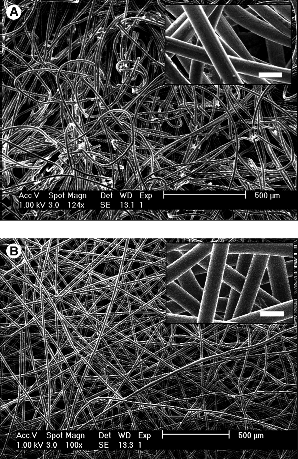
Environmental scanning electron microscopy images of poly(ɛ-caprolactone) group E (
Mechanical properties
Stress-strain curves were obtained from uniaxial tensile tests to assess the mechanical properties of the constructs after 4 weeks of culture. Representative curves of each group are shown in Figure 4, and averaged values are displayed in Table 2. PCL constructs with and without cells were stiffer (higher modulus) and stronger (higher tensile stress at fixed strains) than PGA-P4HB constructs with cells. Because PCL had not degraded after 4 weeks, the polymer dictated the constructs' mechanical response, hence the difference between the PCL and PGA-P4HB constructs. Significantly higher values of the modulus and the stress at fixed strains (5%, 10%, and 15% strain) were found in the PCL scaffolds with cells than in the scaffolds without cells, indicating that the presence of cells significantly improved stiffness and strength. The optimal mechanical properties of the tissue construct are application dependent and can be modulated by changing the fiber diameter of the electrospun scaffold or by varying the intrinsic mechanical properties of the scaffold material. A systematic investigation of the fiber diameter–mechanical property relationship is beyond the scope of this study.

Representative stress–strain curves of tissue-engineered constructs after 4 weeks of culture. The poly (ɛ-caprolactone) (PCL) scaffold was stiffer and stronger than the oly glycolic acid coated with poly 4-hydroxybutyrate construct. The presence of cells improved the stiffness and strength of the PCL scaffold.
Significantly different from PCL with cells (p < 0.01).
Significantly different from PCL without cells (p < 0.01).
Quantitative tissue analysis
Tissue composition (collagen, collagen cross-links, GAGs, and DNA) of all scaffolds was quantified after 4 weeks and is presented in Table 3. No difference was observed in the amount of DNA between the constructs from the PCL and PGA-P4HB scaffold groups. The amount of GAG and Hyp per DNA were approximtely 20% higher in the PCL constructs than in the PGA-P4HB constructs, indicating slightly greater matrix synthesis per cell in the PCL group. However, the number of cross-links per collagen molecule was lower in the PCL constructs than in the PGA-P4HB group.
Significantly different from PCL (p < 0.01).
Qualitative tissue analysis
Images of toluidine blue–stained histological sections of the PCL and PGA-P4HB groups are shown in Figure 5. Tissue formation appeared more homogeneous in the PCL group than in the PGA-P4HB group. Additionally, substantial compaction of PGA-P4HB constructs was observed because of scaffold degradation, in contrast to the unaffected dimensions of PCL constructs.
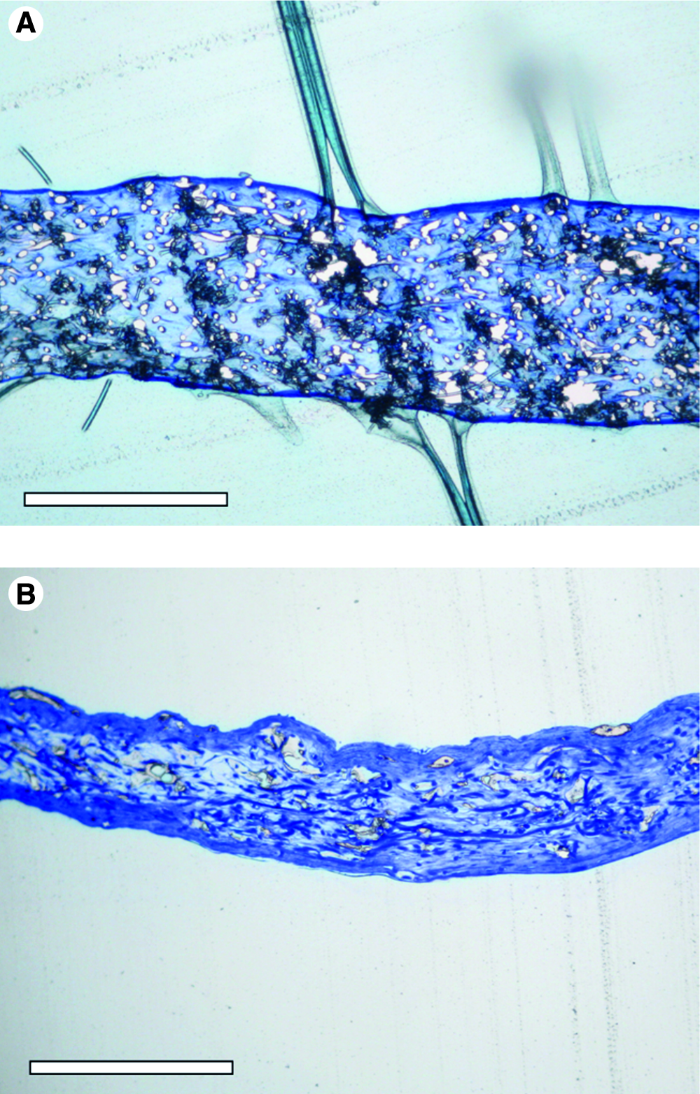
Histological images of toluidine blue stained slides of poly(ɛ-caprolactone) (PCL) group E (
Discussion
The objective of this study was to systematically evaluate how cell delivery can be optimized by tailoring the fiber diameter of electrospun PCL scaffolds for cardiovascular TE purposes. The data presented in this study show that cellular infiltration into electrospun PCL scaffolds depends strongly on the fiber diameter, which is directly related to the pore size of the scaffold.
In this study, human venous myofibroblasts were seeded on various electrospun PCL scaffolds with fiber diameters ranging from 3.4 to 12.1 μm. For this specific cell source, unobstructed cellular infiltration was observed only in the scaffold with average fiber diameter of 12.1 μm. Quantification of cell delivery in five scaffold groups, with fiber diameters ranging from 3.4 up to 12.1 μm, showed a gradual increase in cellular infiltration with increasing fiber diameter.
The results of the cell infiltration study validated the theoretical predictions of Eichhorn's model that pore size increases with increasing fiber diameter. 15 Pham et al. demonstrated high reproducibility and uniformity of electrospun microfiber scaffolds in the same diameter range as were used in the present study. Although cell delivery was not investigated in different size microfiber scaffolds, it was suggested that smaller fibers can impede cellular infiltration. 8 The data presented here confirm this suggestion and showed that even a small change in average fiber diameter can have a significant effect on cellular infiltration. This emphasizes that fiber thickness is an essential parameter for three-dimensional TE applications. Because cell size differs between cell types and species, 23 the optimal electrospun scaffold structure is not a uniform geometry but should be cell-specific.
In the second part of this study, the PCL electrospun scaffold with average fiber diameter of 12.1 μm was evaluated for tissue development and compared with a PGA-P4HB scaffold. The PGA-P4HB model system has produced strong and functional engineered tissues.24,12 The search for an alternative scaffold material and the choice for PCL as a scaffold material were based on two arguments. First, a general shortcoming of the current status of cardiovascular engineered tissues is the lack of elastin. Proper in vivo functioning of vascular tissue-engineered grafts has been unsuccessful because of the lack of elastin biosynthesis in the tissue equivalents. 25 Second, PGA-P4HB degrades within weeks during culture and is therefore not suitable to provide sustained mechanical support in vivo. For particular cardiovascular TE applications (e.g., blood vessels), supported mechanical integrity and elasticity of the scaffold are desired during culture and after implantation. PCL exhibits elastic behavior with only minor permanent deformation and is mechanically stable for 2 to 3 months. 26 Therefore, PCL could function as an elastic substitute, whereas natural elastin is gradually produced to take over this role. It appears to be a promising material for cardiovascular TE, especially when prolonged periods of mechanical support are desired.
With scanning electron microscopy, a high morphological resemblance was observed between the PCL scaffold and the reference PGA-P4HB scaffold before seeding, supporting the potential of the PCL scaffold for TE. After 4 weeks of culture, PGA-P4HB constructs showed a denser tissue structure and more-compact dimensions than PCL constructs. PCL served as a support structure, maintaining its initial dimensions, whereas the PGA-P4HB scaffold degraded and lost its mechanical support, allowing compaction of the construct. Mechanical testing showed that the PCL construct could sustain higher tensile stresses and was stiffer than the PGA-P4HB construct. The PCL scaffold remained fully intact during the 4 weeks of culture, whereas the PGA-P4HB scaffold lost mechanical integrity. The amount of DNA was equal in both groups, although GAG and collagen production of the cells was approximately 20% as high in the PCL constructs as in the PGA-P4HB constructs.
It could be argued that the cells in the two scaffold groups experienced different environmental stimuli, which could account for the difference in ECM production. In the stiff PCL fiber mesh, a rigid environment, which imposes mechanical restraints, surrounds the cells. Alternatively, the PGA-P4HB degraded after 4 weeks of culture, leaving the cells with little mechanical support and no mechanical restraints. Because a mechanically restrained environment can induce myofibroblast differentiation, 27 characterized by increased biosynthesis of ECM components, 28 this might explain the moderately higher levels of matrix components in PCL than in PGA-P4HB. Additionally, scaffold degradation products diffuse into the medium during culture. Therefore, the rapidly degrading PGA-P4HB scaffold produces a considerably higher concentration of acidic degradation products than the slowly degrading PCL scaffold. It has been reported that extracellular pH influences matrix synthesis of GAGs and collagen in chondrocytes. 29 Furthermore, it was demonstrated that high levels of PGA degradation products altered cellular function in terms of proliferation and differentiation. 30 This could account for lower matrix production, which in turn may have promoted collagen cross-linking in the PGA-P4HB constructs, unlike in the PCL constructs.
In summary, successful delivery of cells during seeding on an electrospun scaffold strongly depends on the fiber diameter, and hence pore size, of the electrospun mesh. Despite the attractive features of nanofibrous structures for cell attachment, the data presented here encourage a shift from nano- to microfiber meshes for use in TE. Because cell size varies over a broad range, the optimal electrospun scaffold structure is not generic and should be adapted to the dimensions of the cells to be used.
Footnotes
Acknowledgments
The work of Angelique Balguid formed part of the research program of the Dutch Polymer Institute (project 475). The authors would like to acknowledge Jessica Snabel from TNO Health and Prevention, Leiden, the Netherlands, and Anita van de Loo from Eindhoven University of Technology for their contribution to this work.