




Abstract
Ligaments and tendons have previously been tissue engineered. However, without the bone attachment, implantation of a tissue-engineered ligament would require it to be sutured to the remnant of the injured native tissue. Due to slow repair and remodeling, this would result in a chronically weak tissue that may never return to preinjury function. In contrast, orthopaedic autograft reconstruction of the ligament often uses a bone-to-bone technique for optimal repair. Since bone-to-bone repairs heal better than other methods, implantation of an artificial ligament should also occur from bone-to-bone. The aim of this study was to investigate the use of a poly(ethylene glycol) diacrylate (PEGDA) hydrogel incorporated with hydroxyapatite (HA) and the cell-adhesion peptide RGD (Arg-Gly-Asp) as a material for creating an in vitro tissue interface to engineer intact ligaments (i.e., bone-ligament-bone). Incorporation of HA into PEG hydrogels reduced the swelling ratio but increased mechanical strength and stiffness of the hydrogels. Further, HA addition increased the capacity for cell growth and interface formation. RGD incorporation increased the swelling ratio but decreased mechanical strength and stiffness of the material. Optimum levels of cell attachment were met using a combination of both HA and RGD, but this material had no better mechanical properties than PEG alone. Although adherence of the hydrogels containing HA was achieved, failure occurs at about 4 days with 5% HA. Increasing the proportion of HA improved interface formation; however, with high levels of HA, the PEG HA composite became brittle. This data suggests that HA, by itself or with other materials, might be well suited for engineering the ligament–bone interface.
Introduction
One of the most common clinical applications of impedance matching is in the reconstruction of the anterior cruciate ligament (ACL). Over 100,000 ACL reconstructions are performed annually in the United States. 2 The most common methods of ligament reconstruction use either the middle third of the patellar tendon or the semitendiosis and gracilis tendons to replace the failed ACL. To minimize the impedance mismatch at the interface between the ligament and the bone, a small portion of the patella and tibia is retained in the patellar graft, and the hamstring can be sutured to a bone block harvested from the tibia 3 to permit the repair of ACL from bone-to-bone. 4 Bone-to-bone repair is used both to minimize impedance mismatch of the graft and to promote rapid healing. During the bone-to-bone repair, the composite graft is implanted into a bone tunnel and held in place using interference fit screws made from metal or, more recently, biodegradable materials impregnated with factors to accelerate bone healing. 4 The graft fixation method used is extremely important, as it must provide a firm attachment to permit rehabilitation and promote repair after ligament reconstruction. 4 In the absence of a bone plug, the healing of the tendinous tissue to bone can be impaired. 5
Both of the autografting techniques described above have complications at the site of tissue harvest. Using the middle third of patellar tendon increases the incidence of rupture at the donor site, 6 and leads to chronic anterior knee pain, 7 while using the hamstring graft results in decreased hamstring strength8,9 making it less likely that the patient returns to their previous activity levels. 10 As a result of this donor site morbidity, novel sources of ligamentous materials for reconstruction are continually pursued.
A number of tissue-engineered tendons and/or ligaments have been described.11–15 While the tissues that have been produced are similar to embryonic tendon/ligament in their cellular constituents and function,12,14 they lack the interface between the ligament and the bone, instead they use woven silk sutures, steel mesh, or porous plastic to anchor the growing tissue.15–17 These synthetic interfaces are prone to mechanical failure at the tissue–synthetic interface (Fig. 1A). Mechanical failure is likely due to stress or strain concentrations at the tissue–synthetic interface resulting from the mismatch in mechanical impedance described above.12,18 To overcome this problem, adult, embryonic, and engineered tendons have been used as an interface material resulting in improved function of the tissue–anchor interface. 18 However, any ligament construct that lacks a bony interface would have to be fixed either to the soft-tissue remnants of the injured ligament or by direct attachment of the engineered ligament to bone, which is more prone to failure due to ligament slippage and lack of bone–ligament interface formation. Therefore, engineering and implanting ligaments and tendons from bone-to-bone, to mimic the current and most successful methods of clinical repair, would accelerate functional recovery after implantation and be more clinically relevant.
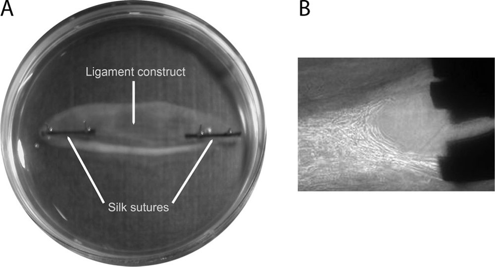
Current method of engineering musculoskeletal constructs. (
In order for engineered tendons and ligaments to be implanted in association with a portion of a bone substitute, a functional interface between the two tissues would have to be developed in vitro. This has not been extensively studied because up until now the focus has been on the development of the functional tendon/ligament itself. With advances in the development of functional tendons/ligaments, it is now prudent to begin to develop the tissue transition that will be required for this technology to reach the clinic.
Bone is a composite material, with mineral hydroxyapatite (HA), deposited as crystals within a collagen matrix. 19 The dense collagen matrix gives the bone its tensile strength and promotes the arrangement of the HA, increasing the torsional strength and stiffness of the tissue. HA is a calcium phosphate ceramic commonly used as a material in bone tissue engineering,20–22 and crystals of HA can be used to engineer a variety of composite materials.
Poly(ethylene glycol) (PEG) hydrogel scaffolds are widely used in tissue engineering due to their hydrophilic nature and controllable, reproducible chemistry. 23 This, in turn, allows particular properties such as the molecular weight, structure, degradation rate, crosslinking density, mechanical strength, and stiffness to be controlled.23,24 Incorporating the RGD (Arg-Gly-Asp) peptide sequence can further increase the functionality of a PEG hydrogel by improving cellular attachment and growth.25–28 The result is a scaffold with controllable matrix density, cell binding, and mechanical strength that can be used on its own or in composite materials.
The aim of this study was to test a composite of a PEG hydrogel embedded with HA as a possible material for engineering the osteoligamentous interface to produce an intact, engineered ligament. The material properties of the PEG hydrogel, incorporated with HA and the cell-adhesion peptide RGD to increase cell attachment, were investigated prior to determining the effectiveness of the material as a tissue-engineering anchor with our fibrin-based ligament constructs.
Materials and Methods
PEG anchor manufacture
PEG diacrylate
Poly(ethylene glycol) diacrylate (PEGDA) was produced by dissolving 20 g PEG (8000 Da; Fluka, Gillingham, Dorset, United Kingdom) in 150 mL benzene and azeotropically distilling to remove moisture. One milliliter triethylamine was added to the dissolved PEG under stirring, followed by the dropwise addition of 1.2 mL acryloyl chloride, and the solution was refluxed under argon for 4 h and left to cool overnight. After filtering to remove reaction by-products, the solution was then distilled to reduce its volume by 50%. Finally, the PEGDA was recrystallized in hexane and dried under vacuum.
Twenty percent PEGDA solutions were produced by dissolving 200 mg of the polymer per mL phosphate-buffered saline (PBS). A photoinitiator solution of 600 mg/mL 2,2 dimethyl-2-phenyl-acetophenone in N-vinyl-pyrrolidone was then added to the PEGDA solution to a concentration of 2 μL/mL and stirred. The solution was then filtered using 0.2 μm syringe filters and stored in ultraviolet (UV)–shielded containers. For polymerization to occur, the hydrogels were then irradiated with UV light (365 nm) for 180 s.
Acrylated GRGDS
Gly-Arg-Gly-Asp-Ser (GRGDS) peptide (Bachem, St. Helens, United Kingdom) was acrylated via a PEG spacer arm using an established method. 26 The peptide was dissolved in 50 mM Tris buffer (pH 8.2) at a concentration of 1 mg/mL. Acryloyl-PEG-NHS (AC-PEG-NHS, MW 8000; Nektar Therapeutics, Huntsville, AL) was added to this solution at a molar ratio of 1.5:1. This was left to react under constant agitation for 2 h at room temperature before being frozen at −80°C and lyophilized overnight.
HA incorporation
HA ([Ca5(OH)(PO4)3] x ) (Sigma-Aldrich, Gillingham, Dorset, United Kingdom) was incorporated into PEGDA ± RGD peptide at weight/volume ratios prior to UV light polymerization. It was essential to minimize the time taken between mixing the solution and UV exposure, as sedimentation of the HA occurs over time due to the low viscosity of the PEG solution.
Characterization of hydrogels
Four types of hydrogel were used throughout this study with weight/volume compositions as follows: (1) PEG = 20% PEGDA; (2) PEG HA = 20% PEGDA + 5% HA; (3) PEG RGD = 20% PEGDA + 0.8 mg/mL acrylated GRGDS in PEG solution; and (4) PEG HA RGD = 20% PEGDA + 5% HA + 0.8 mg/mL acrylated GRGDS in PEG solution.
Experiments determining the swelling of hydrogels both at room temperature and at 37°C were conducted prior to determining the final concentrations of PEG, HA, and RGD used in our final experiments. After 7 days in growth media and size measured every day, we observed a mean percentage increase of 41 ± 10.6% in length and 142.1 ± 17.9% in width. For these initial experiments, five different percentages of PEG were used (10–50%), with no difference observed in the final size of our hydrogel anchors. Twenty percent was chosen as it was the lowest percentage of PEG we could use that gave a hydrogel capable of repeated handling. HA concentrations of 1%, 5%, and 10% were used and again showed no difference in final hydrogel size between groups. Five percent was chosen in our final experiments as hydrogels containing 10% HA mechanically failed following repeated handling. RGD concentrations of 0.4, 0.8, and 1.2 mg/mL were chosen in our initial experiments (based on recommendations by Hern and Hubbell 26 ), and again the concentration of RGD peptide used in the hydrogel did not affect the final size of the hydrogel anchors. Our choice to use 0.8 mg/mL in our final experiments was based on pilot cellular attachment and growth studies (K. Donnelly, unpublished observation).
Swelling data
PEG hydrogels with and without HA and/or RGD peptide were cast in wells of a 96-well plate, weighed immediately, and then swollen in distilled water in a 24-well plate at room temperature. Swelling ratio was calculated by dividing the swollen weight by the dry weight as reported previously 29 with slight modifications. Briefly, weight measurements were taken at set time points until equilibrium occurred. Initial weight was taken as weight of hydrogel when first formed as a gel. Dry weight was measured after gels have been allowed to dehydrate at room temperature for a minimum of 72 h.
Mechanical properties
To determine the mechanical properties of the hydrogels, PEG hydrogels with and without HA and/or RGD peptide were formed in borosilicate theta glass capillary tubes (outer diameter 1.5 mm, inner diameter 1 mm, separation 0.2 mm, length 100 mm; Harvard Apparatus, Kenk, United Kingdom), removed following vacuum dehydration in a vacuum oven at 65°C, and swollen in distilled water for 1 h prior to testing. Load to failure tests were conducted on an Instron 3366 Tensile Testing machine (Instron, Bucks, United Kingdom) in specially designed grips at a constant rate of 4 mm/min. The gauge length of the samples remained constant at 10 mm for all samples, as this was determined by the design of the grips used. All tests were conducted in air at room temperature. A 50 N load cell was used, and data were collected using Bluehill Software. Thirty samples were tested with six discarded due to slippage from the grips. All remaining samples (n = 24) used for data analysis failed in the midportion of the hydrogel and not at the hydrogel–grip interface.
Cell attachment
To visualize cells on the surface of the scaffold, PEG HA RGD hydrogels were formed in glass capillary tubes and removed following vacuum dehydration. Small (∼4 mm) samples were cut and hydrated in growth media (F12 Ham supplemented with 20% fetal bovine serum [FBS], 1% ABAM) for 1 h before being seeded with 100 K primary tendon fibroblasts (PFB) and cultured at 37°C for 4 days. Media were changed on day 2. Samples were then frozen in OCT compound (Agar Scientific, Essex, United Kingdom) in liquid nitrogen–cooled isopentane, and 8-μm-thick longitudinal sections were cut using a cryostat (Leica CM3050S Cryostat, Weltzar, Germany). Sections were stained with DAPI (Sigma-Aldrich) at 1:1000 in PBS and with Rhodamine Phalloidin (Invitrogen, Paisley, United Kingdom) at 1:100 in PBS. Images were taken using an Axiocam Zeiss Camera and Axioskop Microscope (Zeiss, Oberkochen, Germany) and analyzed using NIH image.
Cell counting
PEGDA hydrogels with and without HA and/or RGD peptide were formed in glass capillary tubes and removed following vacuum dehydration as described above. Small samples of each gel type (n = 5) were cut to 4 mm in length and rehydrated in 25 K 3T3 fibroblasts suspended in 100 μL of DMEM supplemented with 10% FBS (Invitrogen) and 1% penicillin/streptomycin (Invitrogen). Anchors were incubated at 37°C for 2 h, then moved to a 96-well plate, washed once in PBS (Invitrogen), and fresh growth media added. Cells were quantified using the celltitre glo luminescent assay (Promega, Southhampton, United Kingdom) and luminometer (LjL Analyst; LJL BioSystems, Sunnyvale, CA) at 2, 24, and 48 h to assess attachment and cellular proliferation.
Bone–ligament interface
Isolation of PFB
PFB were isolated from rat Achilles tendon by overnight digestion in a 0.1% collagenase II solution in F12 Ham media supplemented with 20% FBS and 1% ABAM. Cells were collected by centrifugation, rinsed in FBS (Invitrogen), and resuspended in F12 Ham media (Invitrogen) with 20% FBS and 1% ABAM. Cells were grown at 37°C in 5% CO2, 95% air, and used between passages 2–5.
Ligament formation
Thirty-five millimeter Petri dishes were coated with 2 mL of sylgard (type 184 silicone elastomer; Dow Corning, Midland, MI) and left to polymerize for at least a week before use. The plates were sterilized by soaking in 70% ethanol for 20 min. Anchors of all four hydrogel types (PEG, PEG HA, PEG RGD, and PEG HA RGD) (n = 8 for each type) were cut to ∼4 mm in length and allowed to rehydrate in F12 Ham media supplemented with 20% FBS, 1% ABAM, 200 U/mL thrombin (Calbiochem, Nottingham, United Kingdom), 2 μL/mL Aminohexanoic acid (Sigma-Aldrich), and 2 μL/mL Aprotinin (Roche, Hertfordshire, United Kingdom) solution. Five hundred microliters of the same solution was used to coat the sylgard layer, and the rehydrated anchors were pinned onto the dish using cut minutuen insect pins (Fine Science Tools, Cambridge, United Kingdom) approximately 8-mm apart. Two hundred microliters of 20 mg/mL fibrinogen (Sigma-Aldrich) was added dropwise, and the fibrin gel was left to polymerize at 37°C for 1 h. PFB were seeded on top of the gel at a concentration of 100,000 cells/mL.
Attachment of hydrogel anchors
The attachment of the anchors to the ligament construct was assessed by manually removing the pins from the anchor each day and observing if the anchor remained attached to the ligament construct. Attachment was classed as either attached or not attached, and the same person performed the experiment at the same time each day.
Hydrogels in SBF
Small (∼4 mm) samples of dehydrated PEG HA gels were hydrated in simulated body fluid (SBF) (for method see Kokubo and Takadama 30 ) for 4 months to deposit a layer of HA on the surface of the hydrogel. These gels were then dehydrated and rehydrated in thrombin solution as described above and used as hydrogel anchors for fibrin-based constructs.
Statistics
Data are presented as means ± SEM. Differences in mean values were compared within groups, and significant differences were determined by ANOVA with post hoc Tukey–Kramer HSD test using JMP Statistical Software. The significance level was set at p < 0.05.
Results
Effect of addition of HA and RGD peptide to PEG hydrogels
Swelling properties
Hydrogels were cast in theta-shaped capillary tubes to increase the surface area in contact with the ligament construct. Hydrogels were prepared in capillary tubes (see Materials and Methods section) and rehydrated prior to use (Fig. 2A, B). To assess swelling properties of the hydrogels and the effect of the addition of HA and RGD peptide to the polymer, swelling studies were conducted using all four hydrogel types until the materials reached equilibrium (Fig. 2C, D). After 30 min, PEG RGD hydrogels become significantly heavier (p < 0.05) than all other hydrogel types, indicating an increase in water absorption (Fig. 2C). Hydrogels containing HA had a lower swelling ratio (Fig. 2D) than gels without HA, reaching significance (p < 0.05) after 20 min. Further, at 300 min, a significant difference (p < 0.05) in the swelling ratio is observed between PEG and PEG RGD hydrogels, demonstrating a difference in the mechanical properties between these two gel types (Fig. 2D).
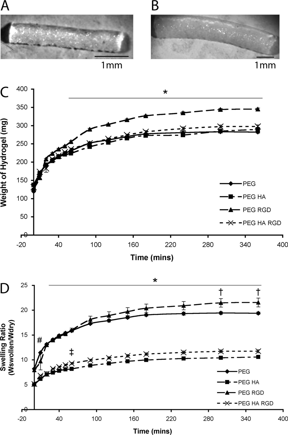
Swelling properties of PEG hydrogels. (
Mechanical data
In conjunction with the assessment of the swelling properties of the hydrogel anchors, the mechanical properties of all four gel types were examined. Table 1 shows the maximum load and extension of the hydrogels, and Figure 3A–C shows the stress, strain, and Young's modulus, respectively. The addition of HA to a PEG hydrogel significantly increases the maximum load, maximum stress, and stiffness of the material. Conversely, the addition of RGD peptide to a PEG hydrogel significantly decreases (p < 0.05) the maximum load, extension, maximum stress, strain, and Young's modulus of the hydrogels. The addition of HA to PEG RGD gel results in the same HA-induced increase in load, stress, and Young's modulus in the PEG HA RGD gels as seen in plain PEG gels with HA addition, confirming that the addition of HA to our hydrogels increases their mechanical strength in both the PEG and PEG RGD gels.
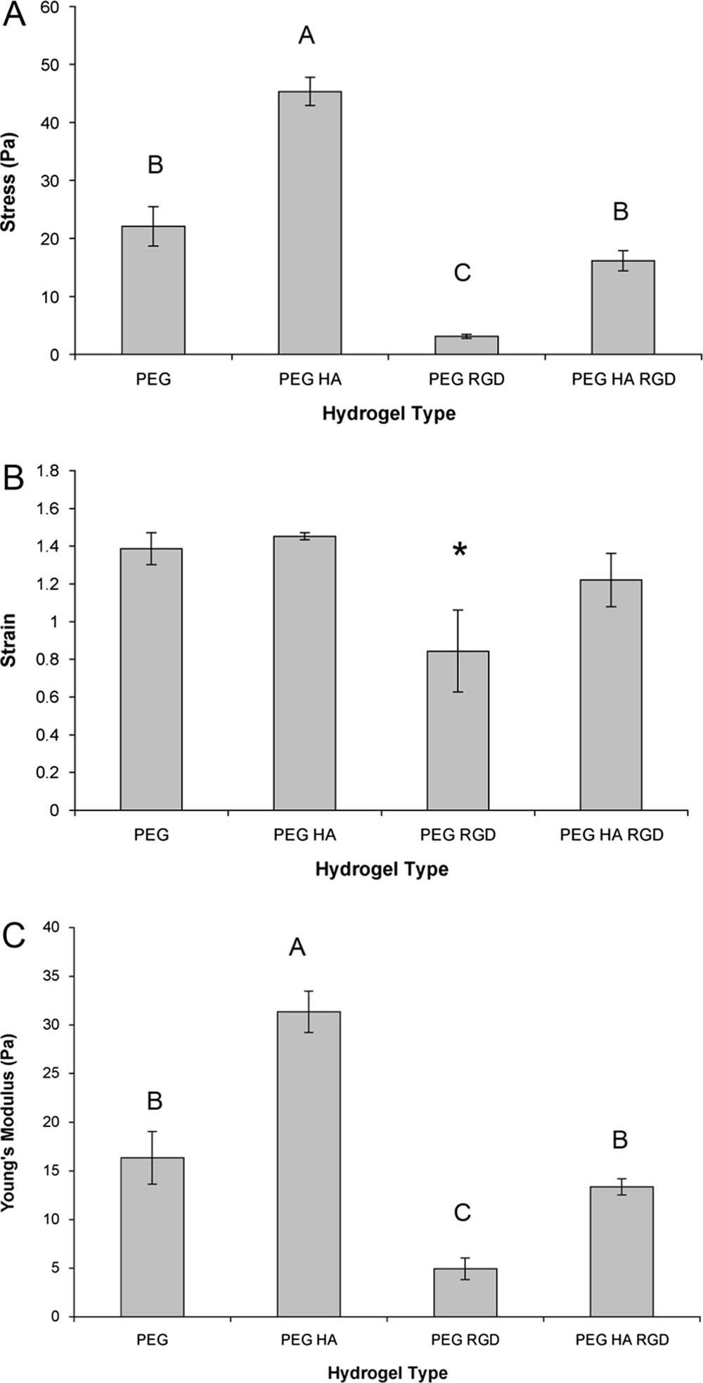
Mechanical properties of PEG hydrogels. (
Results are presented as mean ± SEM for n = 6. Groups not connected by the same letter are significantly different (p < 0.05). HA, hydroxyapatite; PEG, poly(ethylene glycol).
Cell attachment
Images of the PEG HA RGD anchors show that cells adhere to the outside of the scaffold, but do not readily penetrate into the matrix (Fig. 4A–D). To quantify cell attachment and proliferation on the hydrogel anchors, anchors of all four gel types were seeded with 3T3 fibroblasts, and cell numbers were determined at 2, 24, and 48 h (Fig. 4E–H). At 2 h, all hydrogel types showed similar levels of cell attachment (Fig. 4E); however, after 24 h (Fig. 4F) and 48 h (Fig. 4G), significant cell growth is observed on the PEG HA RGD gels (p < 0.05). PEG RGD gels did support cell growth, but due to variability between the samples this was not significantly different than the PEG alone (p > 0.05). As expected, hydrogels without the cell-adhesion peptide do not support significant cell growth. The effect of HA is evident when comparing PEG RGD gels to PEG HA RGD gels, with a threefold increase in cell proliferation at 48 h (Fig. 4G).
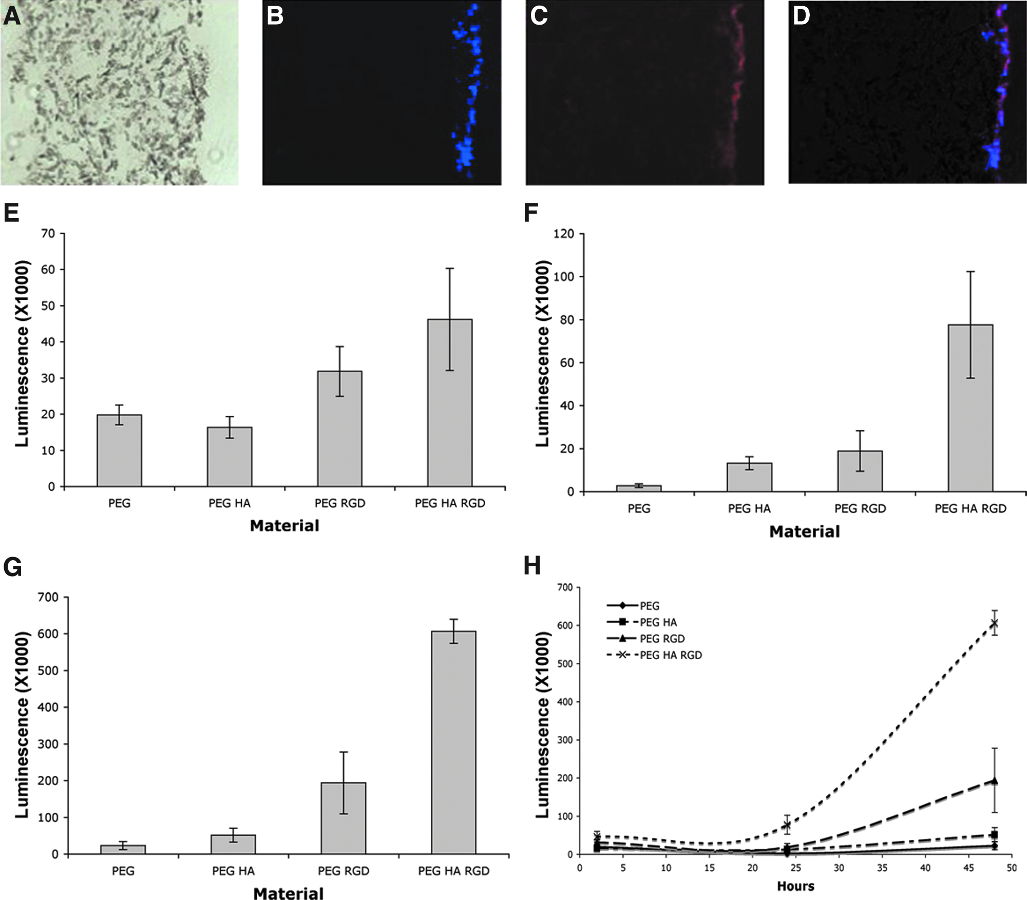
Cell attachment to PEG hydrogels. (
PEG anchor attachment
Our intended model for producing a bone–ligament interface using PEG anchors was to use a cell-seeded scaffold as the anchor, and in doing so, join the anchor and tissue together. Attachment was assessed (see Materials and Methods section) on day 1, and none of the cell-embedded anchors attached to the fibrin gel. The samples were left for 1 week after cell seeding; however, no attachment of the anchors to the tissue was observed when using the cell-seeded anchor method. To create an attachment between the anchor and the ligament, dehydrated hydrogel anchors were hydrated in the thrombin component of fibrin gel prior to polymerization, therefore embedding the anchor into the fibrin gel. As shown in Figure 5B, the incorporation of HA into both PEG and PEG RGD gels significantly increases the attachment time of the anchors to the fibrin gel (p < 0.05). As 5% HA resulted in an increase in attachment time, if the attachment was dependant on HA then adding 2.5% or 10% HA would decrease and increase attachment time, respectively. In support of this hypothesis, a very strong correlation (R 2 = 0.995) between HA concentration and length of attachment time was observed (Fig. 5C). To further investigate the role of HA, PEG HA gels were coated with a further HA layer in SBF. PEG HA SBF anchors had significantly longer attachment time (p < 0.05) than PEG HA gels (Fig. 5D). However, while the HA increased the attachment time of PEG hydrogels, the standard method of using silk sutures as anchors was still significantly better (p < 0.05) than any of our PEG hydrogels, even with higher percentages of HA (Fig. 5D).
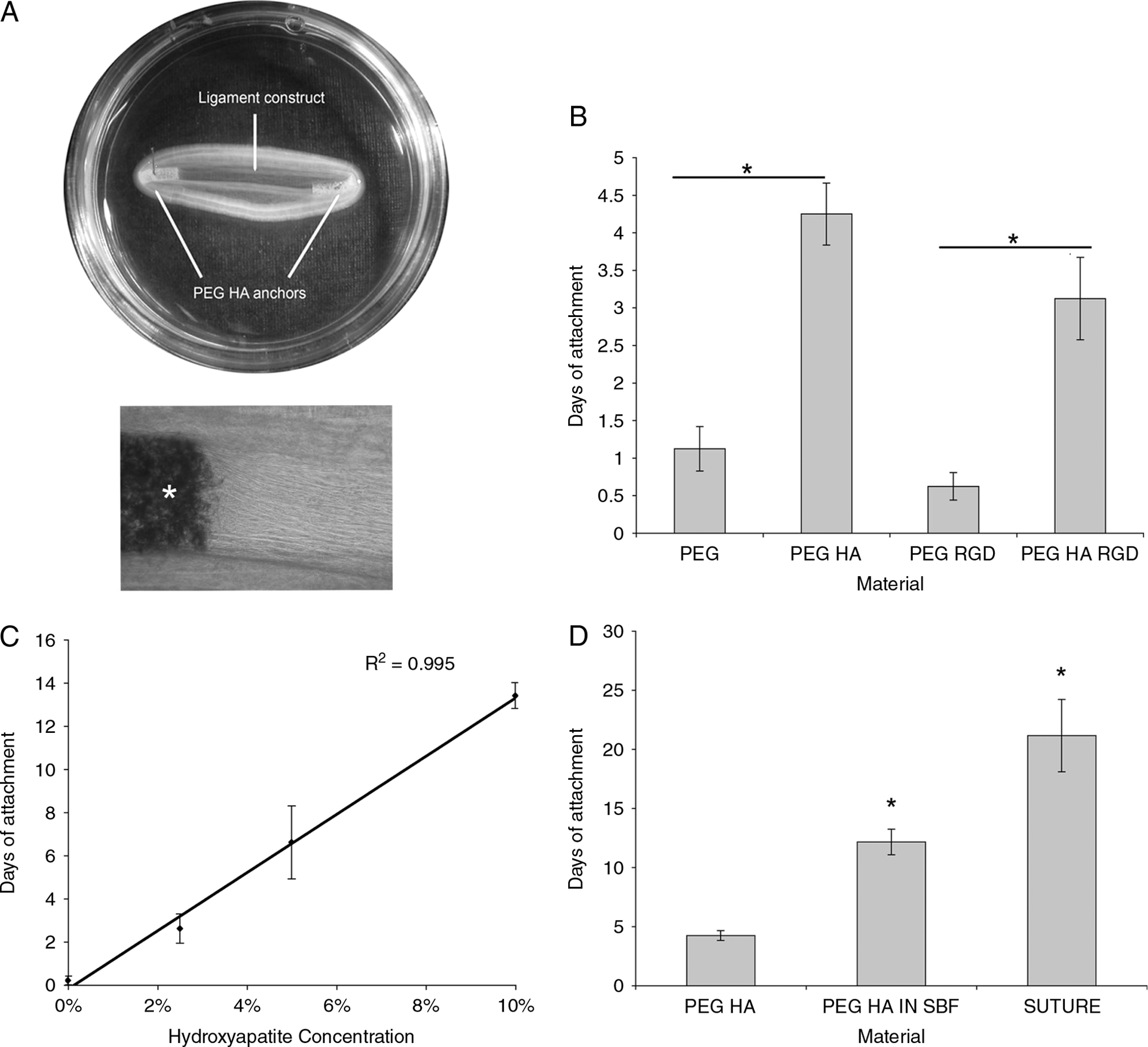
Attachment of anchors to ligament constructs. (
Discussion
Here we report the properties of a PEG hydrogel, whose properties can be altered with the use of HA or RGD peptide incorporation. The addition of HA increases the maximum load, maximum stress, and Young's modulus of PEG hydrogels. Further, HA also increases the capacity of PEG to bind cells and adhere to a fibrin-based construct, promoting the formation of a bone–ligament interface. However, while the addition of HA improves the mechanical properties of the PEG hydrogels, the combination of HA and RGD is required to optimize cellular attachment. Unfortunately, this material has no better mechanical properties than PEG alone.
PEG's success as a scaffold material is largely due to the fact that it is resistant to protein adsorption; therefore, all nonspecific interactions with the scaffold are restricted.24,27,31 The incorporation of cell-adhesion peptides into PEG-based gels during polymerization allows for specific cell–extracellular interactions to take place.26,28,31 As such, a PEG-based hydrogel can present a blank structure on which cell-adhesion peptides can be incorporated to control both the identity and concentration of peptides. As expected, the incorporation of RGD peptide into our hydrogels resulted in a material with a greater capacity for cell adhesion. Peptide incorporation also produced a material with a higher weight, and swelling ratio measurements when compared PEG alone. Further, the mechanical properties of the hydrogel are altered when incorporated with RGD peptide, resulting in a significant decrease in the load, extension, stress, strain, and Young's modulus values when compared to a plain PEG hydrogel. The change in the mechanical properties of PEG RGD is likely explained by the fact that the crosslinking of PEG molecules is impaired by the fact that the RGD peptide cannot bind to another PEG molecule in the way that acrylated PEG molecules can. The result is a decrease in PEG crosslinking and a resultant weaker matrix. This is further substantiated by analysis of the swelling data, where hydrogels containing the RGD peptide have a higher swelling ratio than non-RGD–containing gels, reaching significance at 5 h between PEG and PEG RGD gels. We have also noted an increase in the weight of water absorbed in RGD-containing gels when compared to non-RGD gels (data not shown).
The combination of HA powder with the PEG polymer results in a material that is stronger and stiffer than PEG alone. Varying the amount of HA used within the composite alters the mechanical properties of the hydrogel structures, a property which may be of interest to bone tissue engineers. A 5% weight/volume ratio of HA to PEG was chosen for the current experiments, although we have successfully produced hydrogels containing up to 20% HA. The limiting factor in the current study was the increasing viscosity of the solution with increasing HA concentrations, as pipetting the solution became difficult and the solution could not fill the glass capillary tubes used to make the anchors. In theory, the properties of the hydrogels could be tailored to suit specific applications by altering factors such as the porosity and molecular weight of PEG used, 29 as well as the concentration of RGD and HA.
Unexpectedly, the combination of HA with RGD peptide resulted in a hydrogel with mechanical properties no different than PEG alone. This effect appears to be the result of the mechanical effects of incorporating the RGD peptide into the PEG hydrogels, since the addition of RGD resulted in an 85% decrease in PEG stress. The addition of HA to the PEG RGD increased the relative strength and stiffness of PEG RGD hydrogels to an even greater extent than the PEG hydrogels (5.2-fold vs. ∼2-fold). However, in absolute terms, the 85% decrease in strength and stiffness caused by RGD incorporation means that HA incorporation only improves the mechanical properties of the anchors to levels similar to that of PEG alone. As discussed above, this is likely due to the wider matrix produced by RGD incorporation. We hypothesize that the wider matrix with the addition of RGD is less able to entrap HA during the crosslinking procedure resulting in lower strength and stiffness. This results in the lower strength and stiffness in the PEG HA RGD than the PEG HA gels.
The ability of HA to increase the cellular attachment level of PEG hydrogels is not surprising, since many calcium phosphate scaffolds are used in bone tissue engineering.22,32,33 In the current study, an increase in cell attachment and growth was observed on the PEG HA RGD gels when compared to PEG RGD alone (p < 0.05). For optimum levels of cell attachment to our PEG hydrogels, the addition of cell-adhesion peptides, in this case RGD, is required in combination with HA. At 48 h, there is a threefold increase in cell number when comparing PEG RGD to PEG HA RGD gels. However, increasing the concentration of HA could increase the capacity for cell attachment to the scaffolds, such that the RGD incorporation may not be required.
Incorporating HA into PEG hydrogels increases the capacity for the hydrogel to attach to a fibrin-based scaffold (Fig. 5B–D). This was not completely unexpected because HA is commonly used as a coating in orthopaedic implants, to improve bonding of the tissue and the metal implants, 34 and the use of HA in developing an in vivo neoenthesis has been reported. 35 Although a significant increase in interface longevity is evident when comparing PEG to PEG HA gels, and PEG RGD to PEG HA RGD gels, failure of the bone–ligament interface occurred at approximately day 4 in the 5% HA anchors (Fig. 5C). Increasing the HA concentration to 10% allowed an in vitro interface to be established for approximately 2 weeks, similar to the time observed when using 5% PEG HA hydrogels coated in HA particles by submersion in SBF. Although altering the HA concentration within a PEG hydrogel does increase the length of time, the anchors remain attached to our fibrin constructs; the attachment of the PEG anchors is still significantly less than the silk sutures currently in use and is not sufficient if these constructs are to be implanted.
In vivo, the transition between tissues with different mechanical properties, that is, muscle/tendon or tendon/ligament to bone, is mediated through the intrinsic properties of these tissues and as such, impedance mismatch between the tissues is reduced. The osteotendinous junction or enthesis is designed to ensure the smooth transfer of force from the tendon to the bone.36–38 As tendons develop from fibrous outgrowths of the cartilaginous primordial bone prior to its ossification, 37 the resulting transition from tendon to bone is a zonal arrangement, comprising four separate regions: tendon, fibrocartilage, mineralized fibrocartilage, and mineralized bone.37,38 The outer limit of calcification is demarcated by a tidemark, signifying the transition between the calcified and noncalcified regions of the enthesis.39,40 The zone of calcified fibrocartilage interdigitates with the bone and greatly increases the surface area for attachment of tendon/ligament to bone, resulting in a decrease in strain concentrations and removing the impedance mismatch present between the compliant tendon/ligament and stiffer bone.36–38 It has been reported previously that tendon and ligament failure at fibrocartilaginous entheses actually occurs at the site of the subchondral bone and not at the interface between the two tissues. 40 This strongly implies that the bone itself is weaker than the transition and importantly, that the zonal arrangement of the enthesis works well at dissipating stress uniformly. The graduated regions of the enthesis result in the mechanical properties differing along its length and, just like the tendon as a whole, 41 it is more compliant at the tendon end than at the bone end. 37 It is therefore possible that a pure HA anchor material with graded porosity, increased surface area for attachment, and graded mechanical properties would be better suited for the development of an in vitro ligament–bone interface.
In summary, the addition of HA to a hydrogel of PEG increases its mechanical strength, capacity to bind cells, and ability to form an interface with biological materials. While HA improves the functionality of PEG hydrogels, the PEG HA hydrogels are not sufficient to produce a functional interface between biological and synthetic materials. The fact that HA promotes cellular adhesion and interface formation suggests that anchors made from a greater proportion of calcium phosphate may provide a better functional interface between engineered ligament and bone.
Footnotes
Acknowledgments
The work was supported by grants from the Engineering and Physical Sciences Research Council (EPSRC–EP/E008925/1) and the Biotechnology and Biological Sciences Research Council (BBSRC–BB/F002084/1).
Disclosure Statement
No competing financial interests exist.