
Abstract
In this study we developed polymer scaffolds intended as anchorage rings for cornea prostheses among other applications, and examined their cell compatibility. In particular, a series of interconnected porous polymer scaffolds with pore sizes from 80 to 110 microns were manufactured varying the ratio of hydrophobic to hydrophilic monomeric units along the polymer chains. Further, the effects of fibronectin precoating, a physiological adhesion molecule, were tested. The interactions between the normal human fibroblast cell line MRC-5 and primary human umbilical vein endothelial cells (HUVECs) with the scaffold surfaces were evaluated. Adhesion and growth of the cells was examined by confocal laser scanning microscopy. Whereas MRC-5 fibroblasts showed adhesion and spreading to the scaffolds without any precoating, HUVECs required a fibronectin precoating for adhesion and spreading. Although both cell types attached and spread on scaffold surfaces with a content of up to a 20% hydrophilic monomers, cell adhesion, spreading, and proliferation increased with increasing hydrophobicity of the substrate. This effect is likely due to better adsorption of serum proteins to hydrophobic substrates, which then facilitate cell adhesion. In fact, atomic force microscopy measurements of fibronectin on surfaces representative of our scaffolds revealed that the amount of fibronectin adsorption correlated directly with the hydrophobicity of the surface. Besides cell adhesion we also examined the inflammatory state of HUVECs in contact with the scaffolds. Typical patterns of platelet/endothelial cell adhesion molecule-1 expression were observed at intercellular boarders. HUVECs adhering on the scaffolds retained their proinflammatory response potential as shown by E-selectin mRNA expression after stimulation with lipopolyssacharide (LPS). The proinflammatory activation occurred in most of the cells, thus confirming the presence of a functionally intact endothelium. Little or no expression of the proinflammatory activation markers in the absence of LPS stimulation was observed for HUVECs growing on scaffolds with up to a 20% of hydrophilic component, whereas activation of these markers was observed after stimulation. In conclusion, scaffolds containing up to 20% hydrophilic monomers exhibited excellent cell compatibility toward human fibroblast cell line MRC-5 and human endothelial cells. Atomic force microscopy confirmed that adsorbed serum proteins such as fibronectin probably accounted for the positive correlation of HUVEC adhesion and surface hydrophobicity.
Introduction
Cell adhesion to polymeric substrates is mediated by extracellular matrix proteins. It has been reported that proteins adsorb better to hydrophobic surfaces than to hydrophilic ones.10,11 As a consequence, adhesion of fibroblasts and other cell lines does not occur readily on hydrophilic polymers such as poly(HEMA) (PHEMA)3,12–14 and poly(HEA) (PHEA),15,16 whereas in the presence of serum-containing medium, spontaneous cell adhesion is observed on hydrophobic polymers such as poly(methyl methacrylate) (PMMA) and poly(ethyl methacrylate) (PEMA).13–16 Arg-Gly-Asp (RGD) sequences within ECM proteins such as fibronectin are recognized by cellular transmembrane integrin receptors. Once integrins bind to fibronectin, they activate a cascade of intercellular signaling pathways. 17 Because the chemical structure of a material plays a central role in determining the adsorption and conformation of fibronectin on its surface, a material's chemical composition and molecular structure also greatly influence cell attachment. Another factor in the design of a biomaterial is degradability. While a large branch of tissue engineering is focused on the development of biodegradable materials, nonbiodegradable materials are necessary for the replacement of some tissues. The anchoring ring of a corneal prosthesis is one application of a nonbiodegradable implant material. The “core and skirt” corneal prosthesis consists of a nonporous transparent lens surrounded by a macroporous ring. 18 The goal is to promote cells of the host stroma to grow into the pore structure of the anchoring ring and fixate the synthetic lens avoiding its extrusion.18,19 Thus, this type of three-dimensional (3D) scaffold construct must have an interconnected porous structure to allow cell invasion of the porous network.
The cornea is a nonvascularized tissue. Oxygen and other nutrients must reach the cells by diffusion through the entire tissue. The diffusion process is highly hindered by the presence of the scaffold, and this can compromise the long-term viability of the regenerated tissue inside the scaffold. A possible strategy in the design and implantation of a corneal prosthesis is to induce angiogenesis in the anchoring ring and thus provide cells with the necessary nutrients to grow and persist in the scaffold. At the same time, the anchoring ring should preserve the core of the prosthesis (the lens) from vascularization. Once a scaffold preseeded with stromal cells from the patient's cornea is implanted, new blood vessels should invade only the stroma that forms in the scaffold's anchoring ring. This implies that the conjunctival vasculature must sprout new blood capillaries, which must then grow into the material and provide a blood supply to the cells on the implant. Vascularization of a scaffold may benefit from preseeding of autologous endothelial cells that join the formation of a functional vasculature after implantation. Endothelial cells on a tissue-engineering construct may even self-assemble and form microcapillary-like structures before connecting with the eye's surrounding vasculature upon implantation. 20
Toward a tissue-engineered corneal implant, the goal of this study was to determine whether and how hydrophilicity affects stromal cell attachment, growth, phenotype, and function in 3D P(EA-co-HEA) polymer scaffolds. Because corneal stromal fibroblasts (i.e., keratocytes) are of relevance in corneal healing, 21 the human fibroblast cell line MRC-5 was used as a model cell type22,23 to study the cytoskeletal arrangement after adhesion to the scaffolds by detection of F-Actin.
Toward preendothelialized/prevascularized implants, primary human endothelial cells were used as a model cell type to test endothelial cell compatibility with different polymer scaffolds. Endothelial cell compatibility was measured in terms of cell viability and proper endothelial differentiation/functioning.
Because the role of a substrate's chemical structure in cell attachment is expected to come from its effect on the conformation of the proteins adsorbed to it, the adsorption and conformation of fibronectin onto the substrates were studied by AFM. The characterization of the conformation of protein molecules on the biomaterial by AFM is extremely difficult on curved surfaces, so we specially prepared 2D P(EA-co-HEA) bulk polymers for these measurements with the same chemical composition as 3D P(EA-co-HEA) polymer scaffolds. As reported below, individual fibronectin molecules adsorbed on the surface became observable this way.
Materials and Methods
P(EA-co-HEA) scaffolds
Poly(methyl methacrylate) microspheres with diameters ranging from 80 to 110 microns (PMMA Colacryl DP300; Lucite International, Newton Aycliffe, Durham, United Kingdom) were used as porogen. The microspheres were introduced into a mold consisting of two glass plates and a rubber ring. The mold was placed into an oven at 160°C to allow PMMA microspheres to soften, which were then compressed. To create desired polymers and copolymers for this study, monomer solutions with varying fractions of EA (96% pure; Aldrich, Sigma-Aldrich Química S.A., Madrid, Spain) and HEA (99% pure; Aldrich) were prepared, always with 2% ethyleneglycol dimethacrylate (EGDMA, 98% pure; Aldrich) as the crosslinking agent and 1% benzoin (98% pure; Scharlau Chemie S.A., Sentmenat, Spain) as the photoinitiator. The EA fractions were 1, 0.9, 0.8, and 0.5. Templates of the sintered microspheres were immersed in the monomer solutions, and polymerization at room temperature was initiated by ultraviolet light. After polymerization, the PMMA microspheres were dissolved with ethyl acetate for 48 h by means of a Soxhlet extractor. Then, ethyl acetate was exchanged with ethanol, ethanol/water, and finally water to allow the scaffolds to contract slowly. Scaffolds were cut into 3–4 mm diameter and washed with ethanol in an ultrasound bath for 3 min. Finally, scaffolds were dried under vacuum at 60°C to constant weight and packaged for sterilization with gamma radiation by 25 kGy (Aragogamma S.A., Barcelona, Spain). Figure 1 shows the different scaffolds used for the cell culture experiments, ranging from P(EA-co-HEA) 100/0, a pure hydrophobic scaffold, to P(EA-co-HEA) 50/50, a half-hydrophobic half-hydrophilic scaffold. Because the size of the porogenic particles and their sintering procedure were identical for all the compositions, physical parameters such as surface topology, volume fraction of pores, and pore interconnectivity were similar among all scaffolds. The water sorption and surface tension properties of the copolymers increase proportionally with HEA content. 24
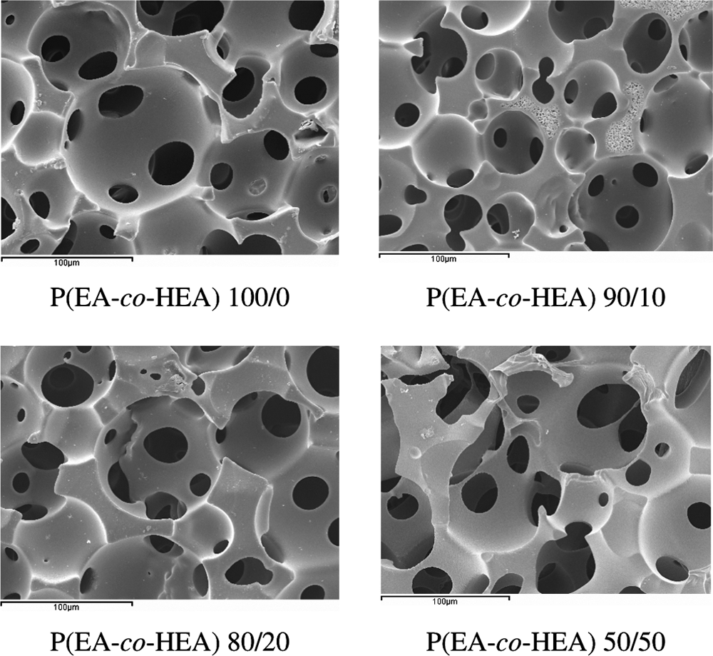
SEM microphotographs of the series of P(EA-co-HEA) scaffolds. Numbers indicate the weight percentage of each component with respect to the total monomeric solution. Scale bar: 100 microns.
Cell culture
The normal human lung fibroblast cell line MRC-5 was cultured in medium Eagle's modified essential medium (EMEM; Sigma-Aldrich, Steinbach, Germany) supplemented with 10% fetal calf serum (FCS; Life Technologies, Karlsruhe, Germany), 2 mM Glutamax I (Life Technologies), and 100 U/100 mg/mL Pen/Strep. Human umbilical vein endothelial cells (HUVECs) were isolated from umbilical cords and cultivated according to a previously published method. 25 The culture medium was M199 (Sigma-Aldrich) supplemented with 20% FCS (Life Technologies), 2 mM Glutamax I (Life Technologies), 100 U/100 mg/mL Pen/Strep, 25 mg/mL sodium heparin (Sigma-Aldrich), and 25 mg/mL endothelial cell growth supplement (ECGS; Becton Dickinson, Bedford, MA).
Preparation of scaffolds for culture
To coat scaffolds with fibronectin, they were incubated in a phosphate buffered saline (PBS), 5 μg/mL solution of fibronectin (Roche, Mannheim, Germany) for 1 h at 37°C. For every type of cell to be seeded, two identical scaffolds each, between 3–4 mm in diameter and 1 mm thick, were glued with medical silicone (Silastic®, Dow Corning Corp., Midland, MI) to individual wells of a 96-well plate. After 15 min at 37°C, the different types of cells were added to each well (3 × 10⁴ cells/well) and incubated in a cell culture incubator at 37°C and 5% CO2. Medium was replaced every 2 to 3 days.
Visualization and characterization of cell growth
MRC-5 cell adhesion and spreading on the scaffolds was assessed by staining for F-actin. At various time points scaffolds seeded with MRC-5 cells were removed from the culture medium and rinsed briefly with PBS. Then, the cells were fixed with 3.7% paraformaldehyde (in PBS) for 15 min at room temperature, washed four times with PBS, and finally permeabilized with 0.2% Triton-X 100 (in PBS) for 10 min. After four subsequent washes with PBS, 1 μg/mL TRITC-conjugated phalloidin (in PBS; Sigma-Aldrich Química S.A.) was added and incubated at room temperature for 30 min. The scaffolds were then washed four times with PBS and incubated with 1 μg/mL Hoechst nuclear stain (Hoechst 33342; Sigma) for 3 min. Scaffolds were then washed with PBS, covered with mounting medium (Gel/Mount, Natutec, Germany), and analyzed by confocal laser scanning microscopy (CLSM) using the Leica TCS SP2 system.
MRC-5 cell viability on the scaffolds was assessed by a live-cell stain. Namely, at various time points after addition of HUVECs to the scaffolds, fresh medium containing 0.1 mM calcein-acetoxymethylester (calcein-AM; Mobitec, Göttingen, Germany) was added and allowed to react for 10–20 min at 37°C.26,27 Calcein-AM is metabolized to calcein, a fluorescent compound, inside viable cells, which consequently fluoresce. Scaffolds with stained cells were then placed onto a microscope slide and examined by CLSM.
Molecular analysis of HUVEC gene expression by reverse-transcription PCR (rtPCR)
HUVECs were stimulated with 100 ng/mL lipopolyssacharide (LPS) for 4 h (Sigma) to assess whether HUVECs maintained their proinflammatory activation as evident by E-selectin expression at the mRNA level. In particular, RNA was isolated from LPS-exposed and untreated control cultures using the RNeasy Micro Kit (Qiagen, Hilden, Germany).
Three identically treated HUVEC-containing scaffolds were placed directly into lysis buffer. Alternatively, cells were removed from tissue culture flasks with trypsin and centrifuged, and the resulting cell pellets were resuspended in lysis buffer. To ensure that the PCR products resulted from cDNA rather than genomic DNA, all RNA samples were treated with DNaseI (RNase-free DNase; Qiagen) during purification. The concentration of isolated RNA was determined using the NanoDrop microspectrophotometer (Nanodrop Technologies, Rockland, DE). Twenty nanogram of RNA was transcribed into cDNA (Omniscript RT Kit; Qiagen) and then stored at −20°C. Amplification of endotheliospecific genes was carried out by PCR on 200 pg cDNA with the reagents supplied in the Taq DNA Polymerase Kit (Qiagen) and with the primers described previously. 28 Briefly, an initial denaturation at 94°C for 2 min was carried out, followed by 35 cycles at 94°C for 30 s, an appropriate annealing temperature (varied for different primer pairs, see Ref. 28) for 30 s, and 72°C for 30 s. The rtPCR was terminated with a final extension step at 72°C for 10 min. The PCR products were resolved by agarose gel (0.8%) electrophoresis in TBE buffer (Sigma) and visualized by ethidium bromide staining.
Analysis of E-selectin and PECAM-1 expression by HUVECs
HUVECs growing on fibronectin-coated scaffolds were rinsed briefly with PBS and then fixed with 3.7% paraformaldehyde (in PBS) for 15 min at room temperature, washed four times with PBS, and then permeabilized with 0.2% Triton-X 100 for 10 min. After another four washes with PBS, antibodies against E-selectin (antibody host, mouse; dilution, 1:100; Monosan, Uden, The Netherlands) or platelet/endothelial cell adhesion molecule-1 (PECAM-1) (antibody host, rabbit; dilution, 1:100; Santa Cruz Biotechnology, Santa Cruz, CA) were added and incubated overnight at 4°C. The scaffolds were then washed four times with PBS, and the following fluorescent secondary antibodies were added at a dilution of 1:1000 and incubated for 1 h at room temperature: an Alexa Fluor 546-coupled anti-mouse antibody to stain for E-selectin and an Alexa Fluor 488-coupled anti-rabbit antibody to stain for PECAM-1 (both from Molecular Probes (Invitrogen), Eugene, OR). This was followed by a final rinse with PBS and a nuclear stain either with 1 μg/mL Hoechst 33342 in PBS or 500 nM propidium iodide in PBS for 3 min. All samples, but scaffold duplicates together, were then washed with PBS, covered with Gel/Mount, and analyzed by CLSM.
Statistical analysis
To evaluate the growth of HUVECs on the different scaffolds, the ratio of green pixels provided by cellular calcein-AM versus total pixels in a CLSM image was used to determine surface coverage by cells. Data were expressed as the mean ± SD, and statistical differences were determined by a nonparametric Kruskal–Wallis test for each material. When significant differences were found between scaffolds, a post hoc Mann–Whitney U-test was performed with paired groups. In the case of MRC-5 cells, the number of nuclei was used to establish the number of cells per square centimeter. Each experiment was performed in duplicate and repeated at least three times using different HUVEC donors, and at least two times using MRC-5 cells.
Fibronectin adsorption onto bulk polymers
Flat P(EA-co-HEA) substrates were prepared in the form of sheets for AFM analysis of fibronectin conformation. These flat substrates were obtained by free radical polymerization of the appropriate mixture of EA (96%; Aldrich) and HEA (99%; Aldrich) using 2% of cross-linking agent ethyleneglycol dimethacrylate (EGDMA, 98% pure; Aldrich) and 1% photoinitiator benzoin (98%; Scharlau). The polymerization took place under ultraviolet light in a mould that consisted of two glass slides with a rubber spacer, resulting in a polymer sheet of approximately 1 mm thickness. Low-molecular-weight species remaining in the sample after polymerization were extracted with boiling ethanol for 24 h, and the samples were then dried at 60°C in vacuo to constant weight. In addition to pure PEA, three different copolymer compositions were obtained, with EA fractions 0.9, 0.8, and 0.5.
Flat substrates were glued to an AFM sample slide and incubated for 15 min at Trm with 1 mL/cm2 of a 5 μg/mL fibronectin solution (Roche) in PBS. After that, the samples were dried under nitrogen flow for a few minutes.
Atomic force microscopy
AFM was performed with a NanoScope III from Digital Instruments operating in tapping mode in air and with the Nanoscope 4.43r8 software version. A silicon cantilever from Veeco was used with a force constant of 2.8 N/m and a resonance frequency of 75 kHz. The phase signal was set to zero at the resonance frequency of the tip. The tapping frequency was 5–10% lower than the resonance frequency. The drive amplitude and amplitude setpoint were 400 mV and 1.48 V, respectively, and the ratio between the amplitude setpoint and the free amplitude was 0.82.
Results
Cell adhesion and growth on scaffolds
Effect of the hydrophobic/hydrophilic ratio
Human fibroblast cell line MRC-5 and HUVECs were seeded onto P(EA-co-HEA) scaffolds with varying hydrophobic/hydrophilic ratios to determine the biological response of these cell types as a function of hydrophobicity.
MRC-5 cell attachment to the different scaffolds was evaluated with and without fibronectin coating. In both cases, MRC-5 cells adhered, spread, and proliferated on scaffolds of pure PEA and of P(EA-co-HEA) 90/10 until a confluent cell layer developed. PEA induced a cellular growth rate slightly higher than P(EA-co-HEA) 90/10, whereas cells on P(EA-co-HEA) 80/20 did not reach confluence (Fig. 2). The main difference between fibronectin-coated and noncoated substrates was the rate of cell proliferation, which appeared slightly higher in the case of fibronectin-coated scaffolds.
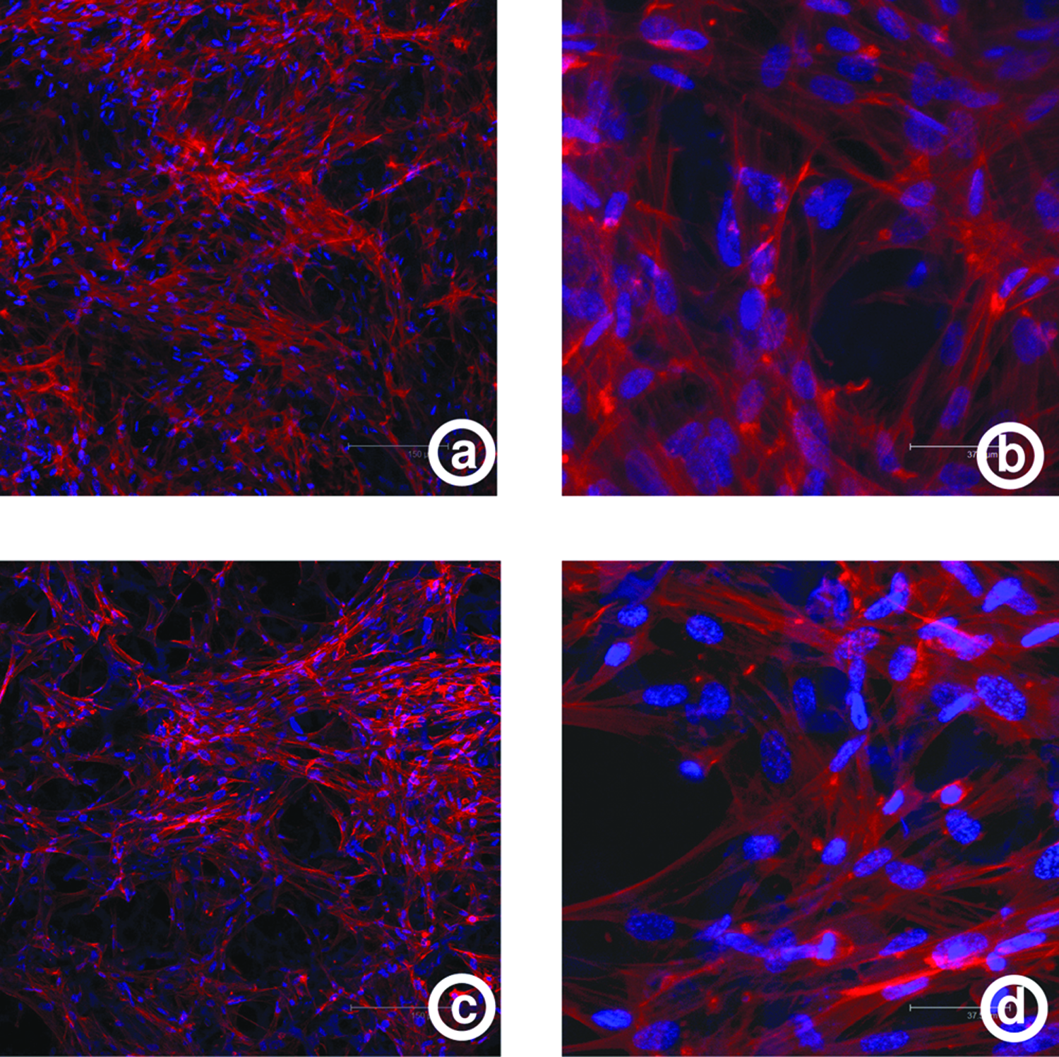
MRC-5 CLSM images of F-Actin and nucleus stained with phalloidin toxin and Hoechst, respectively, 8 days after addition to scaffolds without coating with attachment factors. (
In the absence of fibronectin coating, few HUVECs remained 4 h after addition to the P(EA-co-HEA) scaffolds (data not shown). However, when P(EA-co-HEA) scaffolds were first coated with fibronectin, a significant number of HUVECs attached (Fig. 3). The exception were P(EA-co-HEA) 50/50 scaffolds, on which cell attachment did not occur even after fibronectin coating (data not shown). The results shown in Figure 3 are representative of at least three different HUVEC donors. With increasing culture time, cells seeded onto the fibronectin-coated P(EA-co-HEA) scaffolds started to proliferate but did not reach confluence on the available surface area. Because endothelial cells only grew on fibronectin-coated P(EA-co-HEA) scaffolds, all further experiments were carried out with prior fibronectin coating. Both MRC-5 fibroblasts and HUVECs did not infiltrate the scaffolds during the duration of our cultures.
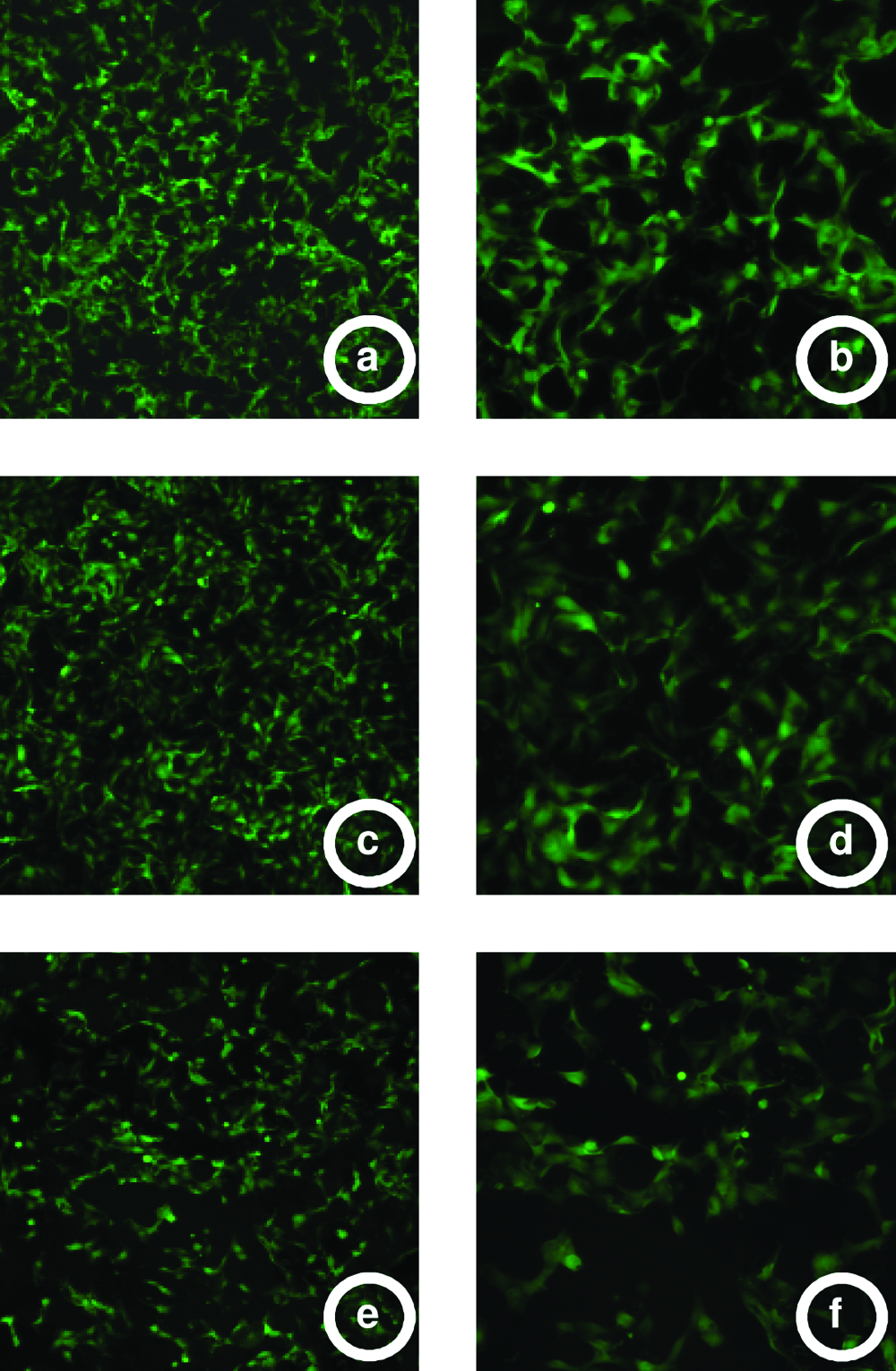
CLSM images of calcein-AM stained adherent HUVEC 5 days after seeded onto the scaffolds previously coated with fibronectin. Fluorescent images were converted to grayscale. (
Kruskal–Wallis analysis of cell populations on different types of surfaces revealed significant differences in the extent of HUVEC proliferation (k = 10.018; p = 0.007). To elucidate differences in cell growth between paired groups, a Mann–Whitney U-test was performed. Results showed differences between PEA and P(EA-co-HEA) 90/10 (U = 2; p = 0.017), and between PEA and P(EA-co-HEA) 80/20 (U = 0; p = 0.004), but no significant differences were found between P(EA-co-HEA) 90/10 and P(EA-co-HEA) 80/20 (U = 9; p = 0.180).
PECAM-1 and E-selectin protein expression by HUVECs on polymer scaffolds
In contrast to MRC-5 fibroblasts, HUVECs did not reach complete confluence on the scaffold surfaces (Fig. 4). To test the proinflammatory activation of endothelial cells, HUVECs were cultivated on fibronectin-coated P(EA-co-HEA) scaffolds and immunostained for E-selectin (red) and counterstained with Hoechst 33342 for cell nuclei (blue) 4 h after LPS stimulation. Nearly all HUVECs were E-selectin positive after LPS exposure as depicted in Figure 4A for the PEA scaffold. Further, after immunostaining for platelet–endothelial cell adhesion molecule 1 (PECAM-1, also known as CD31), cells growing on P(EA-co-HEA) scaffolds showed the characteristic cell–cell contacts typical of endothelial cells in vitro and in vivo (Fig. 4B, C).
Immunofluorescent images of E-selectin–stained (
Endothelial cell gene expression
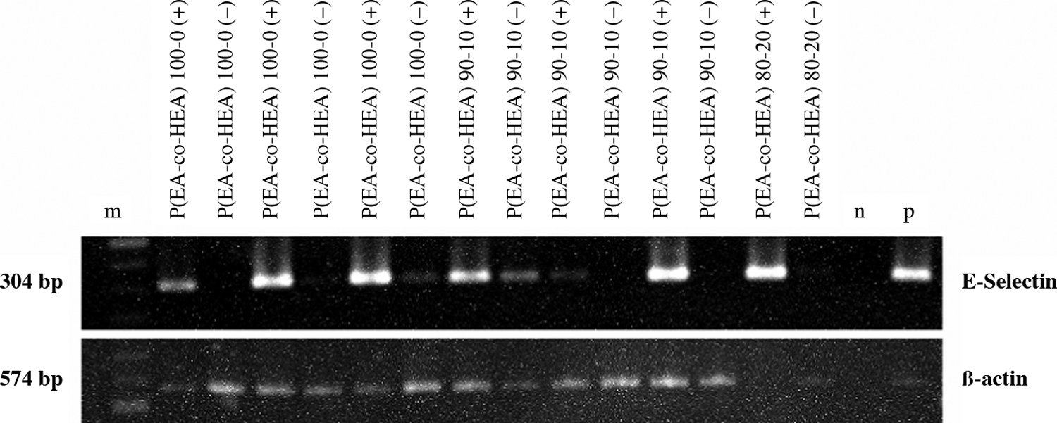
The regulation of endothelial cell gene expression at the mRNA level was examined by rtPCR for HUVECs growing on P(EA-co-HEA) scaffolds. As shown in Figure 5, cells growing on fibronectin-coated P(EA-co-HEA) scaffolds in the absence of LPS (−) exhibited little or no gene expression for the cell adhesion molecule E-selectin, a characteristic marker of inflammation,. In the presence of LPS (+), a strong induction of E-selectin expression was observed as compared to cells in the absence of LPS (−). Thus, not only did the cells growing on P(EA-co-HEA) scaffolds exhibit normal endothelial gene expression in the absence of LPS, but these cells also maintain functional capabilities as reflected in the strong induction of an inflammatory marker in the presence of LPS. This cellular behavior is in agreement with the expression of E-selectin at the protein level, as the majority of HUVECs growing on P(EA-co-HEA) in the presence of LPS were positive for E-selectin by immunostaining (see “PECAM-1 and E-selectin protein expression by HUVECs on polymer scaffolds” section; Fig. 4A). Thus, cells growing on P(EA-co-HEA) scaffolds exhibited a phenotype and cellular behavior very similar to cells growing on standard tissue culture plastic. 29
PCR analysis of endothelial cell gene expression of cells grown on fibronectin-coated scaffolds in the presence or absence of LPS. RNA was isolated from cells grown for 4 h with (+) and without (−) LPS, converted to cDNA, and subjected to PCR as described in Materials and Methods. “m” is molecular base pair (bp) size marker, “p” is positive control, and “n” is the negative water control with no cDNA.
Fibronectin adsorption and conformation on polymer sheets
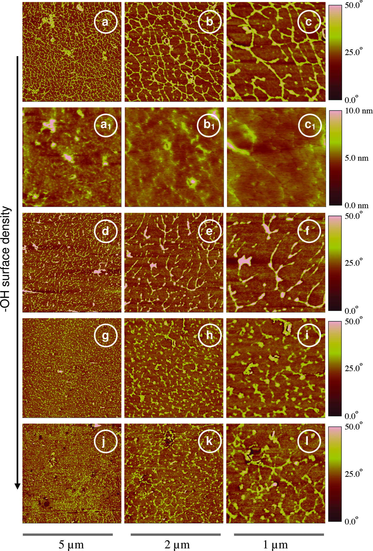
Figure 6 shows the phase magnitude of the different copolymer/fibronectin systems. Fibronectin conformation on the different bulk copolymers was a function of substrate chemistry: in particular, the density of -OH groups in the copolymer surface. Fibronectin assumed an extended conformation on the homopolymer PEA, the most hydrophobic substrate of the series. The morphology of the fibronectin changed to a less extended morphology with increasing surface hydrophilicity, engendered by a higher concentration of –OH groups at the polymer surface. It is remarkable that the formation of a fibronectin network just took place on the PEA homopolymer, whereas no such network arose on the other copolymer substrates. Height images of fibronectin on the PEA homopolymer surface are shown in Figures 6 as a1, b1, and c1. It is worth mentioning that although the roughness of the substrate is small enough to allow detection of the proteins in the height picture, the most appropriate parameter to observe the fibronectin is the phase signal. 30
AFM phase images of fibronectin and the different copolymers at different magnifications. (
Discussion
The polymeric substrates used in this study were macroporous materials with spherical interconnected pores. Considering the physical characteristics of these scaffolds, we expected that cells seeded on them would encounter alternating hydrophilic and hydrophobic nanodomains at the scaffold surface. The chemical structure of the copolymer chains formed by EA and HEA consists of a backbone with –COOCH2CH3 and –COOCH2CH2OH side groups randomly distributed. Modifying the ratio EA/HEA in the copolymers used to create our scaffolds, the surface density of –OH groups was varied, and, accordingly, the surface hydrophobicity. The methodology employed in this work permitted us to characterize the interaction between this macroporous structure and two types of cells—namely, the human fibroblast cell line MRC-5 and HUVECs. Fibronectin coating of the substrates was imperative for HUVEC attachment, whereas no fibronectin coating was needed for MRC-5 adhesion. As in other synthetic materials such as tissue culture polystyrene, the precoating with gelatin, laminin, or fibronectin has significantly increased endothelial cell attachment and proliferation.31–33 Fibronectin coating is frequently required for HUVEC attachment to different materials. 29 In contrast, culture of fibroblasts on different polymeric substrates without prior protein coating (i.e., just with possible adsorption of serum proteins) has been broadly reported in the literature.
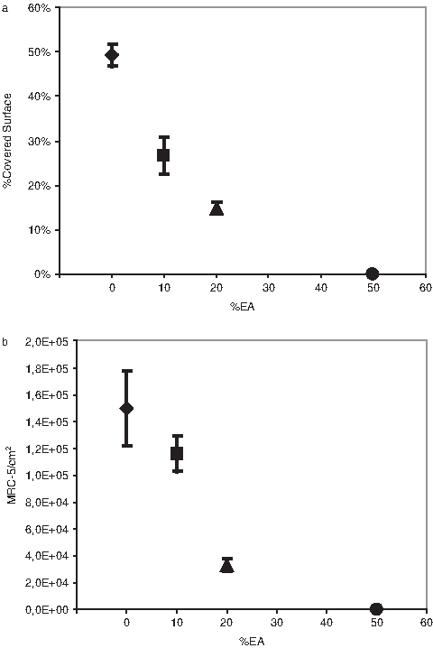
MRC-5 and HUVECs attached to substrates with a content of hydrophilic component of up to 20%. But clearly, the more hydrophobic the scaffold, the better the adhesion, spreading, and proliferation of both cell types. The same correlation was found when the normal human epithelial cell line, IOBA-NHC, was seeded onto bulk polymers with identical chemical composition as reported here. 24 Above this 20% hydrophilicity threshold, as in the case of P(EA-co-HEA) 50/50 no cell attachment was observed even with precoating of adhesion proteins on the surfaces (Fig. 7A, B). Interestingly, other cell types such as chondrocytes, osteoblasts, and neural cells have also been cultured on the polymer substrates described here, but the correlation of hydrophobicity with cell attachment was not observed. In particular, neural cells attached best to P(EA-co-HEA) 50/50. 34
(
To gain a better understanding of the influence of proteins on cell attachment, flat sheets of polymer substrates with identical chemical composition as the corresponding 3D polymer scaffolds were fibronectin coated and visualized by AFM. Only on the PEA homopolymer the formation of a fibronectin network occurred, but less and less with an increase in hydroxyl groups at the surface. This is not what happens with P(EA-co-HEA) copolymers when coated with other ECM proteins such as laminin, where the most extended conformation of the protein and the onset of network formation occur only beyond a certain surface concentration of –OH groups. 30 Nevertheless, biological performance correlated with qualitative measures of the protein conformation on the substrate. 34
The adsorption of proteins onto a surface is driven by the Gibbs free energy of the system. Protein adsorption will take place if the Gibbs free energy in the adsorbed state is lower than the Gibbs free energy of proteins surrounded by water molecules—that is, when proteins do not interact with the substrate. Both the entropy and the enthalpy of the system determine the adsorption and conformation of proteins. Thus, hydrophobic materials should be better substrates for protein adhesion due to the release of water molecules bound to the proteins, which leads to an increase in entropy. 35 On the other hand, enthalpic factors such as van der Waals interactions, ionic bonds, and hydrogen bonds between proteins and the substrate's surface play an important role in the Gibbs free energy.
Both constituents of the Gibbs free energy together will define the equilibrium state—that is, the amount of adsorbed proteins and their conformation. Especially, protein conformation is an important determinant of cell adhesion. The RGD tripeptide is one of the main adhesion domains of fibronectin, 36 and it seems that the more fibronectin extends onto a surface, the better fibronectin's RGD sequence is available to interact with cell membrane receptors. The AFM results of the fibronectin-coated scaffolds are indicative of the correlation between HUVEC adhesion and the conformation of the fibronectin molecules previously deposited on the substrate. The influence of the substrate's chemical structure on cell adhesion is relayed by protein–polymer interactions. Probably, similar principles apply to the case of fibroblasts, although precoating of the surface with fibronectin was not a decisive factor as in the case of HUVECs. Initially, adsorbing proteins of the serum-containing medium and, later, ECM proteins produced by the cells provide adhesion sites to the cells on a synthetic material. 35
Vascularization of the biomaterial is requisite for the cells to obtain nutrients and oxygen for survival. Thus, when the biomaterial is implanted, blood vessels from the host have to grow toward the biomaterial. If a material is seeded with a patient's own endothelial cells, and proper conditions are provided before implantation to preform a microvascular system within the biomaterial, these microvessels could potentially hook-up to the existing vasculature surrounding the implant after implantation. The main goal of our study was to evaluate if normal endothelial phenotypes and functions observed in vivo were maintained also in vitro. HUVECs exhibited normal cell–cell contacts and expression of the proinflammatory activation marker E-selectin upon specific stimulation. In summary, P(EA-co-HEA) scaffolds had no negative effects on endothelial cell function and phenotype, and might thus serve as prevascularized matrices for applications in regenerative medicine, such as corneal implants. In particular, we have developed these biomaterials as a novel anchorage ring for a keratoprosthesis. Therefore, activated stromal fibroblasts (i.e., keratocytes) and endothelial cells forming microvasculature to provide the necessary nutrients should be able to penetrate the scaffold briefly after implantation. Because fibroblasts do not need fibronectin coating, one possibility would be to seed autologous fibroblasts onto the scaffolds, and with time the extracellular matrix and growth factors produced by these cells might recruit the invasion of endothelial cells into the material. Consequently, conditions would exist in which the recipient's vascular system might connect quickly and provide the implant with the necessary nutrients for survival and proper function.
Conclusions
We have manufactured 3D porous scaffolds of P(EA-co-HEA) with varying degrees of hydrophilicity. Normal human fibroblasts (MRC-5 cell line) and human endothelial cells (HUVECs) were cultured on the scaffolds in vitro. Our results indicate that the interaction between these cell types and the materials promises good in vivo performance in terms of cell proliferation, differentiation, and function as long as the content of hydrophilic monomer does not exceed 20%. The degree of hydrophilicity affected the adsorption and conformation of adhesion proteins to the material and, consequently, the adhesion of cells. Such control over cellular behavior on a biomaterial might make it possible one day to create cell-bearing keratoprostheses that promote optimal implant integration and that enable vascularization only in such a manner and special distribution that vision is not impaired, while blood supply to the implant is optimized.
Footnotes
Acknowledgments
This work was supported by the Research Programme FP6 Marie Curie Early Stage Researcher Training through the contract MES-CT-2004-008104. A.J.C.F. would like to acknowledge the generous help and enjoyable discussions with Sabine Fuchs and José Francisco De Lamo Pastor. A.J.C.F., J.L.G.R., and J.M.M.D. acknowledge the support of the Spanish Ministry of Science and Education through MAT2003-5391-C03 project. AFM was performed under the technical guidance of the Microscopy Service at the Universidad Politécnica de Valencia.
Disclosure Statement
No competing financial interests exist.