
Abstract
Despite recent progress, mechanical behavior of tissue-engineered heart valves still needs improvement when native aortic valves are considered as a benchmark. Although it is known that cyclic straining enhances tissue formation, optimal loading protocols have not been defined yet. To obtain a better understanding of the effects of mechanical conditioning on tissue development, mechanical behavior of tissue constructs should be monitored and controlled during culture. However, currently used methods for mechanical characterization (e.g., tensile and indentation tests) are destructive and are only performed at the end-stage of tissue culture. In this study, an inverse experimental–numerical approach was developed that enables a noninvasive and nondestructive assessment of mechanical properties of engineered heart valves. The applied pressure and volumetric deformation of an engineered heart valve were measured during culture, and served as input for the estimation of mechanical properties using a computational model. To validate the method, six heart valves were cultured, and the mechanical properties obtained from the inverse experimental–numerical approach were in good agreement with uniaxial tensile test data. The method provides a real-time, noninvasive and nondestructive functionality and quality check of tissue-engineered heart valves and can be used to monitor and control the evolution of mechanical properties during tissue culture.
Introduction
Research showed that mechanical stimulation has a beneficial effect on tissue properties.4–8 Mechanical stimuli were applied to the developing tissue in bioreactor systems, which are characterized by the type of conditioning. Bioreactors are specified as strain based6,8–12 or flow based,13–16 or as systems in which the physiological environment of the tissue is mimicked.17–25 Most bioreactors applied in heart valve tissue engineering are flow based.17–23,25 However, studies have shown the enhancement of functional tissue formation as a result of cyclic tissue straining of the developing construct.6–11 Hence, as a starting point, the strain-based Diastolic Pulse Duplicator 26 is considered in which dynamic strains are induced in the heart valve leaflets in the closed configuration. In human heart valve culture, this approach gave rise to enhanced tissue formation and nonlinear tissue-like mechanical properties in the strained valves when compared to unloaded valves. 26
The major drawback of the Diastolic Pulse Duplicator and other bioreactor systems is the lack of control during load application. Pressure is applied to the developing heart valve while the mechanical properties of the valve are changing during culture. As a result, the induced tissue deformations are unknown, and vary during tissue culture.
An inverse experimental–numerical approach has been proposed by which leaflet deformation is assessed real-time and noninvasively 27 during culture in a bioreactor system. This technique allows a systematic study of the effects of mechanical straining on tissue development. However, to assess the mechanical properties of the engineered valves, the constructs need to be sacrificed to perform tensile28–32 or indentation tests. 33 To study the mechanical behavior of the engineered constructs during culture and to test the functionality of intact heart valves as a noninvasive quality check, it is desired to assess mechanical properties nondestructively. By further development of the experimental–numerical approach, a noninvasive and nondestructive assessment of mechanical properties of engineered heart valve leaflets is demonstrated here.
Materials and Methods
Numerical model
To relate the combination of applied pressure and induced volumetric deformation of heart valve leaflets to the mechanical properties of the leaflets, a quasi-static numerical model of the heart valve26,34 was employed.
Constitutive law
Assuming incompressibility of the leaflets, the Cauchy stress tensor (
To model nonlinear mechanical behavior, a nonlinear Neo-Hookean model26,34 is used:
The parameter n represents the degree of nonlinearity of the constitutive equation: n > 0 indicates stiffening of the material with increasing strains, whereas n < 0 indicates softening. Note that the classical Neo-Hookean model is obtained for n = 0, with G = G0.
Balance equations
The balance equations that have to be solved are conservation of momentum and mass for an incompressible solid:
Geometry and boundary conditions
The finite element mesh of the leaflets in the closed configuration is symmetric and, therefore, consists of only one half of a leaflet. At the symmetry edge, nodal displacements in the normal direction are suppressed. At the bottom (ventricular) side of the fixed edge, nodal displacements are suppressed in all directions. At the free edge, a contact surface is defined to model coaptation of adjacent leaflets. The radius of the leaflets is set to 12 mm. Pressure is applied to the top surface of the leaflets to model the applied diastolic transvalvular load. Subsequently, volumetric deformations are calculated.
Relationship between applied load, deformation and mechanical properties
The numerical model was employed to obtain the relationship between applied pressure, induced volumetric deformation, and the mechanical properties of the loaded heart valve. The mechanical properties are defined as the product of thickness (t) and shear modulus (G0) of the heart valve, and were used in different combinations as an input for the model. The range of these input parameters was chosen to cover experimental data from previous studies34,35 (Table 1).
For every combination of thickness and shear modulus, physiological pressure differences, ranging from 0 to 15 kPa, were applied to the valve and induced deformations were calculated. To acquire a relation in which every pressure–deformation set will lead to one unique value for the mechanical properties, a polynomial function was fitted through the simulation results. The accuracy of the fit was investigated by plotting the fitted deformation values versus the numerically calculated deformation values.
Effect of inhomogeneity and anisotropy
The effects of inhomogeneity and anisotropy of the heart valve on the estimation of the mechanical properties were investigated. Inhomogeneity was simulated by choosing a gradient in shear moduli from the belly to the commissures of the valve. Three gradients were chosen in which the shear modulus ratio between the belly and commissures is equal to 2, 5, and 100. Modulus values increased from 1 to 2, 0.5 to 2.5, and 0.03 to 3 MPa. Consequently, for every gradient, the mean value of the shear modulus was kept constant at a physiological realistic value of 1.5 MPa. Thickness values were still assumed homogeneous and ranged between 0.35 and 1.0 mm.
To investigate the impact of anisotropy, a structurally based fiber reinforced constitutive model 36 was used. The degree of anisotropy and other input parameters of the model corresponded to the parameters of tissue-engineered heart valves after 2, 3, and 4 weeks of culturing in a previous study. 35
Experimental setup
To assess the mechanical properties of a cultured heart valve using the numerical model, applied pressure and related deformation of the heart valve have to be measured. For this purpose, a previously developed bioreactor setup was used. 27
Description of the bioreactor
The bioreactor system consisted of four main components: a proportional compressed-air valve, a pneumatic pump consisting of a cylinder in which a silicone tube was placed, a bioreactor in which the valve was cultured, and a medium container (Fig. 1). 27 The whole system was filled with 150 mL of culture medium. The silicone tube inside the polycarbonate cylinder, which functions as a pneumatic pump, was compressed and decompressed by pulsatile air pressure from the proportional valve. The amount, frequency, and waveform of air release were controlled via a programmable multi-IO-card using LABVIEW software (National Instruments, Austin, TX). As a result, fluid from the container was injected into the upper chamber of the bioreactor, which caused a dynamic pressure difference over the heart valve. Consecutively, a fluid flow exited the bioreactor from both the lower and the upper chambers. Pressure sensors (P10EZ; BD, Franklin Lakes, NJ) measured the pressure in both chambers. Two flow sensors (Transonic Systems, Ithaca, NY) measured the flow in the tubing through which fluid entered the upper chamber (Fi) and left the lower chamber (Fo) of the bioreactor.

Schematic of the bioreactor setup. The bioreactor setup consists of a compressed air supply (
Pressure and deformation measurement
During pulsatile load application, pressure and flow measurements were performed to assess the pressure difference over the heart valve and the resulting volumetric deformation of the heart valve leaflets. Pressure difference is defined as the pressure measured in the upper chamber of the bioreactor minus the pressure in the lower chamber. In addition, volumetric deformation is represented as the total amount of fluid displaced by the heart valve leaflets when load is applied, and can be distinguished from the leakage through the valve. Relative leakage is defined as the leak flow through the valve divided by the amount of fluid entering the bioreactor.
Tissue culture
To demonstrate and validate the assessment of mechanical properties of a heart valve in culture, six heart valves were tissue engineered, using different conditioning protocols in two successive, but independent experiments. Two valves were cultured in the first experiment, while in the second experiment four valves were produced.
Heart valve scaffold
Stented heart valves were fabricated of anatomically shaped leaflets cut out of nonwoven polyglycolic acid meshes (thickness 1.0 mm; specific gravity 70 mg/cm3; Concordia Manufacturing, Coventry, RI). The dimensions were based on anatomical values measured in human specimens37–39 and were applied in the numerical model. The leaflets were coated with a thin layer of poly-4-hydroxybutyrate (P4HB; molecular weight 1 × 10⁶ Da; TEPHA Inc., Cambridge, MA) and molded in the shape of a trileaflet heart valve. Subsequently, they were attached to a rigid polycarbonate cylinder by sugar leaching of polycaprolactone (molecular weight 8 × 10⁴ Da; Sigma-Aldrich, St.Louis, MO).
Seeding procedure and mechanical conditioning
Cells harvested from the human vena saphena magna were expanded using regular cell culture methods. 40 After expansion, they were counted and seeded onto the scaffold. Cell densities ranged from 2.5 to 4.5 million (passage 7) per cm3 scaffold. Higher cell densities did not improve the mechanical properties of the cultured tissue (unpublished results). Fibrin was used as a cell carrier. 41 The medium to culture these cells consisted of Dulbecco's modified Eagle's medium Advanced (Life Technologies, Carlsbad, CA), supplemented with 10% fetal bovine serum (PAN Biotech GmbH, Aidenbach, Germany), 1% GlutaMax (Life Technologies), and 0.1% gentamycin (PAN Biotech). The medium used for seeding and subsequent tissue culture contained 0.3% gentamycin and additional L-ascorbic acid 2-phosphate (0.25 mg/mL; Sigma) to promote extracelluIar matrix production. 7 Before seeding, the scaffolds were disinfected in 70% ethanol and subsequently immersed in culture medium for 24 h to promote cell attachment.
After seeding, the constructs were placed in the bioreactor system and subjected to culture medium circulation at low speed (4 mL/min) for 5–7 days to allow initial tissue development after seeding. Thereafter, dynamic pressure differences (at 1 Hz) were applied to the heart valve leaflets for 14–21 days (Fig. 2). Total culture time involved the time span in which both (low speed) medium circulation as well as dynamic loading were applied to the engineered valves.

Conditioning protocol in which the applied load is given as a function of culture time for tissue-engineered heart valves 1-1 and 1-2 (
In the first experiment, load was applied stepwise for 9 days, increasing every 3 days. After 14 days, the applied load was decreased and kept constant at the level of days 8–11. Heart valves (valves 1-1 and 1-2) were sacrificed after a total culture time of 3 weeks (Fig. 2A). In the second experiment, pressure was increased more gradually in relatively small 3-day steps until the end of the experiment. Heart valve culture was finished after 3 weeks (valves 2-2 and 2-4) and 4 weeks (valves 2-1 and 2-3) (Fig. 2B).
Estimation of mechanical properties
After heart valve culture and just before sacrifice, tissue-engineered leaflets were subjected to increasing loads in the bioreactor setup. Pressure differences and induced volumetric deformation values were measured to obtain a pressure–deformation (ΔP − ΔV) curve for each valve. This curve is representative for the mechanical properties of the leaflets. Using the method of least-squares, the curve was fitted to the relationship between applied load, deformation, and mechanical properties, discussed in the Numerical model section, to obtain the product of shear modulus and thickness that corresponds to the best fit. Subsequently, the shear modulus of the heart valve was calculated by dividing this product by the average thickness of the heart valve.
Further, correlation coefficients (R2) were calculated that indicate the strength of the relation between the measured and predicted (fitted) pressure–deformation values. In the first experiment, the maximum pressure difference was equal to the applied pressure difference during the final stage of culturing (ΔP = 2.5 kPa). In the second experiment, a higher load was applied, trying to reach pressure values equal to physiological values (ΔP = 11 kPa).
Validation of mechanical properties assessment
To validate the assessment of the mechanical properties of the heart valve leaflets by the experimental–numerical method, cultured heart valves were sacrificed and cut into strips, and then uniaxial tensile tests were performed. For every valve, the strips were cut from the middle part of the leaflets (mainly belly region) in radial (n = 5) and circumferential (n = 5) directions, without including the leaflets free or fixed edges.
First, thickness and width of the tissue samples were measured using a PLμ 2300 noncontact Confocal Imaging Profiler (Sensofar Tech S.L., Terrassa, Spain). After that, the tissue strips were tested until rupture at a constant strain rate (1/60 s−1) using a tensile stage (Kammrath & Weiss GmbH, Dortmund, Germany) equipped with a 20 N load cell. Force and elongation were measured during testing and are converted to Cauchy stress and strain, respectively. The slope of the resulting stress–strain curve in the first 10% strain range is defined as the tangent stiffness of the tissue specimen. For every valve, acquired stiffness values were analyzed for the circumferential and radial directions.
The uniaxial tensile test results, Young's moduli in radial and circumferential directions of the heart valve leaflets, were compared to the mechanical properties estimated by the noninvasive assessment method. Therefore, estimated shear modulus (G) values were rewritten in Young's modulus (E) values. For incompressible materials and small strains, the Young's modulus (E) equals three times the shear modulus (G):
Statistics
The uniaxial tensile test data were not normally distributed and, therefore, tested nonparametric and expressed as median ± 95% confidence interval. The Kruskal-Wallis test does not assume a normal population of the data and was applied for testing equality of population medians among groups. This test was performed to check whether the sample medians were significantly different when p < 0.05. Data were analyzed using the Statgraphics Cemturion XV software package.
Results
Simulations: relationship between applied load, deformation, and mechanical properties
The relation between the transvalvular pressure difference, the induced volumetric deformation, and the mechanical properties of the valve leaflets showed that an increase in deformation at a constant transvalvular pressure difference resulted in a decrease in predicted stiffness of the valve, and vice versa (Fig. 3A). To obtain a smooth relation, the simulation data were described by a polynomial fit, and the correspondence between the original and fitted data was shown in Figure 3B. In this figure, the straight line through the origin with a slope equal to 1 indicates a perfect resemblance between both data sets. It was shown that for small deformation values (ΔV < 0.2 mL) the fitted values were slightly overestimated, while for larger deformations (ΔV > 0.2 mL) deformation values were underestimated.
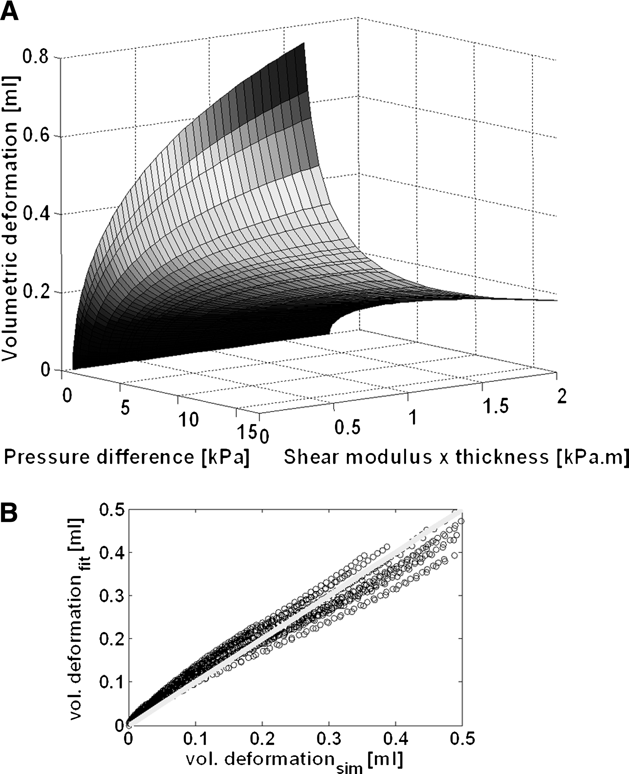
(
Effect of inhomogeneity and anisotropy
The effect of inhomogeneity on the relation between applied load, induced deformation, and mechanical properties of heart valve leaflets was minimal for different degrees of inhomogeneity (Fig. 4).

Numerically obtained relation between the applied pressure difference (kPa) over the heart valve leaflets, induced deformation (mL), and mechanical properties (kPa · m), defined as shear modulus × thickness, of the valve for four different degrees of inhomogeneity. Homogeneous material behavior is represented by “homo,” whereas “inhomo 1,” “inhomo 2,” and “inhomo 3” indicate the increasing degrees of inhomogeneity with shear moduli ranging from 1, 0.5, and 0.03 MPa in the belly to 2, 2.5, and 3 MPa in the commissures, respectively.
The volumetric deformation was relatively insensitive to the degree of anisotropy for practical tissue engineering circumstances. In particular, with anisotropies representative for 2 and 3 weeks of culture, the volumetric deformation was very close to the isotropic case. Anisotropy representative for 4 weeks of culture did have a small effect. In particular, for large pressure differences a small overestimation of the volumetric deformation was observed (Fig. 5).
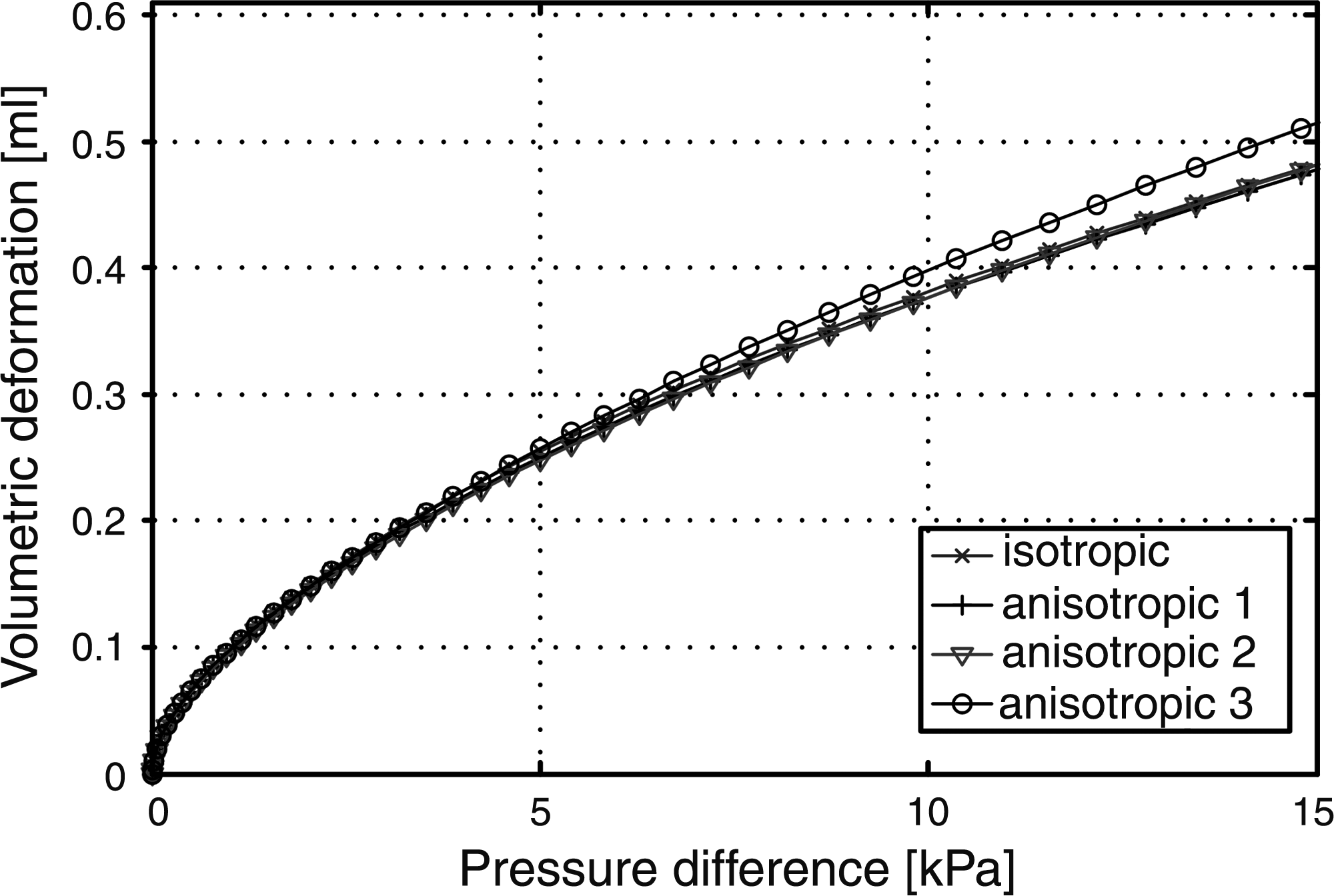
Relationship between applied pressure difference (kPa) and induced volumetric deformation (mL) of the heart valve leaflets, numerically obtained for four different degrees of anisotropy. Isotropic material behavior is represented by “isotropic,” whereas “anisotropic 1,” “anisotropic 2,” and “anisotropic 3” indicate the degrees of anisotropy estimated in a previous study in tissue-engineered heart valves after 2, 3, and 4 weeks of culturing,35 respectively.
Estimation of mechanical properties
The measured pressure–deformation curves and their best fits were depicted in the same chart for every valve (Figs. 6 and 7). In the first experiment, it was shown that measured deformation values did not exceed 0.2 mL, while the maximum applied pressure difference was equal to 2.5 kPa (Fig. 6). Further, a good correlation between measured and fitted data was found for both valves (1-1 and 1-2), whereas the correlation coefficient for valve 1-2 was slightly higher than that for valve 1-1. The relative leakage value was higher for valve 1-1 (10%) than for valve 1-2 (7%), and the estimated mechanical properties indicated values of 0.28 and 0.22 kPa · m for valve 1-1 and 1-2, respectively (Table 2).
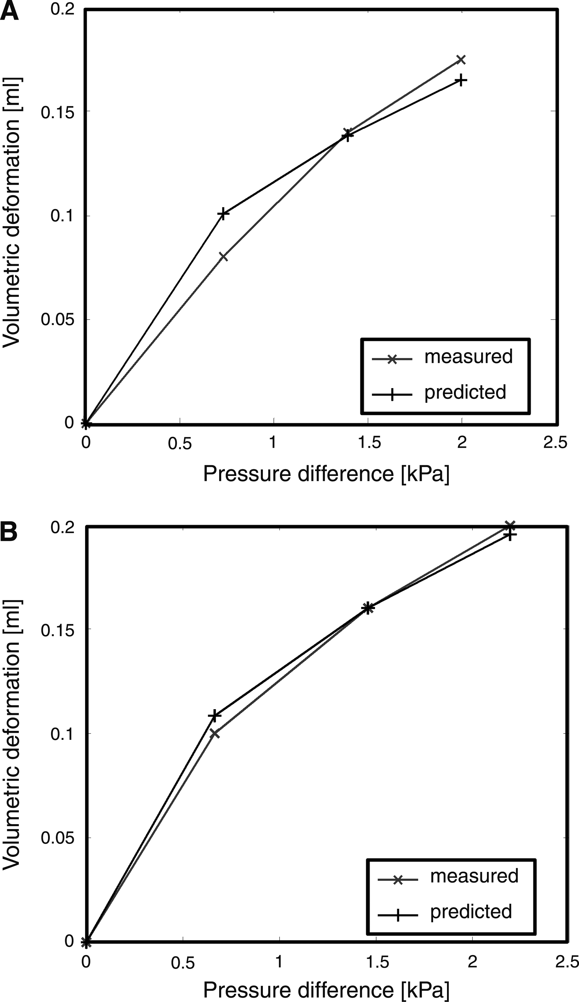
Measured and fitted (predicted) relationship, between applied pressure differences (kPa) and induced volumetric deformation (mL) of the cultured heart valve leaflets in the first tissue engineering experiment. Data are obtained after 3 weeks of culture and just before sacrifice for (

Measured and fitted (predicted) relationship between applied pressure differences (kPa) and induced volumetric deformation (mL) of the cultured heart valve leaflets in the second tissue engineering experiment. Data are obtained just before sacrifice after 3 weeks of culture for (
The relative leakage of the valves (%-leakage), the estimated values of the product of shear modulus and thickness G · t, and the estimation correlation coefficients (R2) are assessed just before, and the mean thickness values after sacrifice of the heart valves. Thereafter, Young's modulus values are calculated from the estimated G · t and thickness values.
In the second experiment, physiological pressure differences (ΔP = 12 kPa) were imposed on three valves (2-1, 2-3, and 2-4), while for valve 2-2 physiological pressure buildup was not possible. Due to the high leak flow, the maximum applied load to valve 2-2 was only 5 kPa (Fig. 7). In addition, the correlation between measured and predicted ΔP − ΔV curves was high for these three valves and relatively low for valve 2-2. Relative leakage values varied between 0% (valve 2-3) and 32% (valve 2-2). As seen in our previous study, 27 volumetric deformation measurements are not accurate when relative leakage values exceed 25%. Consequently, a reliable estimation of the mechanical properties of valve 2-2 could not be made. In conclusion, the estimated product of shear modulus and thickness was equal to 0.67, 0.46, and 0.36 kPa · m for valves 2-1, 2-3, and 2-4, respectively (Table 2).
Validation of mechanical properties assessment
Estimated mechanical properties of the cultured valves were compared with the data obtained from the uniaxial tensile tests. Young's moduli of valves 1-1 and 1-2, cultured in the first experiment, had estimated values equal to 0.97 and 0.77 MPa, respectively (Table 2). In the second experiment, the cultured heart valves had higher estimated Young's moduli, ranging from 1.3 MPa (valve 2-4) to 2.2 MPa (valve 2-1) (Table 2). For all valves except valve 2-2, the magnitude of the estimated stiffness was between the stiffness values obtained for the radial and circumferential direction (Fig. 8).
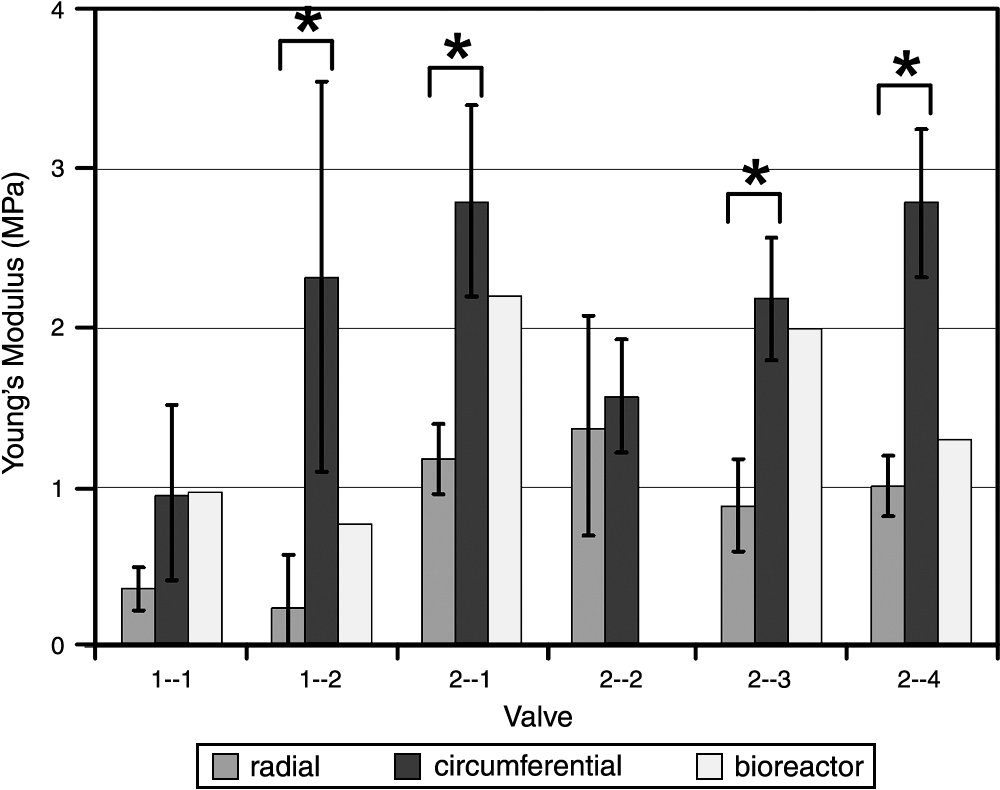
Young's modulus values (MPa) of valves 1-1 to 1-2 and valves 2-1 to 2-4. The medians of the Young's moduli in radial and circumferential directions are represented. The error bars indicate the 95% confidence intervals of the data. Statistically significant differences (p < 0.05) are marked with an asterisk. The bioreactor values represent the Young's moduli assessed by the inverse experimental–numerical estimation method.
Significant differences (p < 0.05) in Young's moduli were found between the radial and circumferential directions of tissue-engineered heart valves 1-2, 2-1, 2-3, and 2-4.
Discussion
In heart valve tissue engineering, the stiffness and strength of cultured heart valves still need additional improvement to reach mechanical characteristics and tissue composition of native aortic valves. One approach to enhance these parameters may be the application of well-defined mechanical stimulation to the developing tissue. Tissue formation and development in cultured heart valves in response to mechanical loading have been evaluated in different ways. Functionality of the engineered constructs has been studied by bioreactor systems in which the physiological environment of the tissue is simulated.3,22,42 Moreover, tissue quality has mainly been studied qualitatively or quantitatively by histology or biochemical assays, respectively.1–3,20,22,23,25,42,43 Additionally, to elucidate the effect of load application on the mechanical characteristics of the tissue-engineered construct, mechanical behavior of heart valve tissue has been evaluated by performing uniaxial or biaxial tensile tests or indentation tests.2,3,22,42 Unfavorably, these methods are destructive and can, therefore, only be performed at the end stage of tissue culture. To study the development of mechanical properties of the tissue-engineered heart valve during culture, it is desired to monitor and control these properties real-time and noninvasively. Hence, the objective of this study was to develop a bioreactor system in which engineered heart valve tissue is mechanically stimulated and in which, during and after culturing, the mechanical properties of the valve leaflets are assessed noninvasively.
The assessment of mechanical properties was performed by an inverse experimental–numerical approach. The relationship between the applied pressure, induced volumetric deformation, and mechanical behavior was described by a polynomial function fit through the deformation values calculated by the model. Good similarity was found between the simulated and fitted volumetric deformation values. The minor deviations observed were probably due to the relatively large influence of thickness on the flexural behavior of the leaflets when the valve deforms. This phenomenon was in particular observed for large deformations and was also seen in our previous study. 27
The effect of inhomogeneity on the mechanical properties estimation method was very small. The maximum gradient in shear moduli was varied by a factor 100 (0.03 MPa in belly region versus 3 MPa in commissures) and resulted in a negligible deviation with respect to the homogeneous shear modulus distribution. The minor effect of inhomogeneity was ascribed to the type of deformation that was considered. The overall deformation of the heart valve leaflets was used, and, therefore, local stiffness differences in the leaflets were counterbalanced. This resulted in average mechanical properties that remained constant. The same phenomenon was observed for the effect of anisotropy. However, anisotropy had a larger influence on the estimation of mechanical properties. The highest degree of anisotropy, which was equal to the degree of anisotropy found in tissue-engineered valves after 4 weeks of culture, 33 resulted in a small overestimation of volumetric deformation values. The insensitiveness of the mechanical properties estimation method for changes in homogeneity or isotropy in the heart valve leaflets implies that the degree of inhomogeneity or anisotropy cannot be assessed using this estimation method.
In this study, six heart valves were tissue engineered of which the mechanical properties were estimated using the measured pressure–deformation (ΔP − ΔV) relation. In the first experiment, the ΔP − ΔV curves of both valves were acquired by applying pressure differences in a relatively small range (ΔPmax = 2.5 kPa), while in the second experiment the maximal applied pressure difference was physiological (ΔPmax = 12 kPa). The latter procedure is favorable in the estimation of mechanical properties. More data points increase the accuracy of the fitting procedure. Further, load application until physiological values provides a quality control to examine whether the cultured heart valve leaflets can sustain a simulated in vivo aortic pressure difference.
The estimated mechanical properties of the engineered heart valve leaflets were validated using uniaxial tensile test data acquired in two orthogonal directions. Despite, uniaxial loading is nonphysiological, the applied numerical model is isotropic. Therefore, it was not necessary to perform biaxial tensile tests to validate the estimation method. In addition, tissue-engineered heart valves showed viscoelastic behavior after culture. However, the influence of preconditioning on the tensile test results was relatively small. The variation in stiffness values due to the presence or absence of preconditioning was smaller than the variation in stiffness values within a tissue-engineerd heart valve. For this reason, preconditioning was not performed. Good agreement was found between both mechanical properties assessment methods when relative leakage values did not exceed 10%. This was also confirmed by the values of the correlation coefficients of the fitting procedure. The coefficients did not have values smaller than 0.97, except for valve 2-2 (R2 = 0.88). An accurate Young's modulus estimation of this valve was not possible because its relative leakage value (32%) exceeded the leakage value (25%) above which volumetric deformation cannot be measured correctly. 27 Consequently, a relative leakage of 10% (valve 1-1) is still acceptable for the estimation of mechanical properties, but 32% (valve 2-2) leakage is not. The high leakage value of valve 2-2 was caused by the inappropriate fabrication of the scaffold of this valve. After culture, the valve leaflets themselves were intact, but the attachment line between one of the leaflets and the polycarbonate wall was leaking. This leakage along the wall hindered pressure build-up, deformation measurement, and, therefore, estimation of mechanical properties.
Further, for all valves, except valve 2-2, estimated stiffness values were larger than the experimentally obtained radial and smaller than circumferential tensile test values. This can be explained by the use of overall volumetric deformation values for the estimation method by which local stiffness differences are evened out.
In this study, the first 10–15% strain range of the measured stress–strain curves was applied to assess Young's moduli of the radial and circumferential oriented heart valve tissue strips. This is in contrast to previous studies in which no strain range was defined, but the steepest part of the stress–strain curve was chosen to determine the mechanical properties.3,7,26,35,42 However, maximum volumetric deformation of the cultured heart valve leaflets under physiological loading did not exceed 0.35 mL (valve 2-4), which corresponds to a mean strain value of approximately 7% in the leaflets. 27 Therefore, the mechanical behavior of the cultured tissue is best characterized by a Young's modulus assessed in the lower 10% strain region of the stress–strain curve, which encompasses in vivo deformation.
Functionality of the cultured heart valve leaflets, except for valve 2-2, was shown by the small leakage values (<10%) and by the sustained physiological diastolic pressure differences (valves 2-1, 2-3, and 2-4). Further, tissue-engineered heart valves examined by uniaxial tensile tests showed some development of anisotropy but did not exhibit the highly anisotropic and nonlinear mechanical behavior seen in native human aortic valves.28,44,45 Young's moduli of the tissue-engineered valves were compared to native human aortic values 45 and appeared to be similar in radial direction (varying between 75% of native value for valve 1-1 to 112% for valve 2-4), but considerably smaller in circumferential direction (from 18% for valve 2-2 to 31% for valve 1-2). To be able to make this comparison, Young's moduli were determined not only from the lower 10% strain region of the obtained strain–stress curves, but also from the steepest part.
Conclusion
The proposed inverse experimental–numerical estimation method allows the assessment of overall mechanical properties of tissue-engineered heart valve leaflets in real-time, noninvasively and nondestructively, during and after culturing. Therefore, this method can serve as a real-time, noninvasive and nondestructive functionality and quality check, and can be used to monitor and control tissue remodeling over time.
Footnotes
Acknowledgments
This research was supported by the Dutch Program for Tissue Engineering.
Disclosure Statement
No competing financial interests exist.