




Abstract
Although calcification remains as the main clinical concern associated with bioprosthetic heart valve replacement surgery, there is evidence that tissue deterioration leads to thromboembolism. In such instances, measures that prevent thrombosis may be beneficial. To minimize thrombosis, endothelialization of the valve surface before implantation has been proposed to facilitate coverage. In this study we aimed to define the optimal flow parameters for the endothelialization of decellularized heart valves using endothelial progenitor cell (EPC)–derived endothelial cells (ECs). We assessed the thrombogenic characteristics of the endothelialized heart valve surface using a bioreactor. EPC-derived ECs were seeded on decellularized porcine valve scaffolds. A computer-controlled bioreactor system was used to determine the optimal flow rates. Successful endothelialization was achieved by preconditioning the cell-seeded valves with stepwise increases in volume flow rate up to 2 L/min for 7 days. We show that decellularized valve scaffolds seeded with EPC-derived ECs improved the anti-thrombotic properties of the valve, whereas the scaffolds without ECs escalated the coagulation process. This study demonstrates that preconditioning of ECs seeded on valve matrices using a bioreactor system is necessary for achieving uniform endothelialization of valve scaffolds, which may reduce thrombotic activity after implantation in vivo.
Introduction
While the concept of using ECs to decrease the rate of thrombosis has been widely accepted, the functional outcome depends primarily on the levels of cell coverage. To achieve full coverage of ECs on tissue implants, various types of bioreactors have been employed to maximize cell coverage. 7 Bioreactors for cardiovascular applications are designed to provide mechanical stimulation that mimics physiological conditions in vitro, as exposure to stimuli such as pulsatile flow and pressure changes has been shown to enhance tissue formation, organization, and function.8–13 The fundamental components of a bioreactor include a pump that exerts a driving force for fluid, a reservoir, and a chamber containing the heart valve implant. 14 These components work in concert to provide an environment that allows to precondition cells on scaffolds in vitro and promote the enhancement of cell–matrix interaction, cellular proliferation, and organization.15,16 It has been suggested that factors such as flow rate, volume, and pressure influence the outcome measures.17,18 However, the optimum conditions that allow for uniform cell coverage have not been defined. In this study we developed a pulsatile bioreactor system that can be manipulated through a computer program to adjust flow parameters to maximize EC coverage on heart valve matrices. We aimed to define the optimal flow parameters for the endothelialization of decellularized heart valves using endothelial progenitor cell (EPC)–derived ECs. We assessed the thrombogenic characteristics of endothelialized heart valve surface using a bioreactor system.
Materials and Methods
EC preparation
Peripheral blood (20 mL per sample) was collected from the jugular vein of sheep. This study was conducted on 10 different animals using 5 independent blood sample collections per animal. The mononuclear cell fraction, which contains EPCs, was isolated using Ficoll-Histopaque-1083 (Sigma, St. Louis, MO) density-gradient centrifugation. The cells were washed with phosphate buffered saline (PBS) to remove contaminating platelets. An aliquot of mononuclear cells was initially cultured in a 35 mm culture dish with Endothelial Cell Growth Medium (EGM-2) supplemented with Single-Quot (Lonza, Walkersville, MD), ascorbic acid, heparin, gentamicin, VEGF, IGF, FGF, EGF, and 2% fetal bovine serum as supplied by the manufacturer for 24 h. Cells were then transferred to fibronectin-coated 35 mm culture plates (Corning, Corning, NY). EC differentiation was initiated with EGM-2. Mononuclear cells were cultured until cells with endothelial morphology were visible and began to proliferate. Cells were further expanded and differentiated into mature ECs. At passage five, the differentiated ECs were characterized by immunohistochemistry using mature EC-specific markers. Further, cells were cultured on Matrigel to confirm their potential to form small capillaries. The cells were successfully grown from every sample collected.
Scaffold preparation and seeding
Porcine pulmonary valves, obtained from a slaughterhouse, were decellularized using 2% Triton X-100 (Sigma-Aldrich, St. Louis, MO) and 0.2% ammonium hydroxide (Fisher Scientific, Fair Lawn, NJ). Valve samples were placed on a mechanical shaker at 4°C, and the detergent solution was changed every 36 h for 4 weeks. After 4 weeks, the remaining valve scaffolds were repeatedly washed with distilled water until all detergents were completely removed. Valve scaffolds were sterilized using γ-irradiation (1 MRad). To test for residual cellularity, decellularized valves were frozen in liquid nitrogen and then mechanically pulverized. The powdered tissue (25 mg) was processed with the QIAamp tissue kit (Qiagen, Hilden, Germany) according to the manufacturer's instructions to isolate any remaining DNA. The samples were run on an agarose gel (0.8%) to identify DNA bands. Samples were also processed for hematoxylin & eosin (H&E) and Movat's pentachrome staining. 19
The decellularized valve matrices were seeded manually at a density of 3 million cells/cm2 on each side of the leaflets, and the cells were allowed to attach for 1 h. This seeding density was decided after multiple densities have been tested, and this one yielded the best coverage. Subsequently, the cell-seeded decellularized porcine valve was transferred to a container filled with EGM-2. The seeding process was repeated on the third day under the same conditions. Culture medium was changed every 2 days for 2 weeks. At that time, the cell-seeded scaffold was placed into a bioreactor chamber filled with fresh EGM-2.
Bioreactor design
The bioreactor system consisted of a valve inlet port, valve mounting post, compliant section, pump, and reservoir (Fig. 1), which was manufactured to be placed in a 37°C CO2 incubator. The valve inlet port was designed with a smooth curve to prevent flow disturbance and maintain laminar flow into the valve. The valve post allowed for varying sized valves to be used by suturing the valves to a skirt material (vascular graft) that fit snugly over the post. A compliant section (capacitance) was located at the outlet port of the valve chamber. This was designed to create a backflow that closes the heart valve. Physiologic flow waveforms were gradually introduced to the valve over time using a digital gear pump (Ismatec, Glattbrugg, Switzerland) controlled by LabView™ software (National Instruments, Austin, Texas), into which the flow protocol was programmed in our laboratory. The digital signal created by the LabView program was converted to an analog signal by an A-to-Z converter, and this analog signal commands the gear pump, enabling the control of flow rate and pulse rate. Flow and pressure were monitored to ensure that physiologic conditions were maintained throughout the experiments. The entire closed loop flow system, which circulates a medium volume of 2.5 L, was constructed from clear polycarbonate, which allowed for direct observation of the valve movement. Polycarbonate is a durable, dimensionally stable, and transparent thermoplastic. Acrylic was used in our previous prototype; however, this material was easily damaged and lost its transparency during sterilization, cleaning, and upon exposure to chemicals.
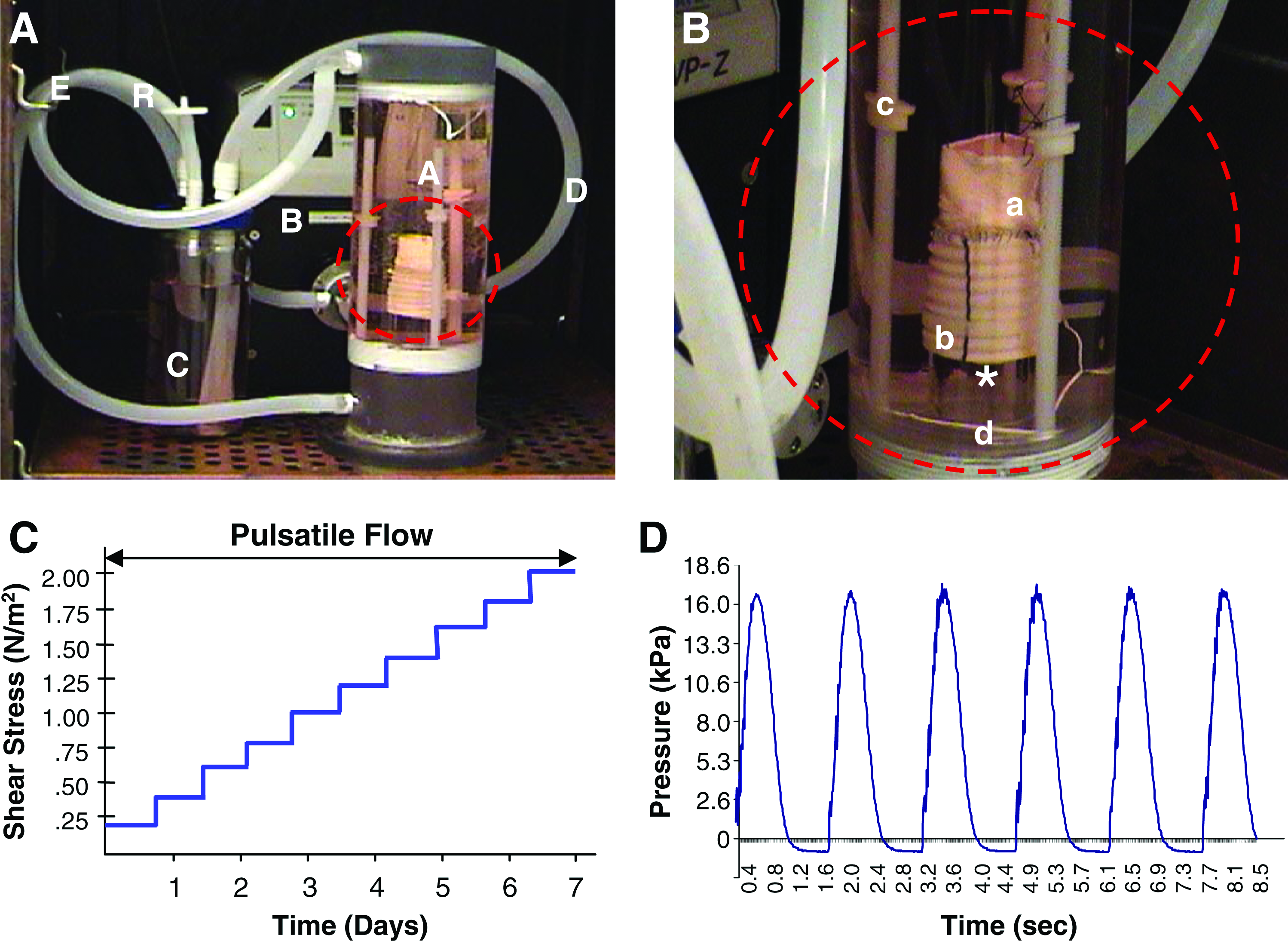
Computer-assisted pulsatile bioreactor system that is capable of controlling flow–pressure curves during preconditioning. (
Control of fluid shear stress and preconditioning
The mean fluid shear stress was determined from velocity measurements of the pulmonary valve. Approximate physiological flow in a juvenile sheep is about 2.0 L/min.
12
This velocity was converted to shear stress values using the Hagen-Poiseuille equation,
After static seeding on decellularized valves, three independent sets of cell-seeded valves were preconditioned initially with continuous flow. Subsequently, the pulsatile flow was applied, and the rate was gradually increased through 10 steps up to 1 L/min (n = 3), 2 L/min (n = 3), or 3 L/min (n = 3). The incremental rate increase of each set was 0.1, 0.2, and 0.3 L/min, respectively. The preconditioning procedure as a whole was done in 7 days, making the duration of each step to be 16.8 h. The pulse rate was maintained at 100 beats/min, and the culture medium was exchanged every 3.5 days. The decellularized valve scaffolds placed in the bioreactor consistently demonstrated opening and closing of the valve leaflets (Fig. 2).
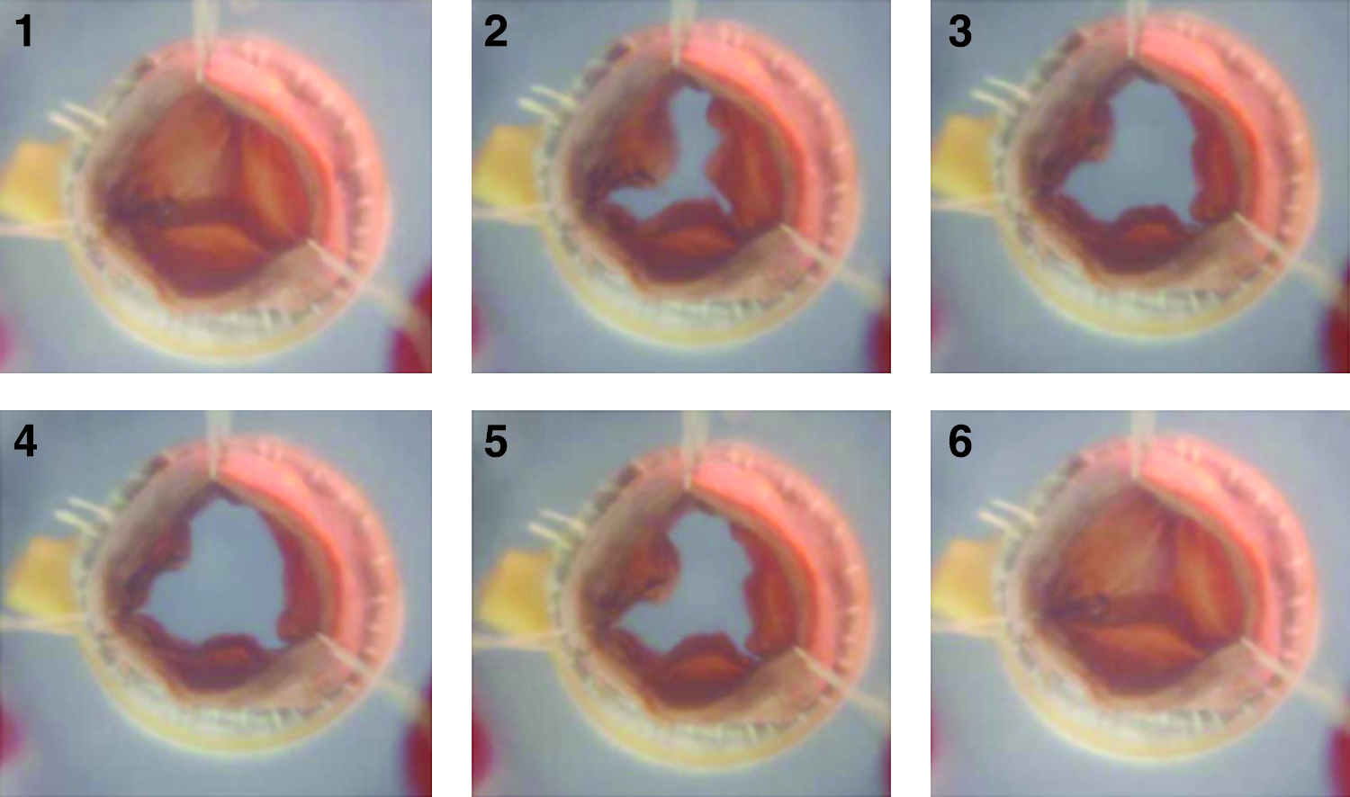
Sequential images showing valve opening and closing in the bioreactor. Medium is removed for enhanced observation. Color images available online at www.liebertonline.com/ten.
Histomorphological evaluation
EPC-derived ECs were plated onto six-well plates for 24 h, fixed with ice-cold methanol, and incubated with primary antibodies, including CD31 (Santa Cruz, Santa Cruz, CA), Flk-1 (Santa Cruz), Ulex-1 (Sigma), von Wilbrand factor (vWf; Dako, Carpinteria, CA), and CD146 (BD Bioscience, San Jose, CA) diluted at 1:50. This was followed by incubation with FITC-conjugated or Texas red–conjugated secondary antibody diluted at 1:300. Samples of cell-seeded valve tissue were embedded in Optimal Cutting Temperature solution (Sakura Finetek, Tokyo, Japan). The whole leaflet with commissure and part of the cusp were included in every block. The frozen tissue block was sectioned at a thickness of 5 μm using a cryotome. H&E staining was performed. For immunohistochemistry, sections of cell-seeded valve tissue were incubated with anti-human vWF (Dako) at room temperature for 2 h, followed by fluorescent-labeled secondary antibodies at room temperature for 30 min. The tissue slide was rinsed, counterstained with 4′-6-Diamidino-2-phenylindole (DAPI), and mounted using Vectashield fluorescent mounting medium (Vector, Burlingame, CA).
Ultrastructural evaluation
For scanning electron microscopy (SEM), the samples were randomly selected from three different regions of the leaflet. The samples were then fixed with 2.5% glutaraldehyde (Gibco Laboratories, Grand Island, NY) for 20 min at room temperature. After thorough washing with PBS, samples were dehydrated in graded ethanol series (50%, 60%, 70%, 80%, 90%, and 100%) and allowed to dry. Samples were then coated with platinum under vacuum and examined by SEM (Model S-2256N; Hitachi, Naka, Japan) with a tilt angle of 45°. Image J image software was used to determine the levels of cell coverage on randomly selected areas of the leaflet.
Platelet adhesion assay
Platelet-rich plasma (PRP) was prepared from fresh human blood by centrifugation at 300 g for 20 min at 4°C. Valve leaflets (both decellularized and seeded with EPC-derived ECs) were placed on 12-well plates and gently rinsed with PBS. PRP was added to the 12-well plates to completely immerse the leaflets, and the plates were incubated for 2 h at 37°C. The leaflets were then gently washed three times with PBS to eliminate nonadherent platelets. Adhered platelets were prepared for SEM imaging as described previously. 20 Platelet density was determined by counting eight random fields and expressing the numbers as average platelets per mm2 of tissue surface area.
Statistical analyses
Data obtained from SEM analysis and platelet adhesion assays were presented as mean ± standard deviation (SD). Unpaired Student's t-test was performed for the data analysis. A value of p < 0.05 was defined as significant.
Results
EPC isolation, differentiation, and characterization
Mononuclear cells were successfully isolated from 20 mL of sheep blood and plated on fibronectin-coated plates. Formation of colonies was observed a few days later (Fig. 3A). Cells grown in differentiation medium showed the typical cobblestone appearance, indicative of ECs (Fig. 3B). At passage 5, which was chosen for all of our experiments based on cell numbers and proper cell characteristics, cells were characterized by immunocytochemistry to demonstrate differentiation into ECs. These cells strongly expressed the endothelial-specific cell markers, including CD31, CD146, Ulex-1, vWF, and Flk-1 (Fig. 3C, D, E, F, and G, respectively). In addition, these cells formed capillary structures typical of ECs when seeded onto Matrigel (Fig. 3H). These data indicate that EPCs from circulating peripheral blood had differentiated into mature ECs in vitro.
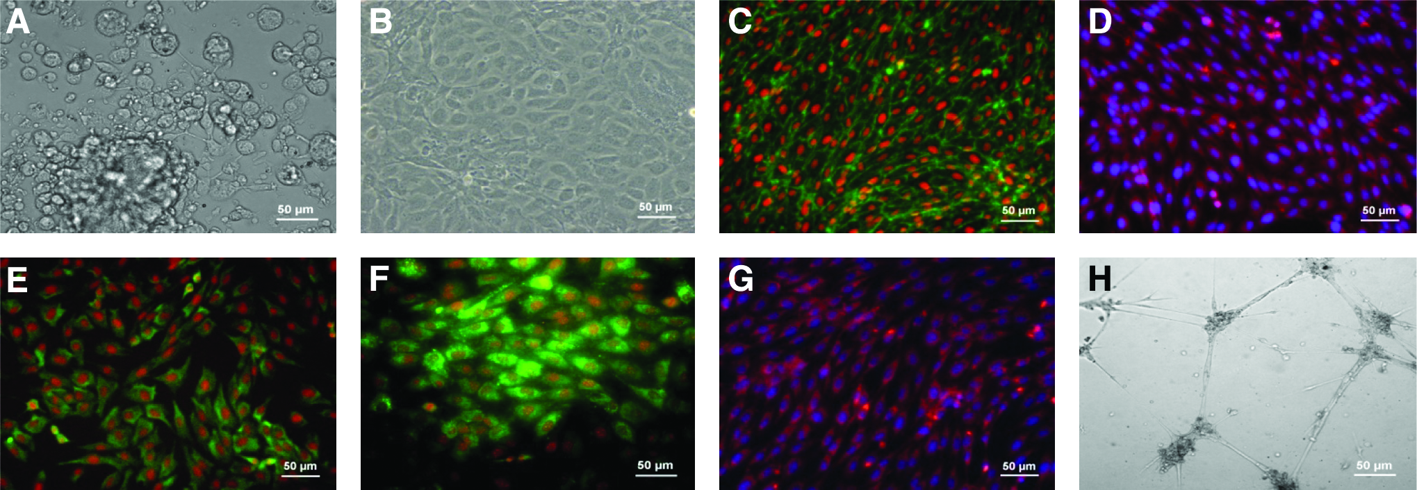
Characterization of endothelial progenitor cell (EPC)-derived endothelial cells (ECs). (
Scaffold preparation and static seeding
Valve matrices were decellularized and tested to determine their suitability to serve as implantable constructs. The decellularized valve matrices maintained their initial structural configuration with intact leaflets (Fig. 4A). No cells were detected in the leaflet after H&E staining (Fig. 4B), and no residual DNA was found in the leaflets, valvular root, or surrounding muscle tissue (Fig. 4C). Further examination using pentachrome staining demonstrated that collagen and elastin fibers were maintained and were distributed throughout the matrix (data not shown). These data indicate that cells were successfully removed from donor valve tissues, while the natural extracellular matrix was preserved. Valve constructs seeded through two stages showed cell attachment on the surface of the leaflets after 2 weeks in culture conditions (Fig. 5A, B). These cells stained positively for vWf (Fig. 5C).
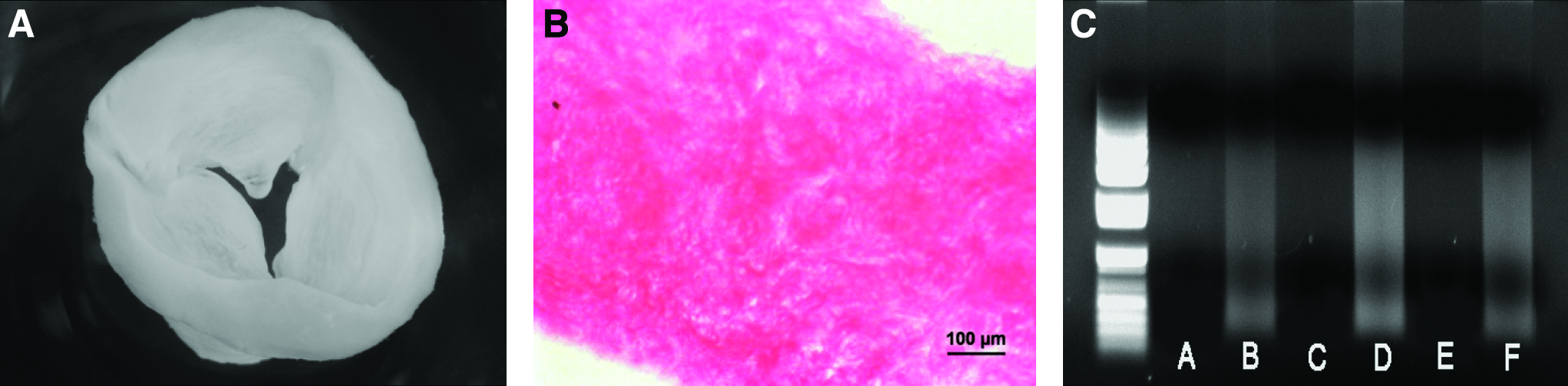
Decellularized porcine pulmonary valve. (
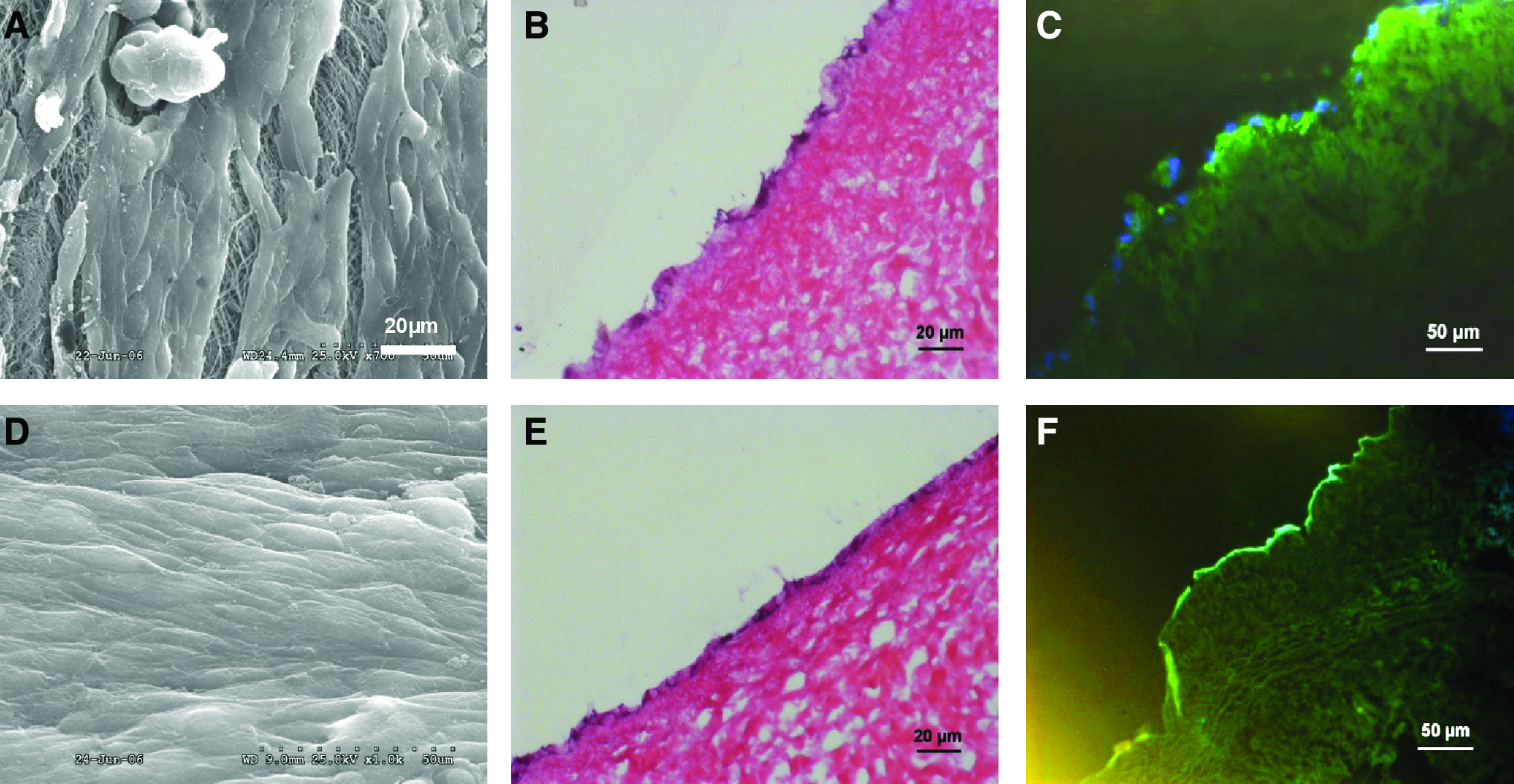
Efficacy of cell seeding was assessed after static culture (
Optimal parameters for bioreactor preconditioning
Uniform EC coverage was evident in valves placed under dynamic pulsatile flow conditions (Fig. 5D–F), whereas incomplete cell coverage was observed on the statically seeded valves (without bioreactor preconditioning) as demonstrated by SEM and H&E (Fig. 5A–C). Under the stepwise increase of flow rate up to 1 L/min, the valve matrix was uniformly covered by ECs (Fig. 5A). However, SEM images showed incomplete coverage of the matrix (∼80%), and the phenotypic appearance of the ECs was elongated along the direction of flow. After further stepwise increments of the flow rate up to 2 L/min, confluent EC layers were observed on the surface of the valve matrix (Fig. 6B, D) and ECs were elongated and aligned toward the direction of flow. However, only a few cells were present on the valve matrix after preconditioning under stepwise increments of the flow rate up to 3 L/min (Fig. 6C, D). Image analysis of the cells present on the preconditioned valve at sL/min flow rate showed significantly higher cell coverage when compared to acellular valves. However, there were no significant differences between this group and normal heart valves.
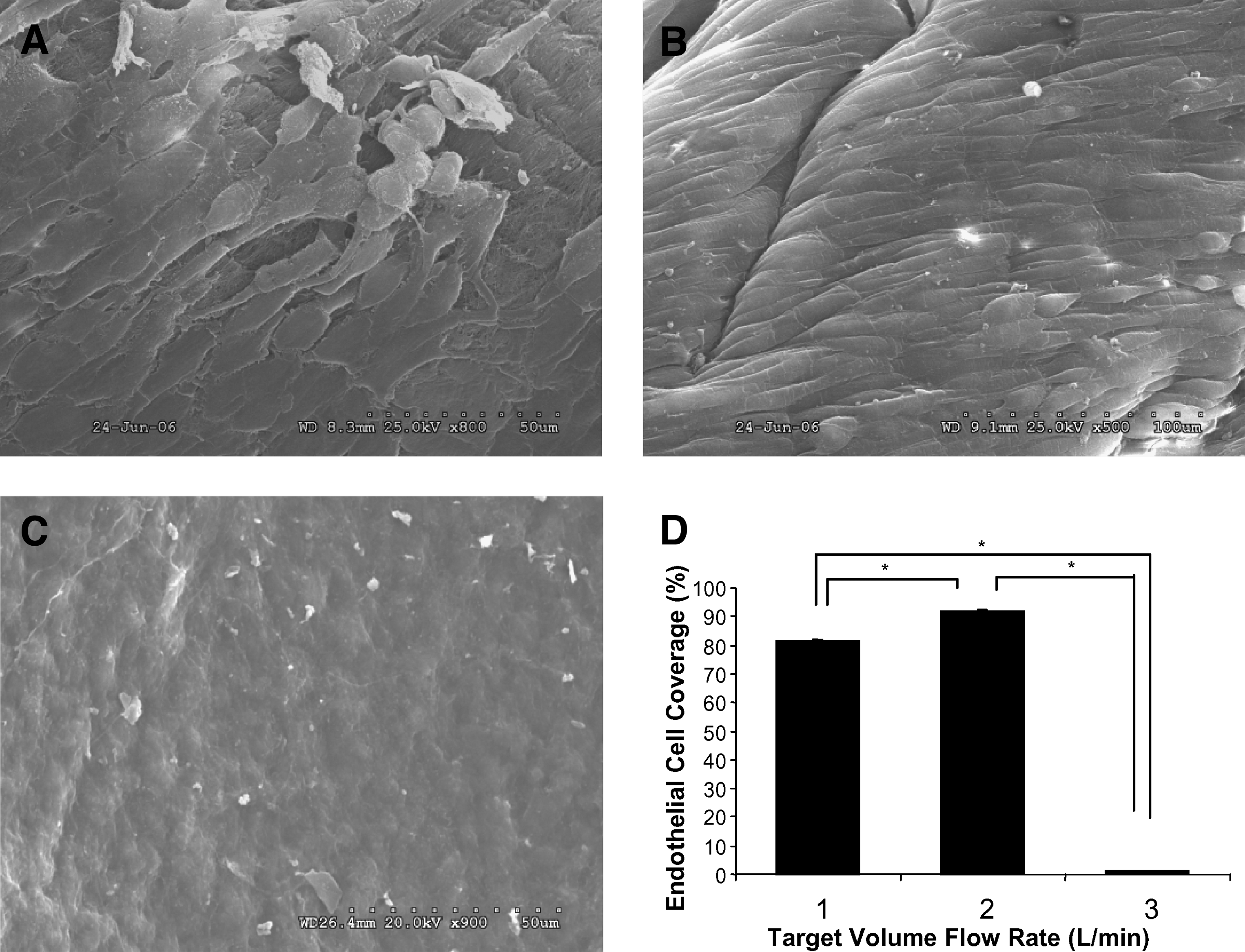
Flow-dependent endothelialization. Gradual increments of pulsatile flow rate up to 1 L/min (
Platelet adhesion assay
To determine whether EC-seeded valve matrices possess anti-thrombogenic properties, anti-platelet properties were assessed. EC-seeded valves demonstrated a significant decrease in platelet aggregation compared to acellular valves (Fig. 7A, B) as evidenced by SEM. Platelet adhesion assays show that 39,200 ± 4249 platelets adhered to unseeded constructs as compared to 6200 ± 1352 platelets on the seeded constructs (Fig. 7C). This significant reduction in platelet adherence may translate to a much needed reduction in the thromboembolic events in vivo.
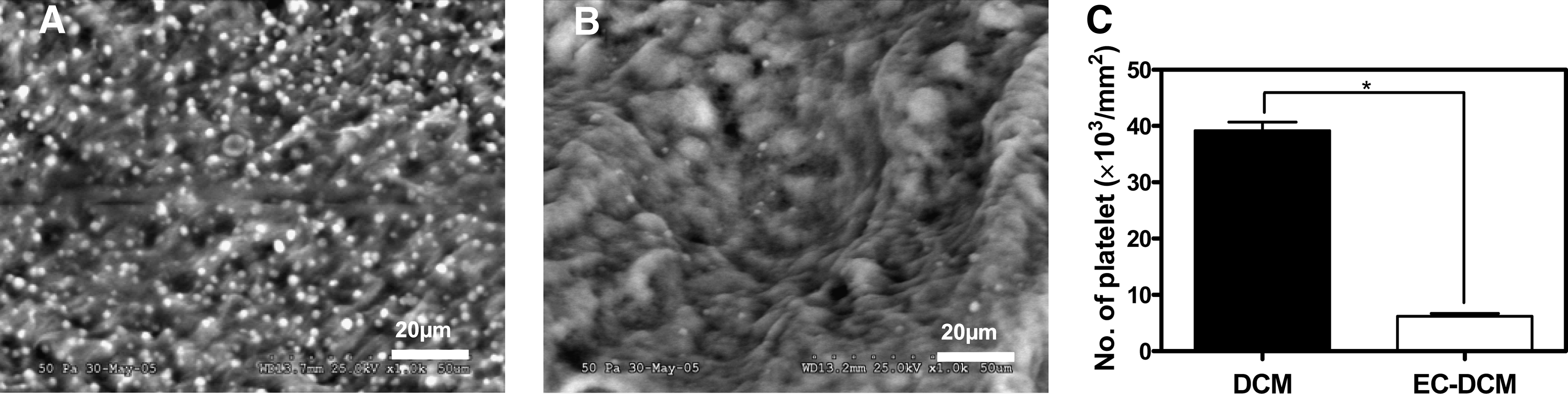
Platelet adhesion assay using SEM. Evaluation of the number of platelets adhered to the decellularized matrix (
Discussion
Heart valve replacement procedures in patients with valve disease are often associated with complications that require additional procedures. Although calcification remains as the main clinical concern associated with bioprosthetic heart valve replacement surgery, there is evidence that tissue deterioration leads to thromboembolism. To reduce the risk of thromboembolism, research efforts have been directed toward developing methods for covering the replacement valve surface with ECs. 21 To effectively coat the valve leaflets with these cells, investigators have used bioreactors to facilitate uniform coverage. 22 However, the optimal conditions that maximize the coverage and maintain cell attachment have not been defined to date. In this study, we developed a computer-assisted heart valve bioreactor system that permits a proper control of flow rate, volume, and pressure to facilitate preconditioning of ECs on the valve surface. In this study we show that endothelialization of a decellularized valve scaffold using ECs derived from circulating EPCs improves the anti-thrombogenic effect of natural valve components using the optimized bioreactor conditions.
Creation of an EC lining that withstands the fluid dynamics of blood flow can be a challenge. It has been shown that abrupt application of high flow on statically seeded ECs in a bioreactor system causes the cells to detach from the valve matrix surface due to the inadequate degrees of adherence. This is especially true near the valve leaflet, where turbulent flow is present. 13 To prevent cell detachment, we have slowly adapted ECs to physiological flow profiles using stepwise, incremental increases in flow rate using a computer-controlled bioreactor system. This approach permitted ECs to maintain attachment and fully adapt to the valve matrix under a high flow environment.
The role of a bioreactor in various tissue engineering applications is to provide an environment that mimics in vivo conditions for enhanced growth of tissue substitutes and to enable systematic studies of the responses of living tissues to various mechanical stimuli. 23 Designing a bioreactor for tissue-engineered heart valves has been a challenge due to the complex nature of the dynamic mechanical environment in which the engineered tissue will reside. The environment of the heart includes bending stress and high shear stress. These unique features were considered in the development of our heart valve bioreactor system, which includes control of flow using the custom designed LabView software.
While the control of hemodynamic parameters is crucial in mimicking physiologic conditions, designing the hardware for a bioreactor system is equally important. In this study, opening of the engineered valve in the bioreactor was controlled with flow generated by the gear pump, and closing was controlled by back pressure created using a silicone compliant section and adjustable constrainer. The bioreactor body was constructed with clear polycarbonate to allow for easy monitoring of valve opening/closing, contamination by color change, and medium circulation. Because sterilization via ethylene oxide or autoclave is common, the material selection criteria must include resistance to heat and chemicals.
In this study circulating EPCs were used to test the feasibility of using autologous EPCs to serve as a cell source in creating tissue-engineered heart valves. We show that small samples of peripheral blood (20 mL) can yield a sufficient number of ECs to cover an entire valve surface. EPC-derived ECs are able to undergo over 10 subculture stages while maintaining cellular characteristics. In this study we have used 3 million cells/cm2 based on our preliminary testing that achieved the best cell coverage by static seeding. The cells were seeded on decellularized valve scaffolds derived from porcine pulmonary valves. The tissue scaffolds were treated with chemicals to remove existing cells leaving tissue matrices that maintain structural integrity, which is similar to native heart valves. The cells on the valve scaffolds attached to the surface and remained viable. However, the cells detached under extreme bioreactor conditions, indicating that flow parameters influence cell attachment.
The pulsatile bioreactor system we developed is designed to provide the control of flow rate (shear stress) on statically seeded valve constructs. The optimal flow rate for cell attachment, adaptation, and proliferation is still unknown and may vary between different bioreactor systems. Therefore, defining the optimal flow rate for maximized endothelialization on valve surfaces is essential for a successful outcome. In this study we increased flow rate incrementally in a linear fashion using a total of 10 steps to achieve the target flow rate. Successful EC coverage was achieved with a volume flow rate up to 2 L/min for 7 days. The shear stress on the seeded ECs was determined at about 20 dyn/cm2. This flow parameter is similar to the physiological pulmonary flow of juvenile sheep, which was our target for subsequent in vivo studies to demonstrate the applicability of this technology. We show that the seeded cells failed to maintain adherence at a higher volume flow rate at 3 L/min.
It is well known that the presence of endothelium on heart valves reduces the risk for platelet aggregation and inflammatory complications.22,23 Therefore, it is likely that tissue-engineered heart valves covered with a layer of ECs would improve functionality. It has been demonstrated that ECs act as a barrier between circulating factors and play an important regulatory role in modulating interactions between the blood and other cells, such as interstitial cells.24,25 In addition, valvular endothelial dysfunction has been associated with inflammatory reactions, calcifications, and thrombosis. 26 These reports further support the need to endothelialize the valve surfaces for successful results. In this study we indirectly tested the degrees of coagulation by performing platelet adhesion assays on the endothelialized valve surface. We show that decellularized valve scaffolds seeded with EPC-derived ECs possessed adequate anti-thrombotic properties, whereas the scaffolds without ECs escalated the coagulation process. The number of platelets adhered on the seeded constructs was much less than the number of platelets adhered to biological and mechanical valves reported in the literature. 27
In this study we sought to define optimal conditions for endothelialization of a heart valve matrix with circulating EPC-derived ECs using a computer-assisted bioreactor system. Successful endothelialization was achieved by preconditioning the cell-seeded valves with stepwise increases in volume flow rate up to 2 L/min in 7 days. This demonstrates that preconditioning of cells seeded on valve matrices using a bioreactor system is necessary for the uniform EC coverage of valve scaffolds, which is a crucial step in reducing thrombotic activity after implantation in vivo. The bioreactor-assisted cell seeding system may be used on mechanical valves to minimize the incidence of thromboembolism. Although thromboembolism is not recognized as the main problem with bioprosthetic valve replacement procedures, further investigation is necessary to determine the role of ECs. Studies are currently being performed to optimize the bioreactor system. In addition, a preclinical animal study is also being conducted to demonstrate the feasibility of using endothelialized valve scaffolds.
Footnotes
Acknowledgments
The authors thank Cindy A. Andrews, Cathy M. Mathis, and Mandy Lockard for technical assistance and Drs. Jennifer Olson and Tamer Aboushwareb for editorial assistance.
Disclosure Statement
No competing financial interests exist.