




Abstract
Tissue vascularization in vitro is necessary for cell transplantation and is a major challenge in tissue engineering. To construct large and regularly vascularized tissue, we focused on the integration of endothelial cell–covered spheroids. Primary rat hepatocytes were cultured on a rotary shaker, and 100–150 μm spheroids were obtained by filtration. The hepatocyte spheroids were coated with collagen by conjugation with a type 1 collagen solution. Collagen-coated hepatocyte spheroids were cocultured with human umbilical vein endothelial cells (HUVECs), and monolayered HUVEC-covered hepatocyte spheroids were constructed. Without a collagen coat, many HUVECs invaded hepatocyte spheroids but did not cover the spheroid surface. To construct regularly vascularized tissue, we packed HUVEC-covered hepatocyte spheroids in hollow fibers used for plasma separation. Packed spheroids attached to each other forming a large cellular tissue with regular distribution of HUVECs. At day 9 after packing, HUVECs invaded the hepatocyte spheroids and a dense vascular network was constructed. Collagen coating of spheroids is useful for the formation of endothelial cell–covered spheroids and subsequent regular vascularized tissue construction.
Introduction
A spheroid is a spherical microtissue assembled from monodispersed cells in vitro. Hepatocyte spheroids have a cell density close to that of native liver tissue and express liver-specific functions at a high level. When hepatocyte spheroids are cultured in vitro, viable spheroid diameter is limited to approximately 100 μm because of their requirements for oxygen and nutrients. 2 Using original culture techniques with different shapes of hepatocyte organoids, we previously demonstrated that hepatocyte tissue should be approximately 100 μm to survive.3–5 Based on this evidence, the arrangement of endothelial cells within hepatocyte tissue at regular 100 μm intervals is a promising structure for larger tissue construction. The endothelial cell–covered hepatocyte spheroid is a candidate tissue unit to align endothelial cells regularly in large hepatic tissue and construct regularly vascularized tissue in vitro. 6
Different types of cells were cocultured with endothelial cells to form vascularized tissue in vitro.7,8 When endothelial cells were added to a nonendothelial spheroid, the behavior of the endothelial cells varied with the combination of cell types.9,10 In most cases, endothelial cells invaded the spheroid but did not cover the spheroid surface. We assumed that extracellular matrix (ECM) molecules are necessary on the surface of the spheroid for an endothelial cell covering. Type 1 collagen is a well-known ECM that affects invasion of endothelial cells. 11 To keep endothelial cells on the surface of the spheroid, we attempted to coat type 1 collagen on the surface of hepatocyte spheroids. Rat hepatocyte spheroids conjugated with a type 1 collagen solution were cocultured with human umbilical vein endothelial cells (HUVECs) (Fig. 1A). As a result, invasion of HUVECs was blocked and hepatocyte spheroids were covered by monolayered HUVECs. After successful construction of HUVEC-covered hepatocyte spheroids, we tried regular alignment of HUVECs within hepatic tissue by packing HUVEC-covered hepatocyte spheroids in hollow fibers used for plasma separation. The concept of coating ECM on the surface of cells or tissue has new possibilities for the arrangement of heterologous cells in tissue culture.

Schematic illustrations. (
Materials and Methods
Cell preparation
HUVECs (Lonza, Basel, Switzerland) were purchased and subcultured in endothelial growth medium-2 (EGM-2; Lonza) at 37°C in a humidified 5% CO2 atmosphere. HUVECs from passage 2 after purchase were used for experiments.
Primary rat hepatocytes were isolated from a male Wistar rat (7–8 weeks old; Kyudo, Kumamoto, Japan) by the collagenase perfusion method. Viable cells (85–95%) were used for spheroid culture. Isolated hepatocytes were suspended in hepatocyte culture medium: Dulbecco's modified Eagle's medium with high glucose (Invitrogen, Carlsbad, CA) supplemented with 50 μg/L epidermal growth factor (EGF, BT-201; Biomedical Technologies, Stoughton, MA), 10 mg/L insulin (Sigma-Aldrich, St. Louis, MO), 60 mg/L proline (Sigma-Aldrich), 7.5 mg/L hydrocortisone (Wako Pure Chemical Industries, Osaka, Japan), 3.7 g/L NaHCO3, 5.985 g/L HEPES, 50 mg/L linoleic acid, 0.1 μm copper (CuSO4·5H2O), 3 nM selenium (H2SeO3), 50 pM zinc (ZnSO4·7H2O), and antibiotics (58.5 mg/L penicillin and 100 mg/L streptomycin).
Spheroid culture
Primary rat hepatocytes were cultured in a six-well plate (Nalge Nunc International KK, Tokyo, Japan) at 1.5 × 10⁶ viable cells/well in 1.5 mL hepatocyte culture medium. Cells were incubated on a rotary shaker at 80 rpm, 37°C in a humidified 5% CO2 atmosphere. Supernatant (1 mL per well) was replaced on day 2, and hepatocyte spheroids were collected on day 4. Collected spheroids were filtered using stainless wire mesh. Hepatocyte spheroids of 100–150 μm sieve fractions were used for experiments.
Image analysis for spheroid diameter profile
Cultured hepatocyte spheroids were sampled and filtered to remove spheroids of less than 45 μm in diameter and were resuspended in 2 mL hepatocyte culture medium. The suspension (150 μL) was transferred on the back of the lid of a 24-well plate (Nalge Nunc International KK). Images were captured with a Leica MZ12 (Leica Microsystems GmbH, Wetzlar, Germany) and a COOLPIX 4500 (Nikon, Tokyo, Japan). Then images were processed using Adobe Photoshop® (Adobe Systems Incorporated, San Jose, CA) to obtain binary images. Binary images were analyzed by ImageJ to measure the projected area of each spheroid. Assuming a true sphere, the diameter was calculated from the projected area. Two individual culture wells were sampled and measured four times, respectively. Error bars indicate the standard deviation of four measurements.
Collagen coating on hepatocyte spheroids
A Collagen Gel Culturing Kit (Nitta Gelatin, Osaka, Japan) was used to coat the surface of hepatocyte spheroids. Hepatocyte spheroids obtained by rotary culture were transferred into a 15 mL centrifuge tube and collected at 40 g, 60 s. Supernatant was removed, and spheroids were resuspended in 1 mL hepatocyte culture medium. Cellmatrix Type I-A was reconstituted by adding 10× culture medium and reconstitution buffer according to the manufacturer's instruction. Reconstituted collagen gel (1 mL) was added to the spheroid suspension and pipetted (final concentration of collagen: 1.2 mg/mL). After adding collagen gel, the spheroid suspension was incubated at 4°C for 1 h and washed twice by adding 10 mL cold hepatocyte culture medium and centrifugation at 40 g, 60 s. The schematic illustration is shown in Figure 1B.
HUVEC-covered hepatocyte spheroid formation
Agarose (4%) was coated on a 100 mm cell culture dish (BD Falcon, Franklin Lakes, NJ) and substituted with EGM-2 before use. Collagen-coated hepatocyte spheroids and HUVECs were suspended in EGM-2. Collagen-coated hepatocyte spheroids (7500–10,000 spheroids) and HUVECs (3 × 10⁶ cells) were plated in 12 mL EGM-2 per dish. Supernatant (10 mL per dish) was replaced every 2 days. For a negative control, hepatocyte spheroids without a collagen coat were also cocultured with HUVECs under the same culture condition.
Vascular network formation using HUVEC-covered hepatocyte spheroids
HUVEC-covered hepatocyte spheroids (collagen-coated hepatocyte spheroids cocultured for 4 days with HUVECs) were collected and inoculated into hollow fibers used for plasma separation (poly ethylene treated with ethylene vinyl alcohol, PE-EVAL; Asahi Kasei Medical, Tokyo, Japan) (inner diameter: 330 μm; outer diameter: 430 μm) (Fig. 1C). HUVEC-covered hepatocyte spheroids in hollow fibers were packed by centrifugation (40 g, 180 s). Approximately 3 cm hollow fibers containing HUVEC-covered hepatocyte spheroids were cultured in a 60 mm Petri dish (BD Falcon) in EGM-2. Media were replaced every 2 days.
Immunofluorescent microscopy
HUVEC-covered hepatocyte spheroids cultured in suspension or in hollow fibers were sampled and embedded in Tissue-Tek® O.C.T. Compound (Sakura Finetechnical, Tokyo, Japan) and frozen in liquid nitrogen. Ten-micron frozen sections were obtained using cryostat (CM1100; Leica Microsystems GmbH).
Sections were fixed in acetone for 10 min at 4°C followed by washing in PBS (5 min, three changes). Sections were incubated in blocking solution (PBS containing 10% skim milk and 6% glycine) for 20 min. Sections were washed in PBS (5 min, three changes) and incubated in blocking solution containing primary antibodies (1:100) for 1 h at room temperature. Rabbit polyclonal anti-human von Willebrand factor (Dako, Glostrup, Denmark ) was used for HUVECs, and goat anti-mouse albumin (BETHYL Laboratories, Montgomery, TX) was used for primary rat hepatocytes (cross reactivity for rat liver was confirmed). For collagen immunostaining, rabbit anti-type 1 collagen (LB-1196; LSL Co., Ltd., Tokyo, Japan) (1:300) was incubated overnight at 4°C. Then, sections were washed in PBS (5 min, three changes) and incubated in blocking solution containing secondary antibodies (1:100) and Hoechst 33342 (1:1000) for 1 h at room temperature. Swine anti-rabbit IgG FITC-conjugated (Dako) and donkey anti-goat IgG rhodamine-conjugated (Millipore, Billerica, MA) antibodies were used for secondary antibodies. Images were captured with a fluorescent microscope (IX71; Olympus, Tokyo, Japan). In some experiments, rabbit anti-goat IgG Rhodamine-conjugated antibody (Millipore) was substituted for donkey anti-goat IgG rhodamine-conjugated antibody (Millipore). Rabbit anti-goat IgG rhodamine-conjugated antibody was used after reaction with swine anti-rabbit IgG FITC conjugated giving a yellow image for von Willebrand factor.
Liver-specific function
We evaluated liver-specific functions of hepatocytes cultured in hollow fibers with or without HUVECs. HUVEC-covered hepatocyte spheroids or collagen-coated hepatocyte spheroids were inoculated into two 3 cm hollow fibers and packed by centrifugation as described above, and then cultured in 2 mL hepatocyte culture medium supplemented with EGM-2 SingleQuots (Lonza) (containing Hydrocortisone, hFGF-B, VEGF, R3-IGF-1, ascorbic acid, heparin, 2% FBS, hEGF, and GA-1000). The albumin concentration of the culture supernatant from the identical culture dish was measured using an enzyme-linked immunosorbent assay. Error bars indicate the standard deviation of two individual culture dishes.
Results
Mass hepatocyte spheroid formation
To form a large number of hepatocyte spheroids, we used rotary culture. Cultured hepatocytes formed spheroids in 2 days. The number of spheroids smaller than 100 μm decreased during rotary culture (Fig. 2). Hepatocytes or hepatocyte spheroids attached to each other resulting in a increase of spheroid diameter. At day 4, spheroids were filtered to obtain approximately 10,000 spheroids of 100–150 μm diameter per six-well plate.
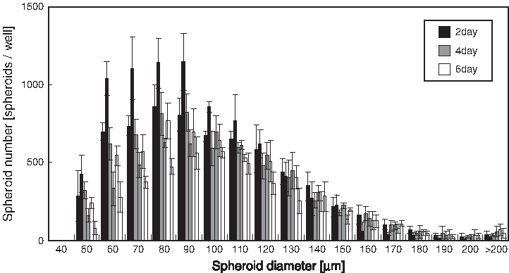
Hepatocyte spheroid diameter profile by rotary culture.
Hepatocyte spheroid–HUVEC coculture
Two days after plating on agarose-coated dishes, some HUVECs formed HUVEC spheroids (Fig. 3A, B). Other HUVECs were attached to the surface of hepatocyte spheroids. Without collagen coating on hepatocyte spheroids, some HUVECs invaded hepatocyte spheroids (Fig. 3B). On day 4 of coculture, many collagen-coated hepatocyte spheroids were covered with single layered HUVECs (Fig. 3C), while HUVECs invaded some hepatocyte spheroids without collagen coating, resulting in irregular distribution (Fig. 3D). To evaluate the rate of HUVEC-covered hepatocyte spheroid formation, we counted a certain number of cross sections of cocultured spheroids by classification according to HUVEC distribution within the spheroid (Table 1). Spheroids with more than 50% coverage of HUVECs were classified as “HUVEC-covered hepatocyte spheroids.” Spheroids with HUVEC invasion were classified as “HUVEC-invaded hepatocyte spheroids.” Spheroids with less than 50% coverage of HUVECs and no HUVEC invasion were classified as “few HUVEC-attached hepatocyte spheroids.” In “HUVEC-covered hepatocyte spheroids,” HUVEC invasion was not found. And in “HUVEC-invaded hepatocyte spheroids,” few HUVECs were on the spheroid surface at less than 50% coverage. Collagen coating did not affect HUVEC attachment to hepatocyte spheroids but did control HUVEC distribution on hepatocyte spheroids (Table 1). This indicates that like a culture dish or a scaffold surface, collagen coating is effective for the cell surface.

Immunofluorescent microscopy of cocultured spheroids stained for albumin (red) and von Willebrand factor (yellow). (
HUVEC, human umbilical vein endothelial cells.
Cyst formation of HUVEC-covered hepatocyte spheroids
We continued to coculture hepatocyte spheroids and HUVECs over 4 days to investigate morphological changes of a single spheroid. We found swelling of spheroids. By day 10 of coculture, clear cyst formation was observed (Fig. 4). Cyst formation was found more frequently in collagen-coated spheroids than in non-collagen-coated spheroid (Fig. 4A, B). This frequency is in accord with the rate of HUVEC-covered hepatocyte spheroids (Table 1). Immunofluorescent microscopy indicated a correlation between cyst formation of spheroids and HUVEC-covered hepatocyte spheroids (Fig. 4C, D). Cyst formation was not found in HUVEC-invaded hepatocyte spheroids (Fig. 4D). HUVEC confluence on the surface of the hepatocyte spheroid and its subsequent proliferation may trigger cyst formation.

Spheroids cocultured for 10 days. HUVEC-covered hepatocyte spheroid tended to swell, forming cyst (white arrows). (
Regular alignment of HUVECs by packing heterospheroids in hollow fiber
After packing HUVEC-covered hepatocyte spheroids in hollow fibers, HUVEC-covered hepatocyte spheroids adhered to each other, organizing into a large tissue in a short culture time (Fig. 5A). HUVECs were distributed in regular intervals of approximately 100 μm. Tubular morphology of HUVECs was observed at the contact area of several heterospheroids. The capillary diameter of a normal liver sinusoid is approximately 10–15 μm, 12 indicating that cavities between HUVEC-covered hepatocyte spheroids could be blood capillaries (Fig. 5B). On day 4 of culture in hollow fibers, HUVECs between HUVEC-covered hepatocyte spheroids tended to diminish and hollow structures composed of HUVECs were observed (Fig. 5C, D). Distribution of nuclei in hollow fibers was not uniform. Some hepatocytes seemed to die (Fig. 5C), while nuclei of living hepatocytes were found in certain regions (Fig. 5D).
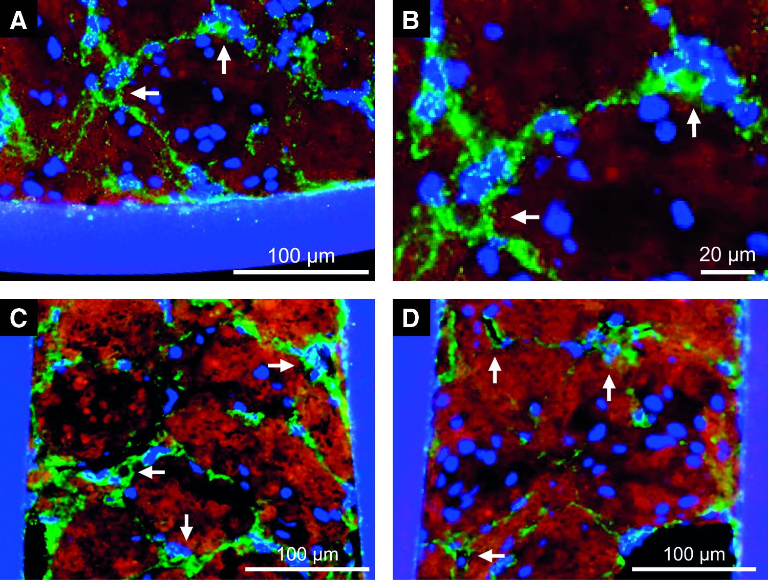
Immunofluorescent microscopy of HUVEC-covered hepatocyte spheroids packed in hollow fibers stained for albumin (red) and von Willebrand factor (green). (
HUVEC morphology changed gradually on day 9 of culture in hollow fibers. A regular network of HUVECs was maintained, and many nuclei were observed at the center of the cellular tissue (Fig. 6A, B). Some HUVECs invaded hepatocyte spheroids forming a dense vascular network (Fig. 6C). Further, tubular morphologies of HUVECs were found more frequently than on early days of culture in hollow fibers (Fig. 6D).
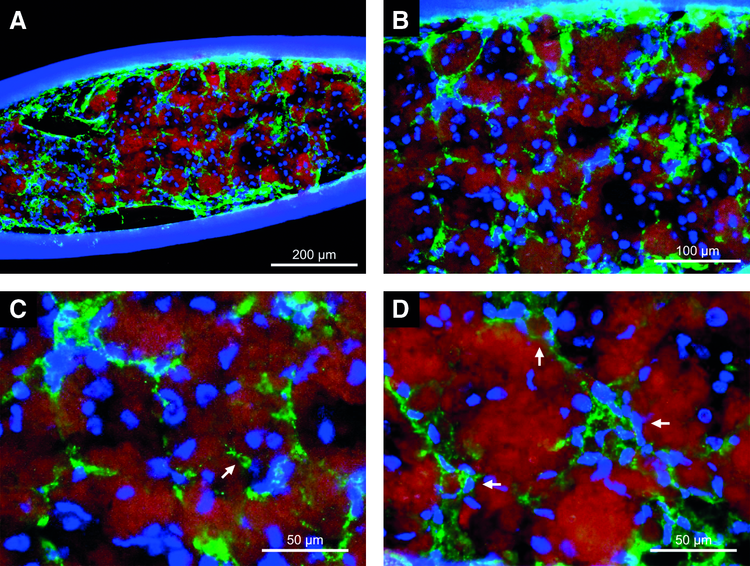
Immunofluorescent microscopy stained for albumin (red) and von Willebrand factor (green). Day 9 of culture of HUVEC-covered hepatocyte spheroids packed in hollow fibers. Sections (20 μm) were stained. (
Albumin secretion rate of HUVEC-covered hepatocyte spheroids inside hollow fibers
We performed another experiment to investigate the effect of coculture with HUVECs on expression of liver-specific functions of hepatocyte spheroids. The rate of albumin secretion of HUVEC-covered hepatocyte spheroids was maintained at a higher level compared to that in hepatocyte spheroids without HUVECs throughout the culture period (Fig. 7).
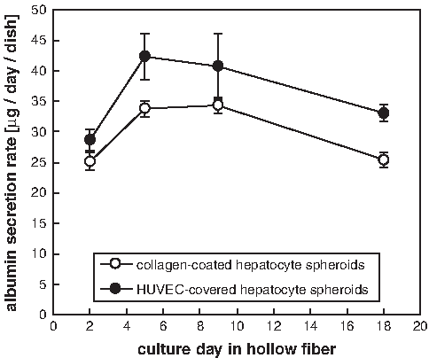
Albumin secretion rate of HUVEC-covered hepatocyte spheroids packed in hollow fibers. As a control, collagen-coated hepatocyte spheroids without HUVECs were packed in hollow fibers.
Discussion
The endothelial cell–covered spheroid is a simple and applicable tissue unit for heterologous cellular tissue culture. HUVEC-covered hepatocyte spheroids of 100–150 μm diameter ensure that HUVECs and hepatocytes coexist within 100–150 μm proximity when they aggregate to form larger tissue. This technique would enhance 3D tissue coculture, especially in terms of regular alignment of heterologous cells on a large scale. Packing endothelial cell–covered spheroids in closed spaces such as the intra capillary space of a hollow fiber would be efficient for rapid vascular formation at contact regions between endothelial cell–covered spheroids. The initial alignment of endothelial cells using endothelial cell–covered spheroids is promising for 100–150 μm regular and continuous vascular network formation in large tissue culture. On the other hand, endothelial invasion, migration, and proliferation are also necessary for dense vascularization.
The endothelial cell–covered spheroid will also provide an in vitro tissue model. All molecules are transported to the endothelial cell–covered spheroid through an endothelial monolayer and ECM barrier. This model will be suitable for studying drug delivery.13,14
A scaffold-free tissue construction method has several advantages or disadvantages over scaffold-based tissue culture. One advantage is high cell density in scaffold-free tissue. This technique meets the requirements for mass cell transplantation, such as liver tissue. This is also suitable for nonproliferative cells that are not able to reach high cell density by cell proliferation during tissue culture or after transplantation. However, they may be fragile for transplant operation.
In this study, HUVECs invaded the hepatocyte spheroid, constructing a dense vascular network after packing in a hollow fiber. A similar phenomenon occurs during liver regeneration after partial hepatectomy. Hepatocytes proliferate to form a cluster after injury; then, stellate cells and sinusoidal endothelial cells invade the hepatocyte cluster.15,16 Regeneration of the liver will be one criterion for liver tissue construction.
Concerning cyst formation of HUVEC-covered hepatocyte spheroids, epithelial cells are known to form cysts when they are cultured in collagen gel. They polarize, forming a closed cystic structure, in which the apical region is on the inner side and the basolateral region is on the outer side of the cyst.17,18 HUVECs covering the surface of the hepatocyte spheroid may affect the polarity of hepatocytes on the surface of a spheroid and then form a closed cystic structure. The typical hepatocyte spheroid forms a bile lumen structure and is considered to be continuous with the outer region.19,20 The HUVEC-covered hepatocyte spheroid may separate bile lumens from the sinusoidal capillaries through tissue construction.
Though we have clarified that the survival thickness of hepatocyte tissue is limited to approximately 100 μm, nuclei were found at the center of the vascularized tissue of 300 μm thickness whose location did not always overlap with the location of HUVECs (Fig. 6). Evaluation of liver-specific function also suggests improvement of hepatocyte viability after coculture with HUVECs (Fig. 7). There are several reports that evaluate liver-specific functions of primary rat hepatocytes cocultured with HUVECs.21,22 Liver-specific function of hepatocytes cocultured with HUVECs varied by spatial distribution of each cells. When hepatocytes and HUVECs were aligned regularly, liver-specific functions improved. 21 This result is in agreement with improvement of liver-specific function by HUVEC-covered hepatocyte spheroids.
We performed immunofluorescent microscopy for type 1 collagen to evaluate collagen distribution of collagen-coated hepatocyte spheroids (Fig. 8). Collagen localization was detected on the spheroid surface, and fluorescence intensity on the spheroid surface varied with location, indicating the nonuniform collagen coat on the hepatocyte spheroid (Fig. 8A). In contrast, intense fluorescence was not detected without a collagen coat (Fig. 8B). Low accumulation of endogenous rat collagen was also found in hepatocyte spheroids that might affect HUVEC invasion (Fig. 8A, B). The collagen coating method needs to be optimized for uniformity on the spheroid surface.
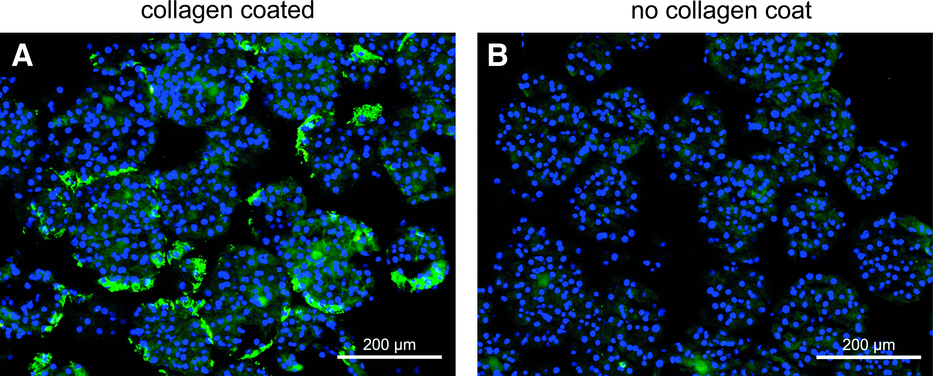
Immunofluorescent microscopy of hepatocyte spheroids stained for type 1 collagen (green). (
Coating collagen on the surface of cells or cell aggregates will serve other merits aside from heterologous cellular distribution control. One advantage is minimum inhibition of molecular transport by ECM. Many types of ECM are barriers to molecular transport. 13 Compared to culture in collagen gel, collagen coats on spheroids lead to less inhibition of exchange of nutrients and growth factors. Another report indicated that ECM coats of hepatocytes improved their adhesion to the liver during cell transplantation. 23
Type 1 collagen is a major ECM for cell culture. Integrin α2β1 is known as an adhesion molecule for type 1 collagen. In addition, collagen affects HUVEC morphology leading to tube formation through binding to integrin.24,25 Type 1 collagen invasion by HUVECs is triggered by matrix metalloproteinases. HUVECs produce matrix metalloproteinases to degrade matrix, including type 1 collagen, and invade. 26 Coating type 1 collagen on the surface of hepatocyte spheroid would serve as a transient anchor and barrier to the invasion by HUVECs.
Different types of ECM were studied in endothelial cells culture and revealed differences in cell morphology. 27 The basement membrane is known for its ECM complex adjacent to endothelial cells. 28 Laminin and type 4 collagen are major components of the basement membrane and are often used for cell culture. When endothelial cells were cultured using laminin gel, they show a static morphology compared to type 1 collagen. 29 Type 4 collagen also has a different effect on endothelial cell morphology than type 1 collagen. 30 Immunohistochemical studies of regenerating rat liver after partial hepatectomy show transient accumulation of laminin.12,31 Coating these ECMs on hepatocyte spheroids would have a different effect than type 1 collagen during vascularization after packing HUVEC-covered hepatocyte spheroids.
In conclusion, a spheroid covered by endothelial cells is a promising tissue unit for vascularized tissue construction. Collagen coating on spheroids is a candidate technique for heterologous cellular distribution control.
Footnotes
Acknowledgments
This study was supported in part by a Grant-in-Aid for Scientific Research (B) 19360375 from the Japan Society for the Promotion of Science, and a grant from the Global Centre of Excellence in Novel Carbon Resource Sciences, Kyushu University.
Disclosure Statement
No competing financial interests exist.