
Abstract
Our aim in this study was to develop a new methodology for constructing a bilayer skin equivalent to create a clinically compliant skin graft composite for the treatment of various skin defects. We utilized tissue-engineered extracellular matrix (TEECM) that maintains the native dermal components, as analogical dermal layer, and gelatin hydrogel containing epidermal growth factor (EGF)–loaded microspheres, as epidermis layer upon TEECM. The bilayer skin equivalent (GG-EGF/TEECM) could be synthesized appropriately and has been hypothesized to provide an enhanced effect on re-epithelization, apart from dermal reconstruction improved by TEECM. Morphological properties, EGF release efficiency, cytotoxicity, and effects on wound repair of this composite were investigated in this study. It was shown that the two layers adhered firmly to each other, and the percentage of cumulative release was approximately 80% by day 14. Moreover, as a biocompatible equivalent, GG-EGF/TEECM improved the healing of the wound area as indicated by significantly earlier re-epithelialization and dermal maturation. These results suggest that GG-EGF/TEECM can provide an optimal microenvironment and proper template for skin tissue engineering.
Introduction
For a more rational design of a smart skin replacement, strategy for construction of artificial skin that shares anatomical and physiological homology with natural skin would be necessarily developed. 2 Previously, we have established a protocol for production of tissue-engineered skin that has been proved to be a clinically effective treatment for various acute skin defects and produces well-contemplated cosmetic results. 11 Based on this, we have developed modified ECM produced from tissue-engineered skin (tissue-engineered extracellular matrix, TEECM) as dermal template, in combination with porous gelatin hydrogel as a scaffold for epidermal layer. Further, we embedded epidermal growth factor (EGF)-incorporated gelatin microspheres in above-mentioned gelatin hydrogels (GG-EGF) that could achieve sustained release and to maintain suitable concentration over an extended time period as proved by our previous study.12–14 It could be hypothesized that the application of microspheres incorporating EGF might substantially improve cell-guided re-epithelialization.
The aim of this study emphasizes the feasibility of synthesizing the composite that contained TEECM and microsphere-incorporated gelatin hydrogel (GG-EGF/TEECM). Further, the properties of this resulted composite were investigated in terms of morphology, EGF release efficiency, and cytotoxicity; especially, the tissue regeneration was surveyed experimentally in a rat model.
Materials and Methods
Preparation of TEECM
Type I collagen (Sigma-Aldrich, St. Louis, MO) was prepared using 8 mg/mL in 0.1% of acetic acid under conditions of ultraviolet irradiation for 45 min on ice. The following mixture of reagents was added by scaling as needed: 10 × Dulbecco's modified Eagle's medium (DMEM, 10:1 vol%; Gibco-BRL, Grand Island, NY), fetal bovine serum (FBS, 10:1 vol%), and 0.1 M NaOH for adjusting the pH to 7.2 ± 0.2. About 106/mL fibroblasts that were isolated from human foreskins obtained during the circumcision of children by sequential trypsin and collagenase digestion (Center for Tissue Engineering, Fourth Military Medical University, Xi'an, China) were rapidly mixed with the above solution. Ten milliliters of the mixture was distributed in wells of 5-cm-diameter culture plates. The plates were placed in an incubator at 37°C and 5% CO2. After gelation for 30 min, 10 mL of the DMEM containing 10% FBS, 2 ng/mL bFGF, 2 ng/mL EGF, and 30 mg/mL vitamin C was added to each well. The gels were kept in culture, and the medium was changed every day. After 7 days of culture, TEECM were freeze–thawed three times under −80°C, each time for 30 min, after which the cells were removed completely. Lastly, the resultant TEECM was lyophilized and sterilized by 60CO.
Construction of bilayered skin substitute
Figure 1 shows the general procedures for the construction of the bilayered skin substitute. After TEECM had been prepared following the steps mentioned above as the under layer, the upper layer containing gelatin hydrogel–incorporated EGF-loaded microspheres (GG-EGF) was constructed integrally, and then the two layers were synthesized as a composite (GG-EGF/TEECM). The detailed processes are described below.
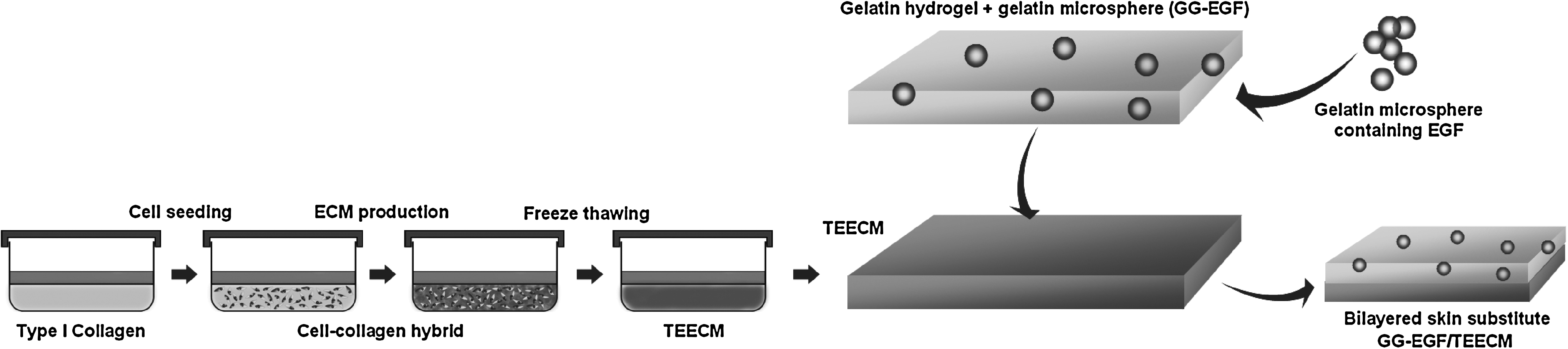
Diagrammatic representation of the procedures for construction of GG-EGF/TEECM. After TEECM had been prepared as the under layer, the upper layer that contained gelatin hydrogel–incorporated, EGF-loaded microspheres (GG-EGF) was constructed integrally, and then the two layers were synthesized as a composite (GG-EGF/TEECM).
Preparation of EGF-loaded microspheres (G-EGF)
Gelatin with an isoelectric point of 5.0 and human recombinant EGF was obtained from Sigma-Aldrich. All other reagents or chemicals were of analytical grade (Shanghai Chemical, Shanghai, China) and were used without any further treatment or purification.
The EGF-loaded microspheres were prepared as described previously. 12 A typical preparation is as follows: 2 mL of 10 wt% gelatin aqueous solution was added dropwise into 30 mL of paraffin oil, while the mixture was mechanically stirred at 800 rpm to form a water-in-oil emulsion. The solution was then rapidly cooled by immersing in ice-water. The formed microspheres were filtered, washed with acetone, and dried at room temperature. The noncrosslinked microspheres prepared were then placed in a 2 wt% glutaraldehyde aqueous solution containing 0.1 wt% Tween 80, and the crosslinking was allowed to proceed at 4°C for 12 h. After crosslinking, the microspheres were placed in 100 mL of 10 mM glycine aqueous solution for 30 min to block any residual unreacted glutaraldehyde. The resulted microspheres were sieved out with 400-screen grit to obtain gelatin microspheres with a diameter smaller than 10 μm for later use. The approach of incorporating EGF into microspheres was as follows: aqueous solution including EGF (50 μg in 1 mL phosphate buffer, pH 7.4) was prepared, the solution was added drop wise to the resulting gelatin microspheres (100 mg), and sorption of the protein to the matrix was allowed, as well as its subsequent sustained release through degradation of the microspheres. This method preserves protein bioactivity; the protein to be released is present in the aqueous solution of gelatin during crosslinking, and its therapeutic effect will most likely be lost because of chemical deactivation.
Preparation of EGF-loaded microsphere-incorporated gelatin hydrogel (GG-EGF)
The gelatin hydrogel was prepared by chemical crosslinking of gelatin. 15 Briefly, 5 wt% aqueous solution of gelatin (1 mL) stirred at about 2000 rpm for 30 min at room temperature, and 0.25 wt% glutaraldehyde solutions were added to form crosslinkings. Then, 0.1 g of EGF-loaded microspheres was added into foaming solutions, and the solutions were poured into round molds, frozen in liquid nitrogen, and freeze-dried for 24 h. The resultant GG-EGF (thickness <2 mm) was exposed to ethylene oxide for 1 h before application to achieve sterilization.
Synthesis of TEECM and GG-EGF
In the present study, bilayer skin substitutes were constructed before crosslinking of gelatin. First, EGF microspheres were dispersed uniformly in 5 wt% aqueous solution of gelatin (1 mL) containing 0.25 wt% of glutaraldehyde, and the mixture was uniformly coated upon TEECM with thickness of 3 cm, followed by gelatin gelation and crosslinking at 24°C overnight. Sequentially, the crosslinked mixture was immersed in 50 mM of glycine aqueous solution under agitation for 1 h to block the residual aldehyde groups of glutaraldehyde, followed by washing twice in 500 mL of double-distilled water at 37°C for 1 h under agitation. The resulting composite was freeze-dried for 48 h and sterilized by ethylene oxide for 24 h. No change in the hydrogel shape was observed before and after freeze-drying and sterilization processes. EGF would probably be lost a little during the preparation, so the addition of microspheres was slightly excessive, and the loss could be overlooked in this study. The samples with free EGF (EGF added in the solution of gelatin) and without EGF were also constructed and sterilized for the following studies. The resulting composites were labeled as GG-EGF/TEECM, G-EGF/TEECM, and GG/TEECM, respectively. The procedures for construction of GG-EGF/TEECM are pictorially illustrated in Figure 1.
In vitro evaluation of bilayered skin substitute
Morphological evaluation of bilayered skin substitute
The typical scanning electron microscopy (SEM) images of GG-EGF/TEECM and TEECM are examined using a scanning electron microscope (Hitachi S3400N; Hitachi, Japan) after coating the samples with a thin layer of gold under vacuum.
Release profile of EGF from bilayered skin substitute
A known amount of GG-EGF/TEECM and G-EGF/TEECM was dispersed in 10 mL of phosphate-buffered saline (PBS, pH 7.4), as the samples can be dispersed easily in aqueous solutions by simple agitation for 2–3 min. The buffer was replaced daily, and the amount of EGF in releasing medium was determined by enzyme-linked immunosorbent assay (ELISA) by Human EGF Quantikine ELISA Kit (R&D Systems, Minneapolis, MN) according to the manufacture's instructions.
Biocompatibility of bilayered skin substitute
The biocompatibility of GG-EGF/TEECM and viability of cells proliferating on GG-EGF/TEECM were confirmed by evaluation using an in vitro cytotoxicity test with fibroblasts (L929) based on growth rates. The cytotoxicity of GG-EGF/TEECM was evaluated by 3-(4,5-dimethylthiazol-2-yl)-2,5-diphenyl-2H-tetrazolium bromide (MTT) assay with 24 h mouse L929 fibroblast (Chinese Academy of Sciences, Beijing, China) cell culture according to the Biological Evaluation of Medical Devices test (GB/T16886.5-2003). Briefly, 3 cm2 of GG-EGF/TEECM was placed in DMEM supplemented with 10% FBS for 24 h while any soluble factors leached into the culture medium. The medium was then removed and placed atop L929 fibroblast cells grown to 70% confluency in a six-well plate. After 24 h, 100 μL MTT (5 mg/mL; Sigma-Aldrich) solution was added to each well. After incubation for 4 h at 37°C, the upper medium was removed carefully, the intracellular formazan was dissolved by adding 350 μL dimethyl sulfoxide to each well, and the cells were continually cultured for another 4 h. After jolted by the shaker for 15 min, the upper solution was transferred to a 96-well plate. The absorbance at 492 nm was recorded under a microplate reader (Bio-Tek, Winooski, VT), and the resultant value is related to the number of living cells. Five samples were used for these measurements to obtain averages and standard deviations.
The potential of GG-EGF/TEECM as skin tissue scaffolds was further assessed in terms of the attachment and the proliferation of human keratinocytes (HaCaT) and human foreskin fibroblasts (HFF) that were cultured on GG-EGF/TEECM for different time points. HaCaT and HFF were purchased from China Center for Type Culture Collection (CCTCC, Wuhan, China). Before cell seeding, GG-EGF/TEECM and both G-EGF/TEECM and TEECM were sterilized by 70% ethanol for 5 min, washed two times with PBS, and then washed once with the culture medium. Subsequently, the specimens were put in empty wells of plates, and 500 μL of the culture medium was pipetted into each well. For the attachment study, 3 × 10⁴ cells/well of HaCaT were seeded and allowed to attach on the surface of GG-EGF/TEECM and both G-EGF/TEECM and TEECM for 2, 6, or 24 h. At each specified seeding time point, the number of the attached cells was quantified by cell counting method. The samples were rinsed with the culture medium and were filtered with a nylon mesh to remove unattached cells. Before harvesting the attached cells by trypsinization, gentle washing with PBS was performed twice. The detached cells were then counted under a hemocytometer. For the proliferation study, 3 × 10⁴ cells/well of the cells were cultured and incubated for 1, 2, or 3 days. At each culturing time point, the number of the cells was determined by cell counting method. Such studies for HFF cells were carried out in the same procedure. All data are averaged from five parallel experiments. Data were analyzed using analysis of variance (ANOVA). Results are reported as mean ± standard deviation (SD).
In vivo studies of bilayered skin substitute
Transplantation surgery
Sixty female SD rats, 8 weeks old, were used for the experiment. Sixty rats were divided into four groups with 15 rats in each group. All the rats were fed with a standard diet and housed singly to prevent fighting and attacks on the wounds in the animal care facilities of the university. The rats were acclimatized to the setting at least a week before using them for in vivo experiments. The animal experiment was conducted according to the committee guidelines of the Fourth Military Medical University for animal experiments, which met the NIH guidelines for the care and use of laboratory animals.
The rats were anesthetized with pentobarbital. After shaving the hair on the back of the rat and sterilizing the skin with baticone alcoholic solution, a full-thickness wound of size 1.5 cm in diameter was created. The wounds were then, respectively, covered with GG-EGF/TEECM, G-EGF/TEECM, and TEECM without cell seeding. Then, nothing but the cotton gauze was applied to the control wound. The animals were given daily injections of ceftazidime intraperitoneally for 7 days after surgery.
Wound area measurements
At 7th, 14th, and 21st days, the wound size (n = 5) was estimated by a simple wound measurements.
16
Assuming that the wound is in the shape of an ellipse (due to skin anisotropy), the following equation for the area of an ellipse was used:
Data are expressed as means ± SD. Analysis of independent samples t-test was used to determine the significant differences among the groups (p-values less than 0.05 were considered significant).
Histological examination
To evaluate the quality of the regenerated tissue, we performed histological examination of the wound-edge tissue 3 weeks postwounding with changing the respective coverings every week. Specimens were fixed in phosphate-buffered 10% formalin, dehydrated in ethanol, cleared in xylene, and embedded in paraffin. They were sectioned and stained with hematoxylin and eosin for histological observation.
Statistical analysis
Data of OD value in MTT assay and wound area calculation were analyzed by independent samples t-test with SPSS v.12.0 software (SPSS, San Rafael, CA). Statistical significance was determined at p < 0.05. All data acquisition and analyses were performed blindly.
Results
In vitro evaluation of bilayered skin substitute
Morphological analysis of GG-EGF/TEECM
SEM micrograph of GG-EGF/TEECM is given in Figure 2. The micrographs indicate that bilayer structure of the skin substitute was distinct, and the two layers adhered firmly to each other (Fig. 2A). The cross-sectional morphology of GG-EGF/TEECM revealed that the bilayered skin substitute was interconnected with pores of size varying from 100 to 150 μm. It was noticed that the microspheres were an integral part of the porous three-dimensional composites, and their incorporation did not significantly affect the porosity and the pore size (Fig. 2B). In the present study, the resulted composite seems to be composed of a loose layer (TEECM; Fig. 2C) and a dense layer (GG-EGF). The dense layer assumed the responsibility of a barrier function to protect the wound and a stimulator to active epithelization, and the loose layer played the role as a guidance to stimulate cell proliferation and granulation tissue formation.

SEM photomicrographs of GG-EGF/TEECM (
Analysis of EGF release kinetics
The in vitro releasing profile was displayed in Figure 3. The release kinetics of EGF was monitored for 14 days. EGF released from GG-EGF/TEECM was in a biphasic fashion, characterized by a fast release phase at initial 1 day followed by a slower one for the remaining days. It was shown that approximately 28% released within the first day and that the cumulative mass of EGF reached a plateau after 2 days. Finally, the percentage of cumulative release was approximately 80% by day 14, indicating an extended time course for complete release, whereas EGF delivered directly (G-EGF/TEECM) almost disappeared after 3 days.
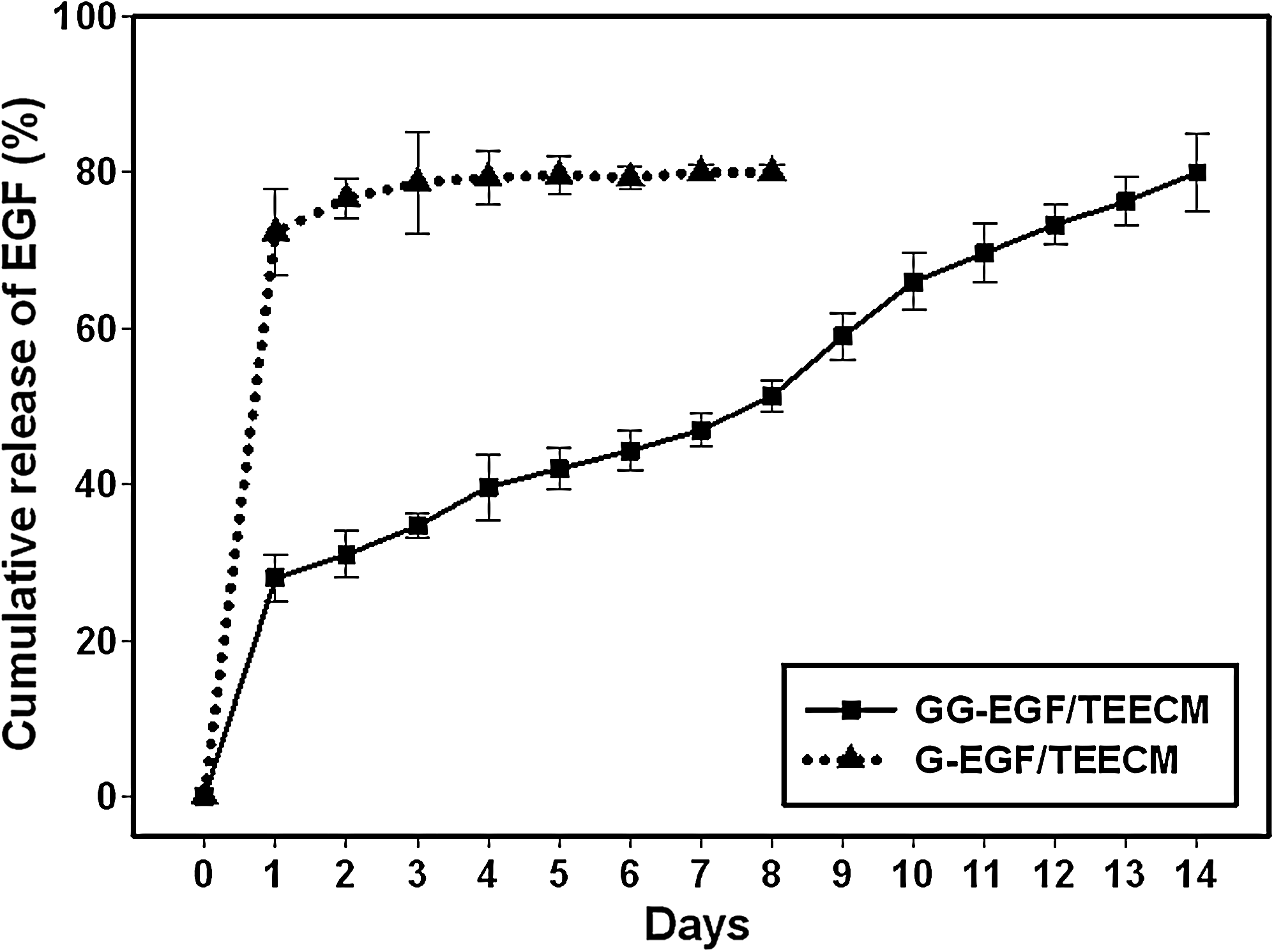
Cumulative release curves of GG-EGF/TEECM and G-EGF/TEECM (ELISA). The release kinetics of EGF was monitored for 14 days. EGF released from GG-EGF/TEECM was in a biphasic fashion: approximately 28% released within the first day, and the cumulative mass of EGF reached a plateau after 2 days. Finally, the percentage of cumulative release was approximately 80% by day 14, indicating an extended time course for complete release, whereas EGF delivered directly (G-EGF/TEECM) almost disappeared after 3 days.
Cell viability analysis
Cytotoxicity evaluation of GG-EGF/TEECM with fibroblasts (L929) by MTT cell viability assays is given in Figure 4A. The MTT absorbance was similar to those of the control (fresh culture medium) throughout the testing period. It revealed that GG-EGF/TEECM was nontoxic and did not release substances harmful to living cells. Both the attachment (at 2–24 h after cell seeding) and the proliferation (at 24–72 h after cell culturing) of HaCaT that were cultured on GG-EGF/TEECM, G-EGF/TEECM, and TEECM are graphically shown in Figure 4B. A similar plot for HFF that were cultured on three groups is graphically shown in Figure 4C.
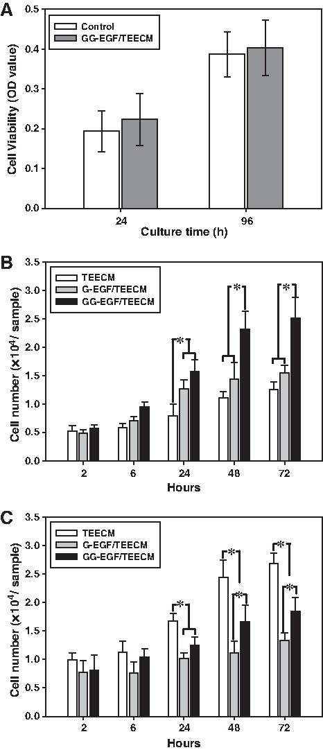
In vitro evaluation of GG-EGF/TEECM. (
At 2 h after cell seeding the number of HaCaT attached was not significantly different among the three groups, while at 24 h after cell culturing, the number of HaCaT attached on GG-EGF/TEECM and G-EGF/TEECM was greater than that on TEECM. However, only GG-EGF/TEECM maintained the superiority until the end. At all of the time points investigated, the number of the cells attached or proliferated on GG-EGF/TEECM was greater than that on G-EGF/TEECM and TEECM. It could be due to sustained release of EGF promoting effects on keratinocytes. For HFF, the number of the cells attached or proliferated on both G-EGF/TEECM and GG-EGF/TEECM, at 24 h after cell culturing, was less than that on TEECM, while a similar tread in terms of the ability of GG-EGF/TEECM to provide better support for the proliferation of the cells than that of G-EGF/TEECM was observed. The general better support for both the attachment and the proliferation of HFF on TEECM over those on both other groups is possibly because that the fibroblasts prefer a more porous and looser surface than keratinocytes do. Consistent with the results described above, the results indicated that GG-EGF/TEECM showed good in vitro biocompatibility and supported attachment and proliferation of both keratinocytes and fibroblasts.
In vivo studies
Figure 5 shows representative animals treated with acellular GG-EGF/TEECM, G-EGF/TEECM, TEECM, and cotton gauze, respectively, after grafting. It was not unexpected, as EGF is known to heal skin wounds efficiently, that there appears a significant improvement on GG-EGF/TEECM–treated wounds, while untreated wounds basically exhibited the slowest healing (p < 0.05 compared with other groups). No significant difference of wound area was found between the G-EGF/TEECM group and TEECM group during healing process. Conversely, 2 weeks after surgery the GG-EGF/TEECM–treated rats exhibited the significantly smaller wound area than other groups (p < 0.05 compared with other groups).
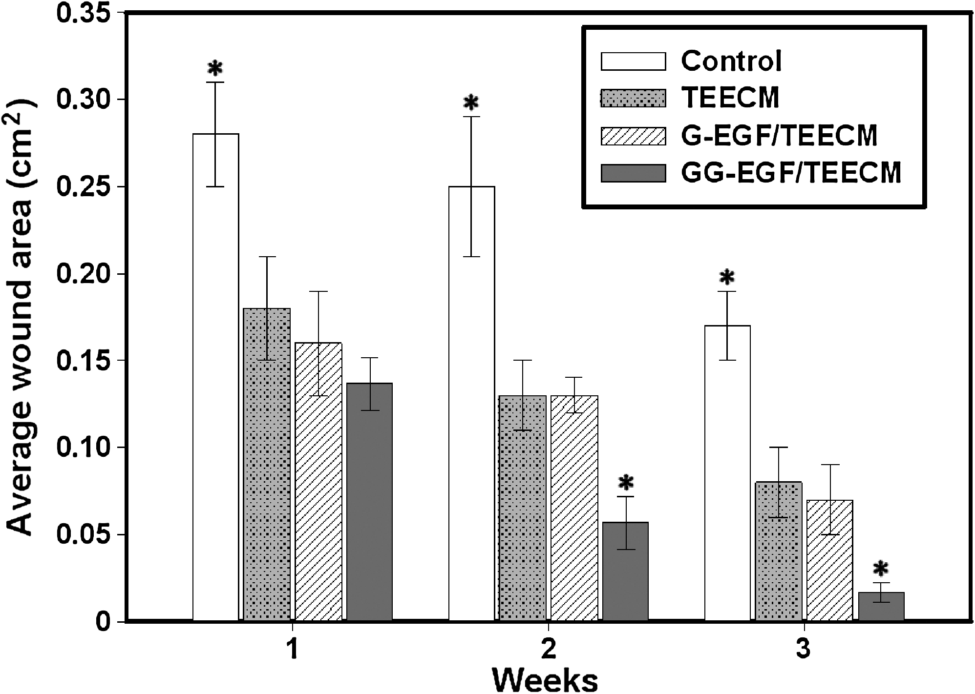
Changes in size of wounds treated with cotton gauze (control), TEECM, G-EGF/TEECM, and GG-EGF/TEECM. Values represent means with SE (n = 5). *p < 0.05 compared with other groups.
Two weeks after the operation, skin defects and crusts were partially observed in the animals of control group and TEECM group, while neither skin defect nor crusts were observed in the groups treated by GG-EGF/TEECM and G-EGF/TEECM. After implantation for 3 weeks, subcutaneous aspect showed grossly normal for the three test samples and there was no evidence of infection, while a superficial skin was still hemorrhagic for control group and also scab was present on the wound bed (Fig. 6A–D). Complete wound closure was induced by the GG-EGF/TEECM treatment after 3 weeks. Meanwhile, the G-EGF/TEECM group displayed distinct wound healing, but the contraction of the wound was more serious than that of GG-EGF/TEECM group. However, the re-epithelization of the TEECM-treated group was not achieved.
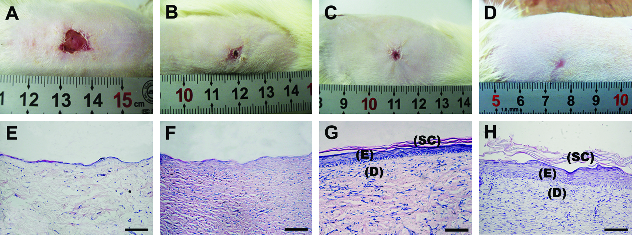
In vivo studies: photographs of macroscopic appearances of wound contraction treated with (
Generally, the wound healing is initiated from inflammatory phase, which is a normal and necessary prerequisite for healing. During early stages of wound healing, it is difficult to assess whether the inflammatory response is part of normal healing process or due to the effect of material. To evaluate the quality of the regenerated tissue, we performed hematoxylin and eosin staining of the wound tissue 3 weeks postwounding. From Figure 6E–H, the focal irregular area of the epithelium noted for some of the test wounds was found only in the control group. One of the main differences between G-EGF/TEECM and TEECM groups was the lack of the epidermal layer. In G-EGF/TEECM group, the continuity of the epithelium was completely restored with some epidermal rete pegs and the desquamation also noted. Conversely, absence of epidermal sheet contributed to a lower percentage of wound closure and re-epithelialization in TEECM group. The result of GG-EGF/TEECM–treated wound was restored almost to normalcy. Figure 6H (GG-EGF/TEECM) shows that the wound was filled with aligned and abundant collagen fiber and fibroproliferative tissues in the dermis. The entire surface of the defect was covered with new stratified epithelium, and the epidermal rete pegs were well developed. Compared to GG-EGF/TEECM group, G-EGF/TEECM group, which lacked controlled releasing EGF, displayed less stratified epidermal layer and looser dermal compartment with irregular collagen fiber. For no cell has been seeded in all groups, the cells presented in the wound healing areas are suggested to immigrate from the host origin. These findings indicated that GG-EGF/TEECM as a wound cover can provide a proper environment for wound healing.
Discussion
It was reported in the literature that skin equivalent requires a barrier protection to prevent infection and desiccation and cell guidance by dermal elements to maximize healing. 17 Therefore, bilayer concept of wound coverage in which both epidermal and dermal analogs are used is widely accepted.18,19 The epidermal layer of such a construct has to have a barrier function to not only protect the wound from bacterial contamination and fluid loss, but also stimulate the cell proliferation and re-epithelialization. Dermal elements are important for cell guidance during granulation tissue formation and re-modeling. Each portion of skin equivalents is essential for restoring normal tissue architecture and for the prevention of scars.
The process of wound repair involves the timed and balanced activity of inflammatory, vascular, and connective tissue, and epithelial cells. 20 All of these components need the ECM to facilitate the healing process. To minimize scar formation and to accelerate healing process, different techniques of skin substitution have been introduced in the last decade.21,22 To achieve this goal, different scaffold materials have been developed, each with different physical properties and each associated with a specific and unique host response when implanted. 23 Recently, scaffolds derived from ECM have been shown to be effective in the repair and reconstruction of several body tissues.4–7 The characteristic of these scaffolds recognized as important for their effectiveness is their ability to induce a host cellular response that supports constructive remodeling rather than default scar tissue formation. 24 In this study, a more effective and controlled extraction method has been introduced for producing ECM that exhibit very low antigenicity and excellent stability while retaining the native dermal structure. Additionally, this method that cells were inoculated before freeze–thawing could contribute to the synthesis of collagen, elastin, and the like; matrix remodeling; collagen fibril alignment; and improvement of mechanical properties.8,9,25,26 The results revealed that TEECM is a potential candidate in wound healing and tissue engineering, and is an analogical dermal layer of bilayer skin equivalent. For epidermal layer, the composite is provided by gelatin hydrogel–incorporated, EGF-loaded microspheres (GG-EGF), which have the hemostatic effect and cell recognition signals of gelatin,27,28 the re-epithelialization promoting feature of GG-EGF,29,30 and the property of water absorptivity, and water vapor transmission coupled with nontoxicity and biodegradability. 31 Due to these advantages, it is a suitable epidermal substitute in this study, and is a successful scaffold in numerous tissue engineering applications.
Our novel approach using this bilayered skin equivalent based on simple techniques offers an advantage by lessening the time required to prepare human skin substitute compared with the previous technique of culturing keratinocytes or fibroblasts on the sheets. 11 The inexistence of cells has the advantage of being cheap, readily available, and easily preserved, and prevents transmission of communicable diseases and immunological reactions. These elements are essential in treating acute clinical cases such as burn injuries.
The stimulation of wound healing by EGF has been confirmed by many researchers as growth of granulation tissue in implants used as inductive matrices.29,30,32 It is believed that the primary mechanism of enhanced wound healing is most likely due to increased proliferation of epidermal cells. The half-life of EGF in the body is, however, too short to exert the biological activity effectively when applied via injection or in free form. It is known that many proteases are activated in the injured tissue, and they easily decompose EGF in the wounded or burned site of skin as soon as it is applied. Therefore, incorporation of EGF into a polymer matrix and its sustained release from this seems as one possible approach to enhance its in vivo efficiency.33–35 In our studies, EGF was incorporated into microspheres to enhance its promoting effects. Further, the existence of gelatin hydrogel made EGF more difficult to diffuse from the matrices, which relatively improved the stability of EGF and prolonged the release time. In present study, 3 weeks after treatment, the histological observations under in vivo assessment revealed that the regenerated epithelium in EGF controlled release group (GG-EGF/TEECM) showed thicker, more developed, and stratified multiple layers, which was similar to that of normal skin, whereas regenerated epitheliums in G-EGF/TEECM group showed thinner epithelial layers due to lower efficiency of EGF.
Taken together, we have successfully produced a composite skin equivalent composed of gelatin hydrogel with EGF-loaded microspheres and tissue engineering EGF, which are known for their beneficial effects on wound healing. The evaluations of morphology, EGF release efficiency, and cytotoxicity of GG-EGF/TEECM showed optimal conditions for maintaining a proper environment conducive for wound healing. Further, these evaluations reveal that growth factors and ECM components in a cell-free matrix are critical for success in biomedical applications. The in vivo studies also showed good biocompatibility and efficiency of GG-EGF/TEECM. The composite GG-EGF/TEECM was found to promote wound healing and induce cell migration and proliferation. In consequence, GG-EGF/TEECM can meet a demand for skin tissue engineering, which is required for their clinical application as a promising strategy in the medical field.
Footnotes
Acknowledgments
This study was supported by funding from National High Technology Research and Development Program of China (2006AA02A119).
Disclosure Statement
No competing financial interests exist.