
Abstract
The shelf life of titanium implant products, that is, a possible time-related change of their bioactivity, has rarely been addressed. The objective of this study was to examine the bioactivity of newly processed and aged titanium surfaces and determine whether ultraviolet (UV) light treatment of the titanium surface restores the possible adverse effects of titanium aging. Titanium disks, either acid-etched or sandblasted, were used immediately after processing (fresh surface) or after storing in dark for 4 weeks (aged surface). Some disks were treated with UV light for 48 h after 4 weeks of storage. Albumin adsorbed to the aged surfaces was only 15% of that adsorbed to the fresh surfaces during 2-h incubation, whereas UV-treated aged surfaces adsorbed equivalent amount of albumin to that for the fresh surfaces. During 24-h incubation, the number of human mesenchymal stem cells attached to the aged surfaces was less than half of that for the fresh surfaces, whereas UV treatment of the aged surfaces increased the number three times. Proliferation, alkaline phosphatase activity, and calcium deposition of the cells were substantially lower on the aged surfaces than on the fresh surfaces, while those on the UV-treated aged surfaces were higher than on the fresh surfaces. The strength of bone–implant integration evaluated at week 2 of healing in a rat femur model was reduced to half after 4 weeks of titanium aging, whereas UV treatment of the aged implants increased the strength to the level equivalent to or even higher than the freshly prepared implants. Fresh and UV-treated aged surfaces were superhydrophilic, while the aged surface was hydrophobic. The data suggest that bioactivity of titanium surfaces degrades with time and that UV treatment of the aged surface increases the bioactivity over the level of the freshly prepared surface.
Introduction
Most orthopedic and dental implant products are packed sterile and sold as storable medical devices at the manufactures' and users' levels. These implant products are, therefore, uncontrollably aged during their inventory and distribution, as well as during the storage at the users' level before use. However, virtually no attention has been paid to the bioactivity of titanium associated with time; the related shelf life of implant products has never been addressed. For instance, there is no information available for users as to when the products were manufactured. In their instruction and product information, there is no time frame indicated for use or storage except for the expiration date of sterilization, which is generally 5 years. If there would be a time-dependent change in bioactivity of titanium, it would be a discovery of novel nature of titanium, leading to a major advancement in implant biological science, and simultaneously, have significant clinical impact in a wide range of therapeutics using titanium-based biomaterials.
Despite advancements in the development of titanium surfaces with better osteoconductivity and improved documentation of the process of osseointegration, the mechanism underlying this unique biological phenomenon is not fully understood. For instance, why the percentage of bone covering implants (bone–implant contact percentage) is far less than the ideal 100% remains unanswered. The reported bone–implant contact percentage is 45 ± 16%, 7 despite the constant implementation of various surface modification technologies. The low bone–implant contact percentage could be attributed to the limited capacity of bone growth around implants and/or the inevitable intervention of unfavorable fibrous soft tissues between newly formed bone and titanium. 4 Unfortunately, a vast majority of implants fail because of bone and implant interfacial failure. 8
Generation of a highly hydrophilic titanium surface by ultraviolet (UV) light treatment was first introduced in 1997. 9 This unique property is ascribed to the altered surface structure of the hydrophilic phase produced by the photocatalytic activity of TiO2. In this model, UV treatment hypothetically creates surface oxygen vacancies at bridging sites, resulting in the conversion of relevant Ti4+ sites to Ti3+ sites, which are favorable for dissociative water adsorption. The hydrophilic nature of material surfaces is a representative marker for surface energy and may play an important role in determining the biocompatibility level of materials through its effect on protein–material–cell interactions.10–12
In this report, several important questions regarding the bioactivity of titanium have been addressed:
Does the bioactivity of titanium relevant to the process of osseointegration change over time after processing? Does UV treatment of titanium improve (in case of a degrading change) or affect aging-like alterations of bioactivity?
The purpose of this study was to compare the bioactivity of several titanium disks of different ages: freshly prepared surfaces, 4-week-old surfaces, and 4-week-old surfaces treated with UV light. We examined the migration, attachment, proliferation, and differentiation of human mesenchymal stem cells (MSCs) on these titanium surfaces, as well as an in vivo ability of osseointegration of theses surfaces in a rat model. Two different surface types of titanium (acid-etched and sandblasted surfaces) were tested in the study.
Materials and Methods
Titanium samples and surface characterization
Disks (20 mm in diameter and 1.5 mm in thickness) and cylindrical rods (1 mm in diameter and 2 mm in length) were made from commercially pure titanium (grade 2). Surfaces of the titanium samples were prepared by either acid etching with 67% (w/w) sulfuric acid (H2SO4) at 120°C for 75 s, or sandblasting with 50 μm Al2O3 particles for 1 min at a pressure of 3 kg/m, followed by rinsing with ultrasonic distilled water for 10 min. The surface morphology was examined by scanning electron microscopy (XL30; Philips, Eindhoven, The Netherlands). Prepared surfaces were used for the experiments either immediately after their fabrication (fresh surface) or after storing them under dark ambient conditions for 4 weeks. Some 4-week-old titanium samples were treated with UV for 48 h under ambient conditions using a 15-W bactericidal lamp (Toshiba, Tokyo, Japan), with intensities of approximately 0.1 mW/cm2 (λ = 360 ± 20 nm) and 2 mW/cm2 (λ = 250 ± 20 nm). Surface energy of each of the surfaces was evaluated by the contact angle of 1 μL H2O using an automatic contact angle–measuring device (DCA-VZ; Kyowa Interface Science, Saitama, Japan).
Measurement of protein adsorption
Bovine serum albumin fraction V (Pierce Biotechnology, Rockford, IL) was used as a model protein. Three hundred milliliters of protein solution (1 mg/mL protein/saline) was spread over a titanium disk using a pipette. After 2, 24, or 72 h of incubation in sterile humidified condition at 37°C, nonadherent protein was removed and washed twice using saline with 0.9% sodium chloride. Aliquots (200 μL) of the initial and removed solutions were mixed with 200 μL of microbicinchoninic acid (Pierce Biotechnology) and incubated at 37°C for 60 min. The amount of protein was quantified using a microplate reader at 562 nm.
Human MSC/osteoblastic cell culture
Human MSCs (Poietics; Cambrex Bio Science, Walkersville, East Rutherford, NJ) were cultured in MSC growth medium consisted of MSC basal medium and growth supplements (SingleQuots; Camrex Bio Science, East Rutherford, NJ). The growth supplements contained fetal bovine serum, L-glutamine, and penicillin/streptomycin. Cells were incubated in a humidified atmosphere with 95% air, 5% CO2 at 37°C. After 2–4 passages, the cells were cultured in the MSC osteogenic induction medium consisting of the MSC basal medium and the osteogenic induction supplement (SingleQuots) that contained fetal bovine serum, L-glutamine, penicillin/streptomycin, dexamethasone, ascorbate, and β-glycerophosphate. At 80% confluency of the last passage, cells were detached using 0.25% trypsin-1 mM ethylenediaminetetraacetic acid (EDTA)-4Na and seeded onto titanium disks at a density of 3 × 10⁴ cells/cm2. The culture medium was renewed every 3 days. Cells with and without osteogenic induction were used in alkaline phosphatase (ALP) activity and mineralization assays, and migration, attachment, cytomorphology, and proliferation assays, respectively.
Migration assay
Migration of human MSCs to titanium surfaces was examined using dual-chamber migration assay (345-024K; Trevigen, Gaithersburg, MD). Cells were seeded into the top chamber in the culture medium. A titanium disk was placed at the bottom of the lower chamber. The percentage of cells that penetrated into the lower chamber after 3 h of incubation at 37°C through a polyester membrane with 8-μm-diameter pores was analyzed using the plate reader after staining with calcein-AM.
Cell attachment assay
Initial attachment of human MSCs was evaluated by measuring the quantity of the cells attached to titanium substrates after 3 and 24 h of incubation. Cells were gently rinsed twice with phosphate-buffered saline and treated with 0.1% collagenase in 300 μL of 0.25% trypsin-1 mM (EDTA)-4Na for 15 min at 37°C. A hematocytometer was used to count the number of detached cells obtained. Scanning electron microscopy was performed to confirm the absence of cell remnants on substrates. The cells were also quantified by 4-[3-(4-iodophenyl)-2-(4-nitrophenyl)-2H-5-tetrazolio]-1,3-benzene disulfate (WST-1)-based colorimetry (WST-1; Roche Applied Science, Mannheim, Germany) at matching time points. The culture well was incubated at 37°C for 4 h with 100 μL of tetrazolium salt (WST-1) reagent. The amount of formazan product was measured using an enzyme-linked immunosorbent assay (ELISA) reader at 420 nm.
Morphology and morphometry of cells
Confocal laser scanning microscopy was performed to examine the morphology and cytoskeletal arrangement of human MSCs. After 3 h of culture, the cells were fixed in 10% formalin, and stained using a fluorescent dye, rhodamine phalloidin (actin filament red color; Molecular Probes, Eugene, OR). The cultures were also immunochemically stained with mouse anti-paxillin monoclonal antibody (Abcam, Cambridge, MA), followed by the adding of fluorescein isothiocyanate–conjugated anti-mouse secondary antibody (Abcam). The cell area, perimeter, and Feret's diameter were quantitatively assessed using an image analyzer (ImageJ; NIH, Bethesda, ML).
Cell proliferation
The proliferative activity of human MSCs was measured by 5-bromo-2′-deoxyuridine (BrdU) incorporation during DNA synthesis. At day 3 of culture, 100 μL of 100 mM BrdU solution (Roche Applied Science) was added to the culture wells and incubated for 10 h. After trypsinizing the cells and denaturing DNAs, the cultures were incubated with anti-BrdU conjugated with peroxidase for 90 min and reacted with tetramethylbenzidine for color development. Absorbance at 370 nm was measured using the ELISA reader. The density of the cells was also evaluated at days 2 and 4 to confirm the proliferative activity.
ALP activity
The ALP activity of cultured human MSCs in the osteogenic induction medium was examined by a colorimetry-based assay. Day 10 cultures were rinsed with ddH2O and added with 250 μL p-nitrophenylphosphate (LabAssay ATP; Wako Pure Chemicals, Richmond, VA), and then incubated at 37°C for 15 min. The ALP activity was evaluated from the amount of nitrophenol released through the enzymatic reaction and measured at a wavelength of 405 nm using the ELISA reader.
Mineralization assay
The mineralization capability of cultured human MSCs was examined by a colorimetric detection of deposited calcium. Cultures at day 25 were washed with phosphate-buffered saline and incubated overnight in 1 mL of 0.5 M HCl solution with gentle shaking. The solution was mixed with o-cresolphthalein complexone in alkaline medium (Calcium Binding and Buffer Reagent; Sigma, St. Louis, MO) to produce a red calcium–cresolphthalein complexone complex. Color intensity was measured by the ELISA reader at 575 nm absorbance.
Animal surgery
Eight-week-old male Sprague-Dawley rats were anesthetized with 1–2% isoflurane inhalation. After their left legs were shaved and scrubbed with 10% providone–iodine solution, the distal aspects of the left femurs were carefully exposed via skin incision and muscle dissection. The flat surfaces of the distal femurs were selected for implant placement. The implant site was prepared 9 mm from the distal edge of the left femur by drilling with a 0.8 mm round burr and enlarged using reamers (#ISO 090 and 100). Profuse irrigation with sterile isotonic saline was used for cooling and cleaning. A titanium cylindrical rod (1 mm in diameter and 2 mm in length) was placed into the prepared hole. Surgical sites were then closed in layers. Muscle and skin were sutured separately with resorbable suture thread. The University of California at Los Angeles Chancellor's Animal Research Committee approved this protocol, and all experimentation was performed in accordance with the U.S. Department of Agriculture guidelines of animal research.
Implant biomechanical push-in test
The established implant biomechanical push-in test was used to assess the biomechanical strength of bone–implant integration.13,14 At week 2 of healing, left femurs containing a cylindrical implant were harvested and embedded into autopolymerizing resin with the top surface of the implant being horizontal. Micro-computed tomography was used to confirm that the implants were free from cortical bone support from the lateral and bottom sides of the implant. The testing machine (Instron 5544 electro-mechanical testing system; Instron, Canton, MA) equipped with a 2000 N load cell and a pushing rod (diameter = 0.8 mm) was used to load the implant vertically downward at a crosshead speed of 1 mm/min. The push-in value was determined by measuring the peak of the load–displacement curve.
Statistical analyses
Three samples were used for the cell culture studies, except for the evaluation of cell morphometry, which required 10 cell samples. Two-way analysis of variance (ANOVA) was performed to examine the effects of culture time and titanium surfaces having different ages, with or without UV treatment. If necessary, a post hoc Bonferroni test was conducted to examine differences among the fresh, 4-week-old, and UV-treated 4-week-old surfaces; p < 0.05 was considered statistically significant. If data were available at only one time point, one-way ANOVA was used to determine the differences among the experimental groups. A total of 30 rats, that is, 5 rats for each of the three surface conditions (fresh, 4-week-old, and UV-treated 4-week-old surfaces) of acid-etched and sandblasted implants, were used for the in vivo biomechanical push-in test (n = 5).
Results
Surface morphology of titanium samples
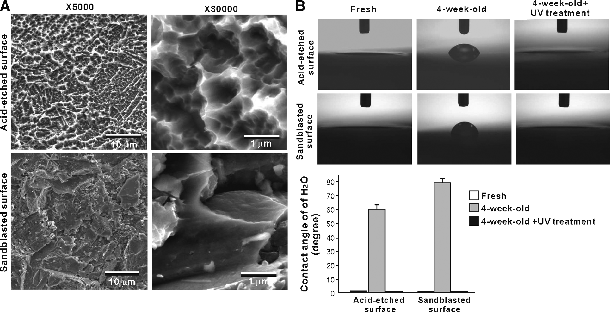
The acid-etched titanium surface presented a microtopographic configuration with uniformly created peaks and pits (Fig. 1A). The size of each compartmental structure ranged from 0.5 to 1.5 μm. The sandblasted surface was also roughened all over the surface. The created roughness was relatively irregular in shape and larger in scale compared to that on the acid-etched surface.
Surface characterization of titanium disks used in this study. (
Superhydrophilicity removed on aged titanium surfaces is restored by UV light treatment
Freshly prepared acid-etched surfaces (immediately after processing) showed a contact angle of H2O that was less than 3°, indicative of their superhydrophilic nature (contact angle <5°) (Fig. 1B). The 4-week-old acid-etched disks (i.e., stored for 4 weeks) showed a contact angle of higher than 55°, indicative of their hydrophobic nature. The contact angle of the 4-week-old acid-etched surface decreased to the level less than 3° after UV light treatment, indicating the restoration of superhydrophilicity. Similar age-related degradation and UV-mediated recovery of hydrophilicity were also found on sandblasted surfaces (Fig. 1B).
Delayed and diminished protein adsorption to aged acid-etched titanium is restored by UV treatment
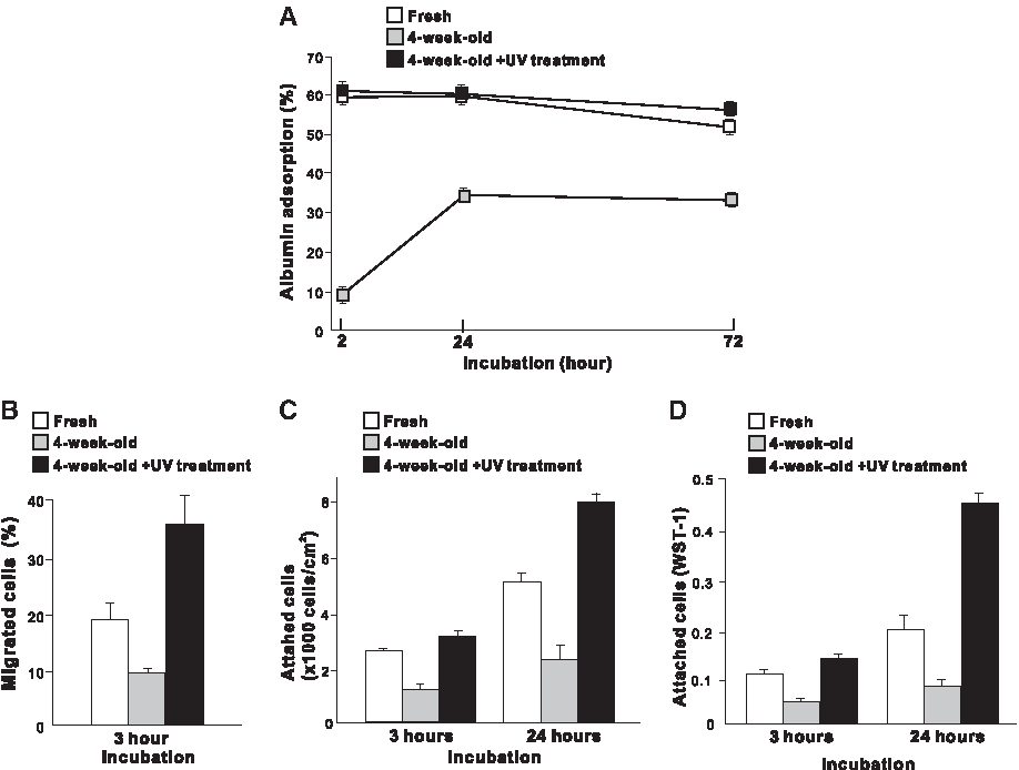
To determine whether there are possible age-related and UV-induced changes in bioactivity of titanium, acid-etched titanium disks were tested at the first setout in a series of in vitro experiments. Two-way ANOVA showed that albumin adsorption varied significantly on the differently conditioned acid-etched surfaces (p < 0.01; Fig. 2A). After 2 h of incubation, only approximately 10% of albumin incubated in the culture was adsorbed to the 4-week-old acid-etched surface, while approximately 60% of albumin adsorbed to the fresh acid-etched surface (p < 0.01; Bonferroni). The amount of albumin adsorption was 40% less for the 4-week-old surface than for the fresh surface even after 72 h of incubation (p < 0.01). The UV-treated 4-week-old surface showed an albumin adsorption level equivalent to that of the fresh surfaces after 2 and 24 h of incubation, and exhibited an even greater level after 72 h (p < 0.05).
Initial bioactivity of acid-etched titanium surfaces with different ages and with or without UV treatment. (
Stem cell migration and attachment diminished on aged acid-etched titanium and excelled on UV-treated acid-etched titanium
The number of human MSCs that had migrated through 8 μm holes varied significantly among the acid-etched surfaces with different age, and with and without UV treatment (p < 0.01, one-way ANOVA; Fig. 2B). The number of cells that migrated to the 4-week-old surface during 3 h of incubation was 50% of the number observed for the fresh surface and 25% of the number for the UV-treated 4-week-old surface (p < 0.01). The UV-treated 4-week-old surfaces showed a twofold greater cellular migration than the fresh surface (p < 0.01).
The number of human MSCs attached to the acid-etched surfaces increased in the following order: UV-treated 4-week-old surface > fresh surface > 4-week-old surface (p < 0.01; two-way ANOVA; Fig. 1C). The number of cells attached to the 4-week-old surface was less than 50% to the fresh surface at both 3-h and 24-h incubation. The UV-treated 4-week-old surface showed a substantially higher cell attachment than the fresh surface at 24 h (p < 0.01). The result from a WST-1 stain confirmed that the cell attachment was considerably reduced on the 4-week-old surface and increased on the UV-treated 4-week-old surface (Fig. 2D).
Delayed cell spread on aged acid-etched titanium is recovered by UV treatment
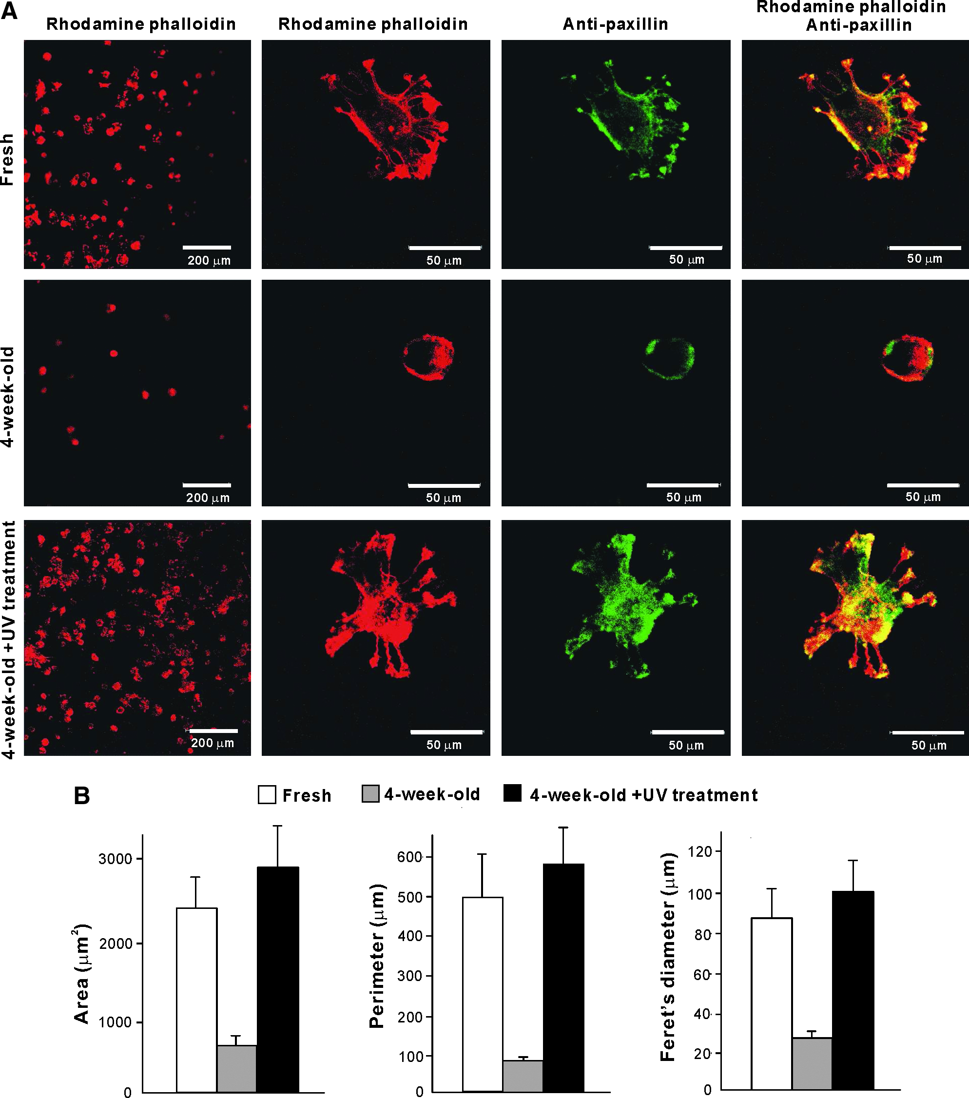
Low-magnification images captured after 3 h of incubation of human MSCs with actin filament staining showed that the number of cells appeared to be greatest on the UV-treated 4-week-old acid-etched surface and lowest on the 4-week-old acid-etched surface, confirming the result from the cell attachment assays (Fig. 3A). High-magnification images with actin stain revealed that cells were clearly larger with their processes spread in multiple directions on the fresh and UV-treated 4-week-old surfaces, whereas cells remained in rounded form with little cytoskeletal development on the 4-week-old surface. Intensive localization of paxillin along the cellular configuration was observed in the cells on the fresh and UV-treated 4-week-old surfaces. In particular, the dense cytoplasmic positive stain was seen in the cells seeded on the UV-treated 4-week-old surface.
Initial spread and cytoskeletal arrangement of human MSCs 3 h after seeding onto differently conditioned acid-etched titanium surfaces: fresh surface, 4-week-old surface, and UV-treated 4-week-old surface. (
Cytomorphometric evaluations of the area, perimeter, and Feret's diameter demonstrated significant differences in these parameters among the three different conditions of acid-etched surfaces (ANOVA, p < 0.01; Fig. 3B). These parameters were five- to eightfold greater for the fresh and the UV-treated surfaces than for the 4-week-old surface (Bonferroni, p < 0.01). There were no significant differences between the fresh and UV-treated surfaces.
Reduced cellular proliferation on aged acid-etched titanium and its UV-mediated restoration and further enhancement
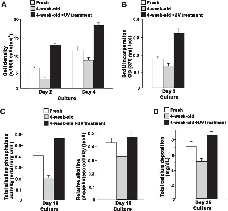
The number of human MSCs propagated on the acid-etched surfaces at days 2 and 4 varied significantly among the three different surface conditions (two-way ANOVA, p < 0.01; Fig. 4A). At day 4, the fresh surface exhibited 30% more cells than the 4-week-old surface (p < 0.05), while the UV-treated 4-week-old surface exhibited 150% more cells than the 4-week-old surface (p < 0.01). The BrdU incorporation per cell was also significantly greater in the following order: UV-treated 4-week-old surfaces, fresh surfaces, and 4-week-old surfaces (p < 0.01; Fig. 4B), confirming the likewise modulated rate of proliferation.
Cell proliferation and functional phenotypes of human MSCs/osteoblastic cells compromised on aged acid-etched titanium disks and enhanced on UV-treated acid-etched titanium disks. Differently conditioned acid-etched titanium surfaces are used: fresh surface, 4-week-old surface, and UV-treated 4-week-old surface. (
Delayed and impaired osteoblastic differentiation and mineralization on aged acid-etched titanium and its UV-mediated restoration and further promotion
Total ALP activity measured at culture day 10 in human MSCs/osteoblastic cells was significantly lower on the 4-week-old acid-etched surface than on the fresh acid-etched surface (p < 0.01; Fig. 4C). The ALP activity on the UV-treated 4-week-old acid-etched surface was even higher than on the fresh surface, which was over 2.5 times greater than on the 4-week-old surface. An analysis of ALP activity relative to the cell number also showed a significant contrast among the cultures. The ALP activity in the cells on the fresh and UV-treated 4-week-old surfaces was significantly higher than that on the 4-week-old surface (p < 0.05).
Total calcium deposition was greatest on the UV-treated 4-week-old surface and lowest on the 4-week-old surface (p < 0.05; Fig. 4D). The calcium deposition on the UV-treated 4-week-old surface increased by 70% compared with that on the 4-week-old surface.
Titanium age-related abatement of cell function and its restoration by UV found on sandblasted titanium
A series of in vitro experiments were also performed on sandblasted titanium disks to determine whether the similar age- and UV-induced phenomena occur on titanium surfaces other than the acid-etched surface. Like the results from the acid-etched surface, the cell density and proliferative activity were consistently lower on the 4-week-old surface than on the fresh surfaces (p < 0.01; Fig. 5A, B). The cell density at day 4 and cell proliferation at day 3 were significantly higher on the UV-treated 4-week-old surface than on the fresh surface (p < 0.05). The level of ALP activity with and without the standardization relative to cell number was higher for the fresh and UV-treated 4-week-old surfaces than for the 4-week-old surface (p < 0.01), whereas there was no significant differences between the fresh and UV-treated surfaces (Fig. 5C). The total calcium deposition decreased considerably on the 4-week-old sandblasted surface compared with the fresh sandblasted surface (p < 0.01; Fig. 5D). The calcium deposition on the UV-treated surface was significantly higher than that on the fresh surface (p < 0.05).

Age-related degradation and UV-induced enhancement in bioactivity proved on sandblasted titanium surfaces. Human MSCs/osteoblastic cells were cultured on differently conditioned sandblasted titanium disks: fresh surface, 4-week-old surface, and UV-treated 4-week-old surface. (
In vivo strength of osseointegration compromised by titanium aging, and restored and even surmounted by UV treatment
Titanium implants with freshly prepared acid-etched surfaces exhibited a two time greater push-in value than those with the 4-week-old surfaces (p < 0.01; Fig. 6). The UV treatment of the 4-week-old surface increased the push-in value to the level equivalent to that of the fresh surface. The push-in value for 4-week-old sandblasted implants was also reduced to half of that for the freshly prepared matching surface (p < 0.05). Notably, the implants with UV-treated 4-week-old sandblasted surfaces showed an even higher push-in value than the implants with freshly prepared sandblasted surfaces (p < 0.05).
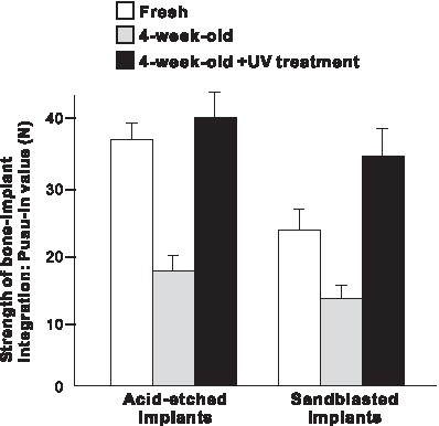
The effect of different age of titanium implants and of UV pretreatment on the strength of bone–implant integration evaluated by biomechanical push-in test. Titanium implants with two different surface topographies were tested: acid-etched and sandblasted surfaces. Data are shown as the mean ± SD (n = 5).
Discussion
This study consisting of a series of in vitro and in vivo experiments revealed two major findings: (1) the freshly prepared titanium surface showed substantially higher bioactivity than the 4-week-old surface and (2) the UV-treated 4-week-old surface showed higher bioactivity than the fresh surface. These findings indicate the following: (1) bioactivity of titanium degrades with time during storage and (2) UV treatment restores and even enhances the bioactivity beyond the maximal level that the surface inherently possesses. If the former phenomenon could be considered to be an aging-like change of titanium bioactivity, the latter could be considered to be titanium surface modification or biofunctionalization beyond rejuvenation-like reactivation.
The aging-like effect, that is, the attenuated bioactivity of the 4-week-old titanium surface compared to the freshly prepared surface, was substantial. Protein adsorption and cell attachment within 24 h of incubation was reduced to 50% or less compared with the fresh surface. Cell proliferation and representation of mature-stage osteoblastic phenotypes were also impaired substantially. Further, although designing additional in vivo studies is required, including histology, the push-in test performed in this study using the rat femur model demonstrated that there was a significant in vivo effect of titanium aging. Notably, the biomechanical strength of implant fixation was reduced to half after storing the implants for 4 weeks. These substantial biological deteriorations were found similarly for two different surface types of titanium (acid-etched and sandblasted surfaces).
Protein adsorption and cellular attachment are key biological steps in initiating the cascade of osteogenic processes around biomaterials. For instance, cell-surface receptors, integrins, interact with proteins adsorbed to the materials by binding their amino acid sequence arginine-glycine-aspartic acid (RGD in the one-letter amino acid code).15–17 This process plays a crucial role in cell attachment and subsequently regulates the spread, proliferation, and other functions of the cells. 17 These two processes behaved similarly in the present results; protein adsorption and attachment of human MSCs were reduced on 4-week-old titanium surfaces, whereas they increased on UV-treated surfaces. Spreading behavior of human MSCs was substantially delayed on 4-week-old surfaces, whereas it was expedited on the UV-treated surfaces. The development of a cellular skeleton and extension of cellular processes, which are the necessary steps for the initiation of cellular function and intracellular signaling, were remarkably contrasted between the fresh and aged titanium surfaces. In fact, the expression of paxillin, an adaptor protein that regulates focal adhesion and subsequent cellular function through its initiation of signal transduction, was downregulated on the 4-week-aged surface and, in contrast, upregulated on the UV-treated aged surface. This study does not provide evidence of links or interactions among the different biological processes of cell attachment, proliferation, differentiation, and in vivo osteogenesis. Further studies will determine whether and how the initial protein and cellular affinity environment compromised on aged titanium and enhanced on UV-treated titanium affect the subsequent biological events and eventually the level of osseointegration.
The deterioration in bioactivity of the aged titanium was associated with the removal of superhydrophilicity on both acid-etched and sandblasted surfaces, while the UV-induced recovery of bioactivity was associated with their revitalized superhydrophilicity. Superhydrophilic nature seemed to have a positive role in increasing the bioactivity of titanium. However, it is immature to conclude the existence of a direct link between the superhydrophilicity and high bioactivity. In fact, regardless of surface processing methods of acid etching and sandblasting, fresh surfaces and UV-treated 4-week-old surfaces, both exhibiting similar superhydrophilic nature, yielded distinct levels of bioactivity, indicating the requirement for further exploration of the change in other properties of titanium during aging and UV treatment. Association of substrate wettability with cellular behavior and response has been contentious and may be dependent upon the type of cells, substrates, and the range of wettability change. The increased cellular attachment is not necessarily obtained with an increase of surface wettability of biomaterial surfaces.18–20 Moreover, increased surface hydrophilicity does not necessarily result in the promoted osteogenesis of osteogenic cells. 21 The interpretation from these studies, however, is difficult because of the following: (1) the surface topography and composition of the substrates, together with the change in the hydrophilicity, varied significantly in the literature, (2) the ranges of the previously tested change in hydrophilicity were relatively small, and the superhydrophilic nature (<5° of contact angle) of the substrate was not tested, and (3) most importantly, the effects of titanium wettability properties on osteoblastic behavior and response have rarely been studied. The present study created the substantial change of wettability from hydrophobic (>50° contact angle) to superhydrophilic (<5° of water contact angle), while maintaining an identical surface topography of the substrates. Further studies implementing gradually changing levels of hydrophilicity, as well as greater levels of titanium aging, should be conducted to explore their biological effects.
The present results regarding the degrading bioactivity of titanium over time may have two potential significance. First, as mentioned earlier, titanium implant products, regardless of dental or orthopedic use, have been considered biologically stable to deliver consistent clinical performance. Currently, there is no time frame indication or expiration for titanium-based implant products. Commercially circulated products can be assumed to be uncontrollably and substantially aged. In light of this current mechanism of circulation, it could be possible that the bioactivity level of implant products available in the market is significantly lower than the ideal bioactivity that the products are expected to generate at the time of manufacture. The time-related degrading bioactivity of titanium, as demonstrated in this study using human cells and an in vivo animal model, should draw an immediate attention for further investigation and potential clinical implication. The second potential impact is regarding the research design. To the best of our knowledge, the age of titanium surfaces has never been standardized in previous in vitro or in vivo research. This study uncovered that titanium substrates with identical surface topographies but with different ages have been found to exert a substantial difference in their in vitro bioactivities and in vivo osseointegration ability. We confirmed that this phenomenon occurred very similarly on different surface topographies tested, implying a potential applicability of this phenomenon to other titanium-based materials. Future studies comparing different titanium surfaces may require the standardization of their age, or an otherwise careful interpretation of the research outcome.
Conclusion
The present in vitro and in vivo study examined possible changes in bioactivity of titanium surfaces during their aging and the effect of UV treatment on the aged titanium surfaces. Two different surface processing techniques of acid etching and sandblasting were employed. The rates of attachment, proliferation, and differentiation of human MSCs on titanium surfaces that were stored for 4 weeks after processing were substantially lower than those on freshly processed titanium surfaces, in association with the removal of superhydrophilicity. In vivo strength of osseointegration obtained by the 4-week-old implants was only half of that of the freshly prepared implants. However, UV light treatment of the 4-week-aged titanium surfaces increased the bioactivity to the level equal to or even higher than the freshly prepared surfaces, in association with the regeneration of superhydrophilicity. These results indicate an aging-like time-related degradation of titanium bioactivity and a novel rejuvenation-like photo-functionalization of titanium to overcome the age-related biological degradation.
Footnotes
Acknowledgment
This work was supported by JAMSEA and Implant-Perio Study Group.
Disclosure Statement
No competing financial interests exist.