
Abstract
We have previously shown that administration of platelet-rich plasma–impregnated gelatin hydrogel microspheres (PRP-GHMs) into a degenerated intervertebral disc (IVD) markedly suppresses progression of IVD degeneration. In the current study, we characterized the in vivo effects of PRP-GHM treatment in a degenerated IVD model in rabbit. On magnetic resonance images, the IVD height was significantly greater after treatment with PRP-GHMs compared with phosphate-buffered saline–impregnated GHMs, PRP without GHMs, and needle puncture only. Water content was also preserved in PRP-GHM–treated IVDs. Consistent with this observation, the mRNA expression of proteoglycan core protein and type II collagen was significantly higher after PRP-GHM treatment compared with other treatment groups. No proliferating cells were found in the nucleus pulposus and inner annulus fibrosus in any groups, but the number of apoptotic cells in the nucleus pulposus after PRP-GHM treatment was significantly lower than that after other treatments. These results provide an improved understanding of the therapeutic effects of PRP-GHM treatment of degenerated IVDs.
Introduction
Platelet-rich plasma (PRP) contains mixed autologous growth factors such as TGF-β1, platelet-derived growth factor (PDGF), and insulin-like growth factor (IGF)-1,15–19 and is easily purified from the patient's peripheral blood with a centrifugation apparatus, making it free from concerns of transmissible diseases. Biodegradable gelatin hydrogel microspheres (GHMs) have been used as a drug delivery system for administration of PRP to degenerated IVDs in vivo. We previously showed that IVD degeneration was markedly inhibited after administration of PRP-impregnated GHMs (PRP-GHMs), whereas IVD degeneration progressed after administration of PRP without GHM. 20 Tabata et al. reported that GHM forms a polyion complex with various growth factors, and that growth factors immobilized in GHM are released continuously with maintenance of biological activity by degradation of GHM in vivo. 21 Our previous histological and immunohistochemical studies revealed that the structure and accumulation of PGs in IVDs is well preserved in rabbits injected with PRP-GHMs, 20 but the detailed characterization of deceleration of IVD degeneration is unknown. This approach may be important for treatment of degenerated IVDs in humans, and therefore the objective of the study was to characterize the therapeutic effects of PRP-GHMs in vivo.
Materials and Methods
Biodegradable GHMs
Biodegradable GHMs were created by crosslinking an aqueous solution of gelatin dispersed in an oil phase using glutaraldehyde (Wako Pure Chemical Industries, Osaka, Japan).20,21 The gelatin (Nitta Gelatin, Osaka, Japan) used in this procedure had an isoelectric point of 5.0 and was isolated from bovine bone type I collagen using an alkaline process. The diameter of the GHMs ranged from 10 to 20 μm in a swollen state in phosphate-buffered saline (PBS), and the water content was 99 vol%.
IVD degeneration model
All the experiments were approved by the Experimental Animal Committee of the Kyoto Prefectural University of Medicine. IVD degeneration models were created by aspirating 0.005 to 0.008 g of the NP in noncontiguous discs (L2-3, L4-5, and L6-7) in 128 male Japanese white rabbits (weighing 2.5 to 2.8 kg) under inhalation anesthesia.20,22,23 The rabbits were obtained from Oriental Bio Service (Kyoto, Japan).
Preparation of PRP and intradiscal administration
PRP was prepared as previously described. 20 Briefly, 20 mL of fresh blood was obtained from the PRP-GHM group and PRP-only group under inhalation anesthesia. Acid citrate dextrose-A solution (2 mL) was added to the blood to prevent coagulation, and the mixture was centrifuged for 10 min with a centrifugation apparatus (KN70; Kubota, Tokyo, Japan) at 1500 rpm. The collected supernatant was centrifuged again at 3000 rpm for 10 min, and precipitated platelets were collected, including 300 μL of supernatant, to prepare PRP. To impregnate GHMs with PRP, 60 μL of PRP was immediately dropped onto 3.0 mg of sterilized GHMs, and the mixture was incubated for 1 h at 37°C (PRP-GHM group). The same procedure was performed using PBS instead of PRP (PBS-GHM group). Two weeks after partial aspiration of the NP, the impregnated GHMs were diluted by adding 180 μL of PBS, and 20 μL was then injected into the NP of the respective degenerated IVDs (L2-3, L4-5, and L6-7) using a 27-gauge insulin injector under inhalation anesthesia. For rabbits treated with PRP without GHM (PRP-only group), 5 μL of PRP was mixed with 15 μL of PBS and injected. For negative control group (Puncture-only group), only needle puncture was performed without injecting any sample. The rabbits were euthanized with pentobarbital (120 mg/kg) (Abbott Laboratories, North Chicago, IL) at 2, 4, and 8 weeks after injection, and the following analyses were performed.
Magnetic resonance imaging
To evaluate the effect of PRP on the degenerated IVDs using magnetic resonance (MR) imaging, the spinal columns were removed en bloc with the peripheral soft tissue at 2, 4, and 8 weeks after injection (n = 3 for each group at each time point). Mid-sagittal T1-weighted and T2-weighted images were taken between L1-2 and L6-7 using an INOVA 300 (7.05 Tesla; Varian, Palo Alto, CA), with 0.1 mM MnCl2 solution used as the phantom. The T1-weighted images were obtained using the following conditions: repetition time/echo time, 500/10 ms; field of view, 5 × 3 cm; matrix, 512 × 256; slice thickness, 2 mm; number of transitions, 2. The T2-weighted images were obtained as follows: repetition time/echo time, 2000/120 ms; field of view, 5 × 3 cm; matrix, 512 × 512; slice thickness, 2 mm; number of transitions, 2. The mean values of the anterior and posterior disc height were evaluated on the T1-weighted images.24,25 The mean values of IVD heights were also measured in healthy controls (n = 3). The signal intensity within the IVDs was evaluated using the T2-weighted images. IVD degeneration was classified using the Pfirrmann classification, with grades I to V scored from 1 to 5. Grade I: structure, homogeneous bright white; distinction between NP and AF, clear; signal intensity, hyperintense or isointense to cerebrospinal fluid; disc height, normal. Grade II: structure, inhomogeneous with or without horizontal bands; distinction, clear; signal intensity, hyperintense or isointense to cerebrospinal fluid; disc height, normal. Grade III: structure, inhomogeneous, gray; distinction, unclear; signal intensity, intermediate; disc height, normal to slightly decreased. Grade IV: structure, inhomogeneous, gray to black; distinction, lost; signal intensity, intermediate to hypointense; disc height, normal to moderately decreased. Grade V: structure, inhomogeneous, black; distinction, lost; signal intensity, hypointense; disc height, collapsed disc space. 26
Analysis of gene expression of PG core protein and type II collagen
A real-time polymerase chain reaction (PCR) was performed to analyze gene expression in NPs.27,28 The NP was isolated from IVDs (L2-3, L4-5, and L6-7) at 2, 4, and 8 weeks after injection and immediately frozen with liquid nitrogen (n = 3 for each group at each time point). mRNA was extracted from the frozen NP using an RNeasy Micro kit (Qiagen, Hilden, Germany). The concentration of mRNA in each sample was measured using an Ultrospec 3300 pro spectrophotometer (Amersham Pharmacia Biotech, Cambridge, United Kingdom). Complementary DNA was synthesized from the extracted mRNA (0.5 μg) using an oligo (dT) primer (Toyobo, Osaka, Japan). A Biosystem 7300 sequencer (Applied Biosystems, Foster City, CA) was used for real-time PCR. Increases in reporter fluorescence were monitored using TaqMan probes for PG core protein and type II collagen. The primers and TaqMan probes for the PCR were designed by the Assays-by-Design Service (Applied Biosystems). In a 25 μL PCR, 2 μL of complementary DNA was amplified using 50 nM of forward and reverse primers, 200 nM of ribosomal RNA (rRNA) probe (VIC-TAMRA label), and TaqMan 2 × Universal PCR Master Mix (Applied Biosystems) for the internal control. For the target gene, a TaqMan probe (FAM-TAMRA label) and a TaqMan 2 × Universal PCR Master Mix were used. Gene expression of PG core protein was measured over 40 cycles at 95°C for 15 s, followed by 60°C for 1 min; type II collagen expression was measured over 45 cycles at 95°C for 15 s, followed by 60°C for 1 min. The following gene-specific primers and TaqMan probes were used29,30: PG core protein: forward 5′-CGC CTA CCA GGA CAA GGT-3′, reverse 5′-GCG CAG GCT CTG GAT CTC-3′, and probe 5′-[FAM]-TCG CTG CCC AAC TAC-[TAMRA]-3′ type II collagen: forward 5′-CCT GTG CGA CGA CAT AAT CTG T-3′, reverse 5′-GCA GTG GCG AGG TCA GTA G-3′, and probe 5′-[FAM]-CAG TCC TTG GTG TCT TC-[TAMRA]-3′. For quantification of the changes in gene expression, the comparative threshold cycle (Ct) method was used to calculate the relative fold changes normalized against rRNA.29,30 Values were the means of nine samples (n = 3, nine IVDs), and each sample was assayed in duplicate. Gene expression was quantified as the difference between the Ct value of the sample for the target gene and the mean Ct value for the endogenous control (rRNA) in that sample. Relative expression was calculated as the difference between the Ct values of the test and control samples. Ct values in healthy rabbits were used as control data (n = 3).
Proliferating cell nuclear antigen immunohistochemistry
To evaluate the proliferative activity of the IVD cells, immunohistochemistry for proliferating cell nuclear antigen (PCNA) was performed on sections at 2, 4, and 8 weeks after injection. 31 Vertebral body–disc–vertebral body units from each group were fixed with 10% neutral formalin (Nacalai Tesque, Kyoto, Japan) (n = 3 for each group at each time point) and decalcified with 0.5 M ethylenediaminetetraacetic acid (pH 7.5). The IVDs were then embedded in 80% alcohol paraffin to create sections of 5 μm thickness in the midsagittal plane. An anti-rat PCNA mouse monoclonal antibody (1:400 dilution) (Dako A/S, Produktionsvej, Denmark; cat. no. M0879) was used as the primary antibody. All procedures were performed according to the recommendations of the manufacturers. Randomly selected dewaxed sections were processed in a warm bath with distilled water to repair antigens. For inactivation of endogenous peroxidase, the sections were treated with 3% hydrogen peroxide. Simple Stain Rat MAX-PO (M) (Nichirei, Tokyo, Japan) was used as the secondary antibody, with 3′,3′-diaminobenzidine solution used to elicit a color change. Finally, the sections were counterstained with hematoxylin. The distribution of cells with positive staining for PCNA was observed by light microscopy.
Terminal deoxynucleotidyl transferase–mediated dUTP-biotin nick-end labeling staining
Terminal deoxynucleotidyl transferase–mediated dUTP-biotin nick-end labeling (TUNEL) staining for detection of apoptotic cells was conducted on similar sections (n = 3 for each group at each time point) to those described above.32,33 ApopTag (Chemicon International, Temecula, CA; cat. no. S7100) reagent was used in the assay, and all procedures were performed following the manufacturer's instructions. Randomly selected dewaxed sections were treated with proteinase K (Dako A/S) and with 3% hydrogen peroxide for inactivation of endogenous peroxidase. The sections were incubated for 1 h at 37°C with TdT enzyme, followed by treatment with anti-digoxigenin-peroxidase. 3′,3′-diaminobenzidine solution was used to elicit a color change, and the sections were counterstained with hematoxylin. The distribution of TUNEL-positive cells was observed by light microscopy. The total numbers of NP and TUNEL-positive cells were counted, and the percentage of TUNEL-positive cells among the NP cells was calculated for each group. An orthopedic resident blinded to the group identities examined the MR images and counted the PCNA-positive and TUNEL-positive cells.
Kinetic analysis of TGF-β release in IVDs
An in vivo release test for 125I-labeled human recombinant TGF-β1 (PerkinElmer, Boston, MA) in the IVDs was performed to examine the efficacy of GHMs as a release matrix for PRP growth factors. 34 Labeled TGF-β solution (15 μL, 1.56 MBq/mL) mixed with 45 μL of PBS was impregnated into the prepared GHMs (3 mg). The impregnated GHMs were diluted by adding 180 μL of PBS, and 20 μL was then injected into the degenerated IVDs (TGF-β GHM group). As a control group, a solution with the same labeled amount of TGF-β was administered to the IVDs (TGF-β–only group). Rabbits were sacrificed at 1, 3, 7, 14, and 28 days after injection (n = 2, six IVDs per day), and the persistent radioactivity in the IVDs was measured using a Cobra II gamma counter (Packard, Meriden, CT). 29 To detect the remaining radioactivity through 28 days after injection, the relatively higher concentration of labeled TGF-β was selected in comparison with that of TGF-β in PRP. 20
Statistical procedure
Data for grading of MR images and TUNEL staining were analyzed using a Mann–Whitney U-test and a Kruskal–Wallis test. Gene expression and kinetic data were analyzed using a Student's t-test. p-Values < 0.05 were considered to indicate statistical significance. All results are expressed as mean values ± standard error of the mean.
Results
Analysis of MR images
MR images of spinal columns from each group were examined for evaluation of the effect of PRP on degenerated IVDs. The disc height was measured, and the Pfirrmann classification was used for comparison of the images. There were no differences among the respective images (L2-3, L4-5, and L6-7) in each groups. In the PRP-GHM group, the signal intensity in T2-weighted images was higher up to 8 weeks after the injection relative to the other groups. The distinction between NP and AF, and the signal intensity of NPs were preserved in PRP-GHM–treated IVDs, but lost in other groups (Fig. 1). The mean disc height in the healthy rabbits was 2.68 ± 0.06 mm. The mean disc height in the T1-weighted images of the IVDs was significantly higher in the PRP-GHM group at 8 weeks after the injection (2.49 ± 0.13 mm at 2 weeks, 2.44 ± 0.13 at 4 weeks, and 2.36 ± 0.07 at 8 weeks) compared with the PBS-GHM group (2.34 ± 0.12, 2.27 ± 0.12, and 2.00 ± 0.10, respectively), PRP-only group (2.41 ± 0.07, 2.35 ± 0.04, and 1.88 ± 0.09, respectively), and Puncture-only group (2.23 ± 0.04, 2.20 ± 0.09, and 1.87 ± 0.13, respectively) (Fig. 2). The mean MR image grading score in the PRP-GHM group was 2.11 ± 0.11, 2.44 ± 0.24, and 2.67 ± 0.28 at 2, 4, and 8 weeks, which were significantly lower through 8 weeks than the PBS-GHM group (2.78 ± 0.15, 3.33 ± 0.17, and 3.67 ± 0.71, respectively), PRP-only group (2.67 ± 0.24, 3.33 ± 0.24, and 3.44 ± 0.29, respectively), and Puncture-only group (2.67 ± 0.24, 3.22 ± 0.32, and 3.44 ± 0.73, respectively) (p < 0.05) (Fig. 3).
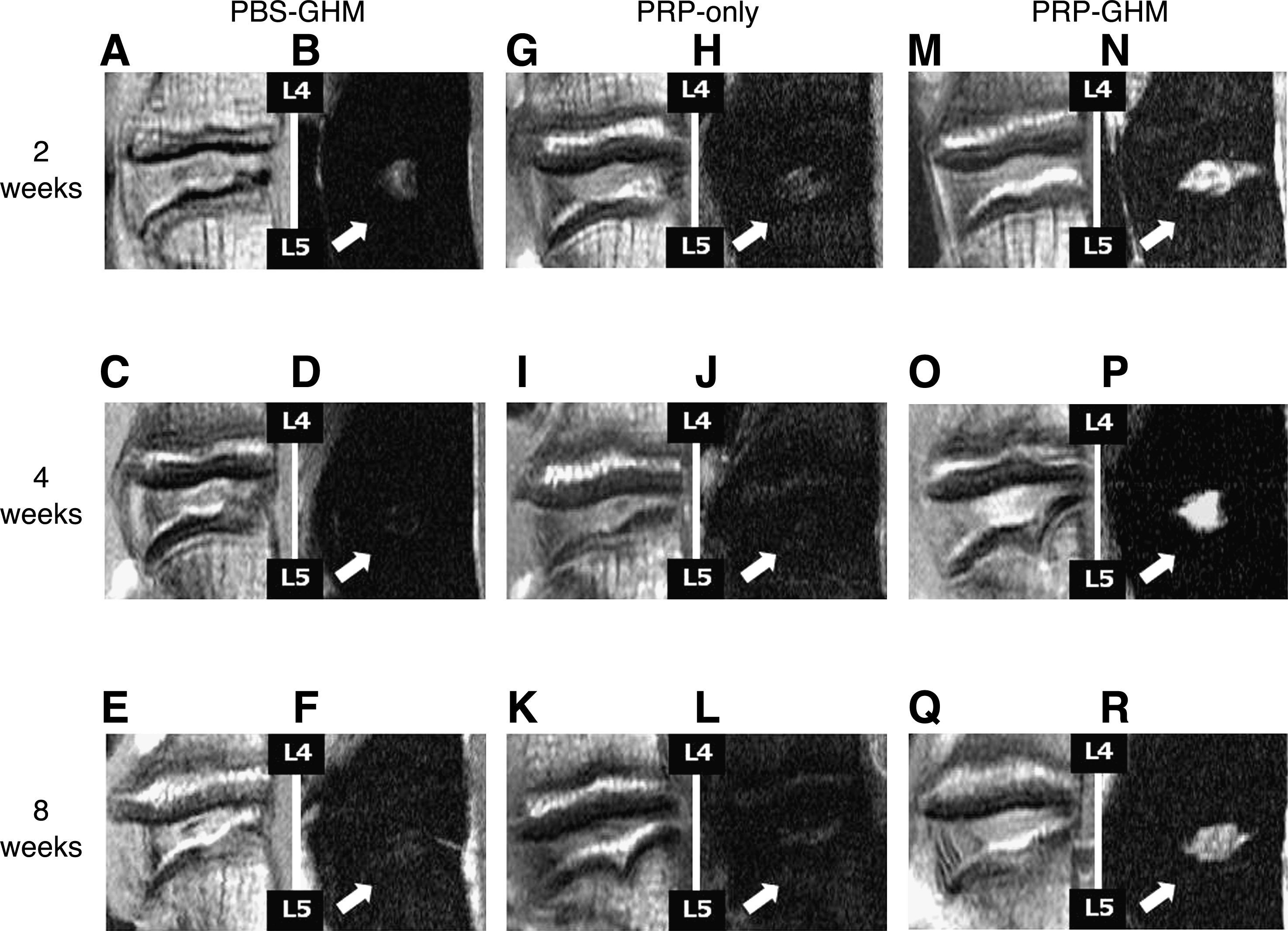
Magnetic resonance images after injection of phosphate-buffered saline (PBS)-gelatin hydrogel microspheres (GHMs) (
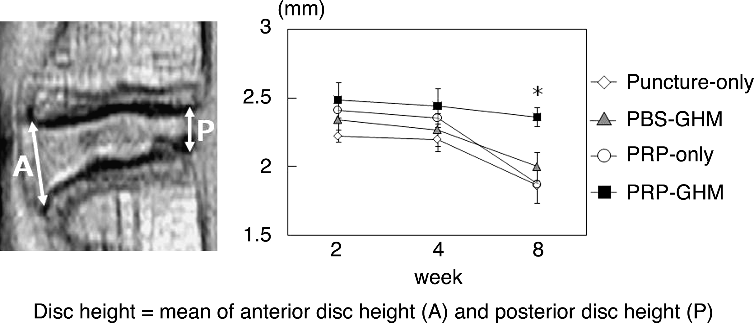
Mean values of anterior and posterior disc heights in T1-weighted images. Each value is expressed as the mean ± standard error. *p < 0.05 versus the PRP-GHM group.

Mean magnetic resonance image grading score using the Pfirrmann classification. Each value is expressed as the mean ± standard error. *p < 0.05 versus the PRP-GHM group.
Gene expression analysis for PG core protein and type II collagen
To investigate the effect of PRP on the main ECM component genes in NPs, the levels of PG core protein and type II collagen mRNA were compared using real-time PCR. The PG core protein mRNA in the PRP-GHM group was significantly higher at 2 weeks after the injection (0.84 ± 0.11 at 2 weeks, 0.51 ± 0.09 at 4 weeks, and 0.42 ± 0.09 at 8 weeks), but not at 4 and 8 weeks than in the PBS-GHM group (0.46 ± 0.12, 0.39 ± 0.10, and 0.29 ± 0.08, respectively), PRP-only group (0.46 ± 0.07, 0.36 ± 0.07, and 0.34 ± 0.04, respectively), and Puncture-only group (0.52 ± 0.07, 0.49 ± 0.04, and 0.28 ± 0.04, respectively) (p < 0.05) (Fig. 4). Similarly, the type II collagen mRNA in the PRP-GHM group was significantly higher at 2 weeks after the injection (0.73 ± 0.12 at 2 weeks, 0.35 ± 0.07 at 4 weeks, and 0.10 ± 0.03 at 8 weeks), but not at 4 and 8 weeks than in the PBS-GHM group (0.33 ± 0.10, 0.11 ± 0.03, and undetectable, respectively), PRP-only group (0.39 ± 0.08, 0.23 ± 0.05, and 0.08 ± 0.02, respectively), and Puncture-only group (0.38 ± 0.06, 0.10 ± 0.03, and 0.04 ± 0.01, respectively) (p < 0.05) (Fig. 5).
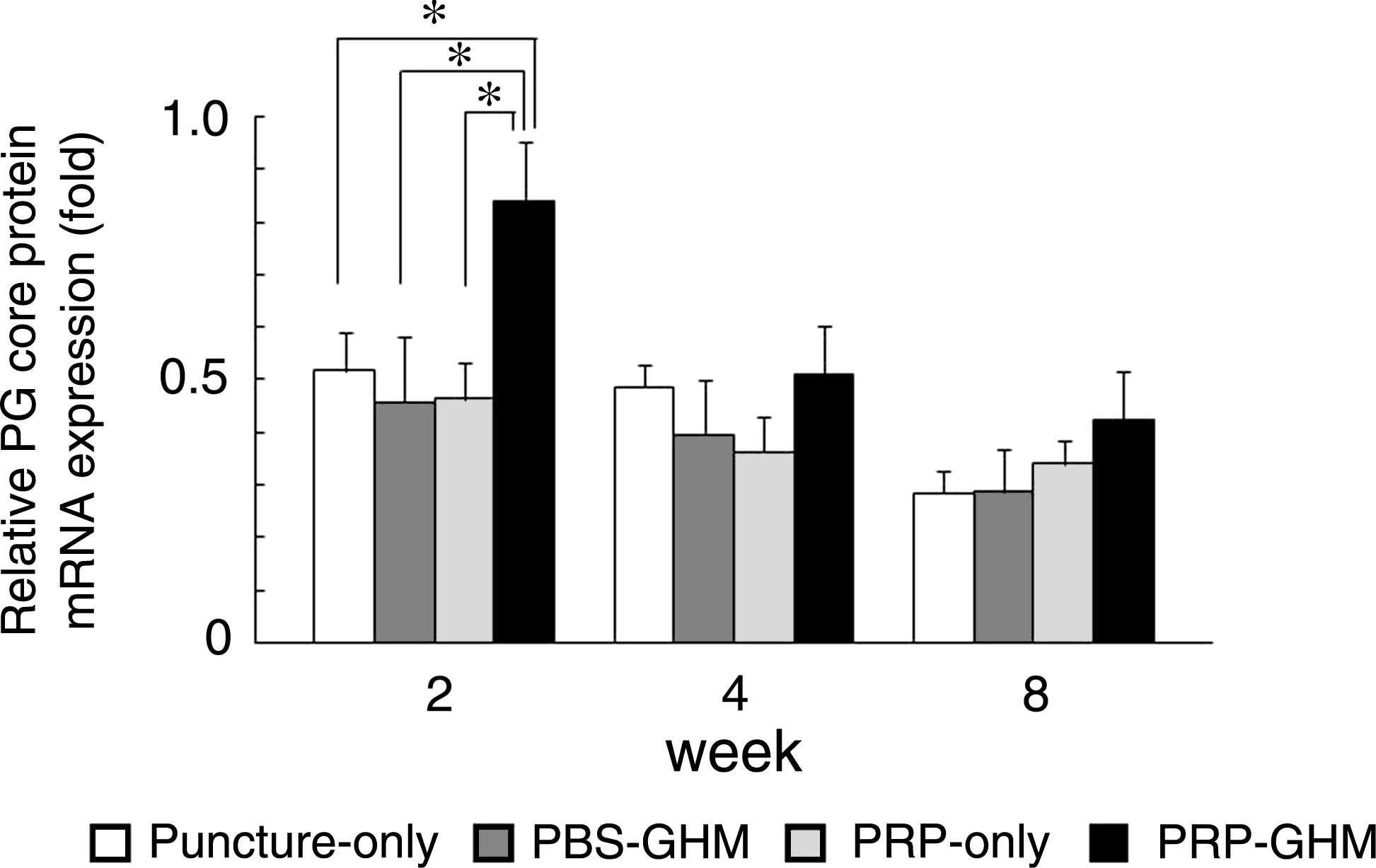
Gene expression of proteoglycans (PG) core protein in the NP determined by real-time polymerase chain reaction. Each value is expressed as the mean ± standard error. *p < 0.05 versus the PRP-GHM group.
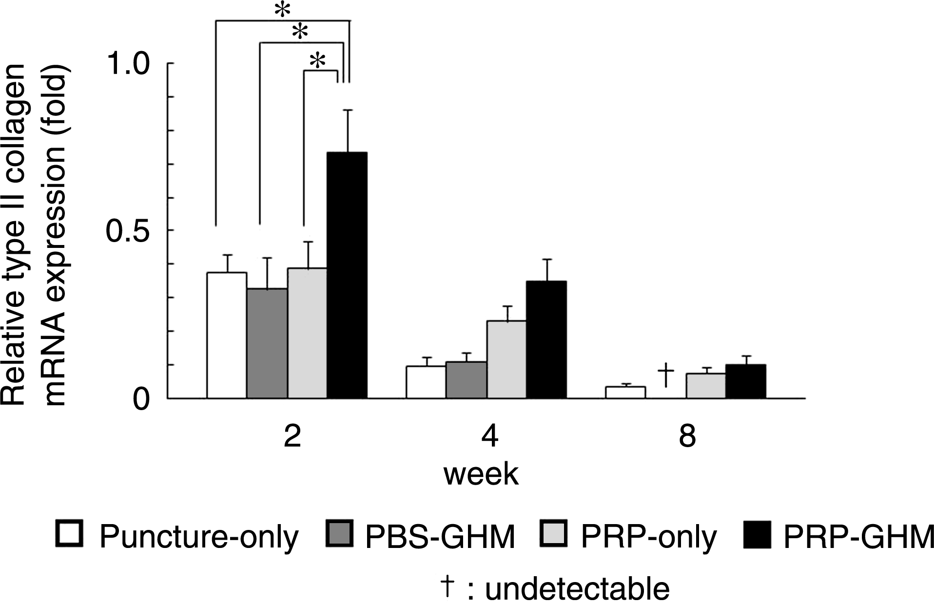
Gene expression of type II collagen in the NP determined by real-time polymerase chain reaction. Each value is expressed as the mean ± standard error. *p < 0.05 versus the PRP-GHM group. †Undetectable.
Evaluation of proliferating cells in IVDs
Immunohistochemistry for PCNA was performed to evaluate the proliferative effect of PRP in IVD cells. There were many PCNA-positive cells in the marrow of vertebrae in all groups, and PCNA was localized in the nucleus (Fig. 6A, E). PCNA-positive cells were also found in the anterior longitudinal ligament and part of the outer AF at the puncture site in each group (Fig. 6F). No PCNA-positive cells were found in the NP, including the periphery (Fig. 6B, G, H), the inner AF (Fig. 6C), and the outer AF at the nonpunctured site (Fig. 6D) in all groups through 8 weeks after injection.
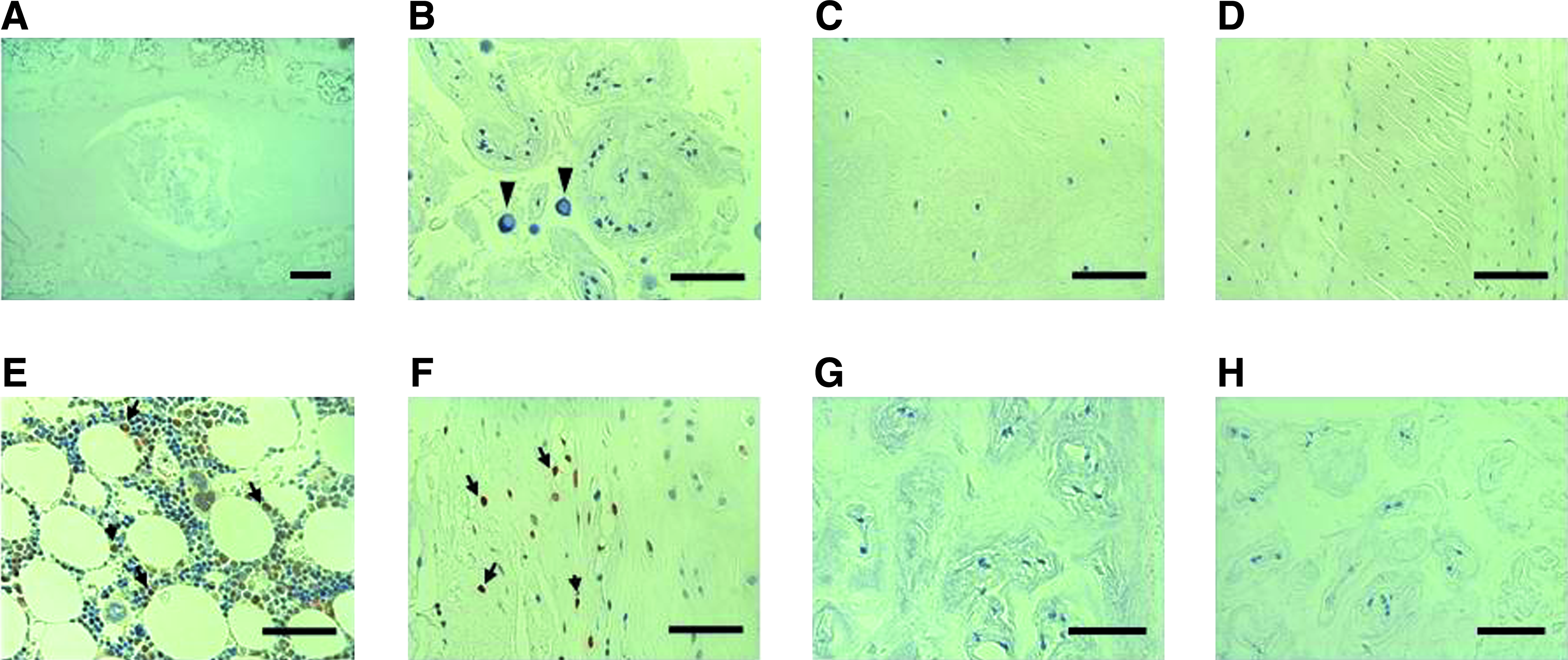
Immunohistochemistry for proliferating cell nuclear antigen after PRP-GHMs treatment. Intervertebral disc (IVD) (
Evaluation of apoptosis in IVDs
TUNEL staining was conducted to investigate the effect of PRP on apoptosis in NP cells. TUNEL-positive cells were observed in the NPs (Fig. 7A, E, F), inner AF (Fig. 7B), outer AF (Fig. 7C), and marrow of vertebrae (Fig. 7D) in all groups at 2 weeks after injection. At this time, the percentage of TUNEL-positive cells in the PRP-GHM group was 19.4 ±2.8%, which was significantly lower than 31.7 ± 3.3, 26.3 ± 4.2, and 27.1 ± 1.1 in the PBS-GHM, PRP-only, and Puncture-only groups, respectively (p < 0.05) (Fig. 8). At 4 and 8 weeks after injection, almost all NP cells could not be distinguished from connective tissues in the PBS-GHM (Fig. 7H), PRP-only, and Puncture-only groups because of the severe IVD degeneration. The percentages of TUNEL-positive cells in the PRP-GHM group were 18.1 ± 2.3 and 17.0 ± 5.4 at 4 and 8 weeks after injection (Fig. 7G).

Terminal deoxynucleotidyl transferase–mediated dUTP-biotin nick-end labeling (TUNEL) staining in IVDs after treatment with PRP-GHMs (
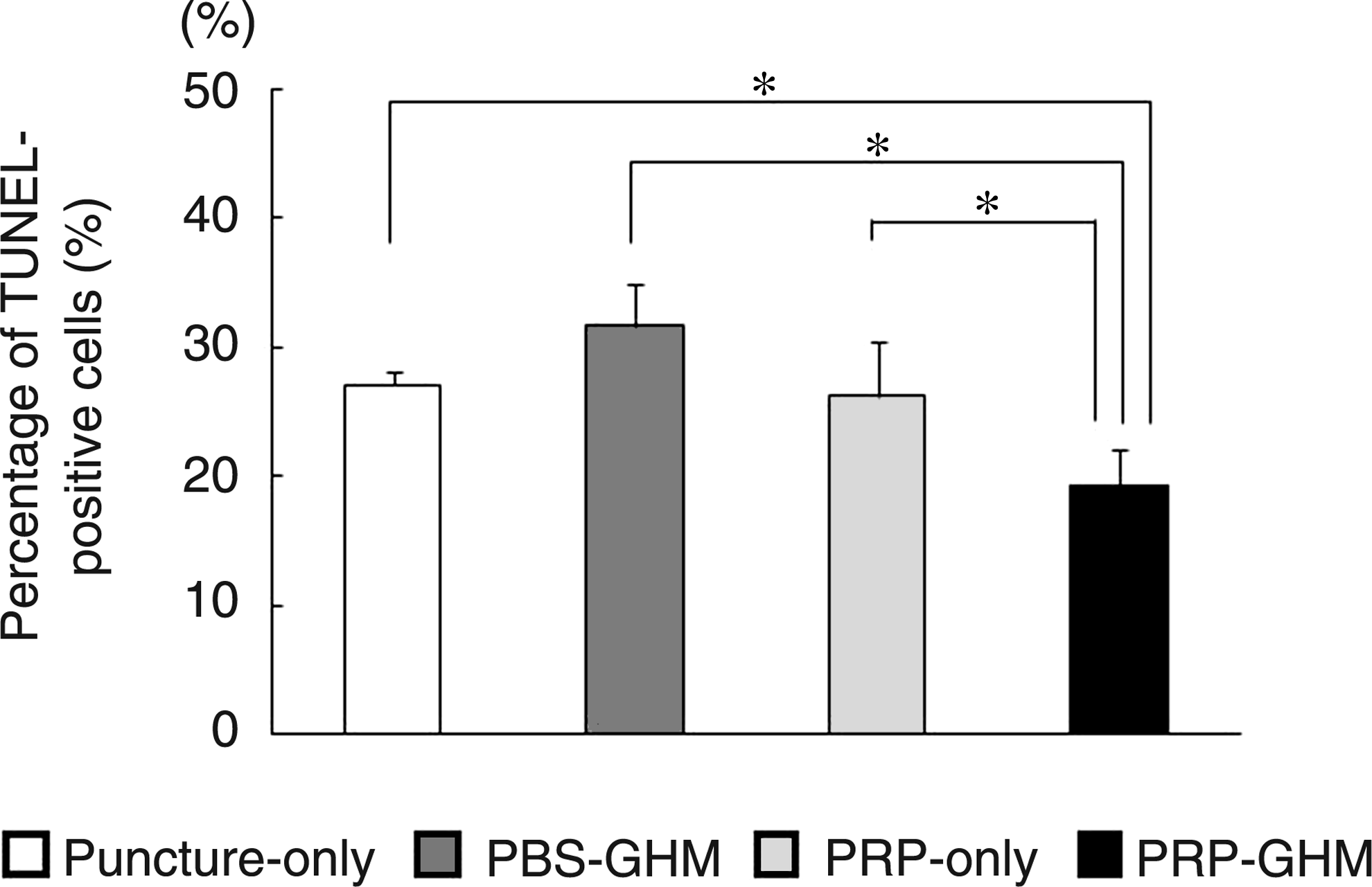
Percentage of TUNEL-positive cells among the total number of NP cells at 2 weeks after injection. Each value is expressed as the mean ± standard error. *p < 0.05 versus the PRP-GHM group.
Controlled release of TGF-β in the IVD of rabbits
Residual radiation was measured with a gamma counter for evaluation of the controlled release profile of TGF-β in IVDs. The mean residual radiation in the TGF-β GHM group was 29.7 ± 0.9%, 23.5 ± 2.2, 20.9 ± 2.3, 8.3 ± 2.2, and 1.2 ± 0.2 at 1, 3, 7, 14, and 28 days, which were significantly higher at 1, 3, 7, and 14 days than the TGF-β–only group (7.4 ± 0.8, 5.2 ± 1.3, 4.2 ± 1.0, 3.5 ± 0.6, and 1.1 ± 0.3 respectively) (Fig. 9).
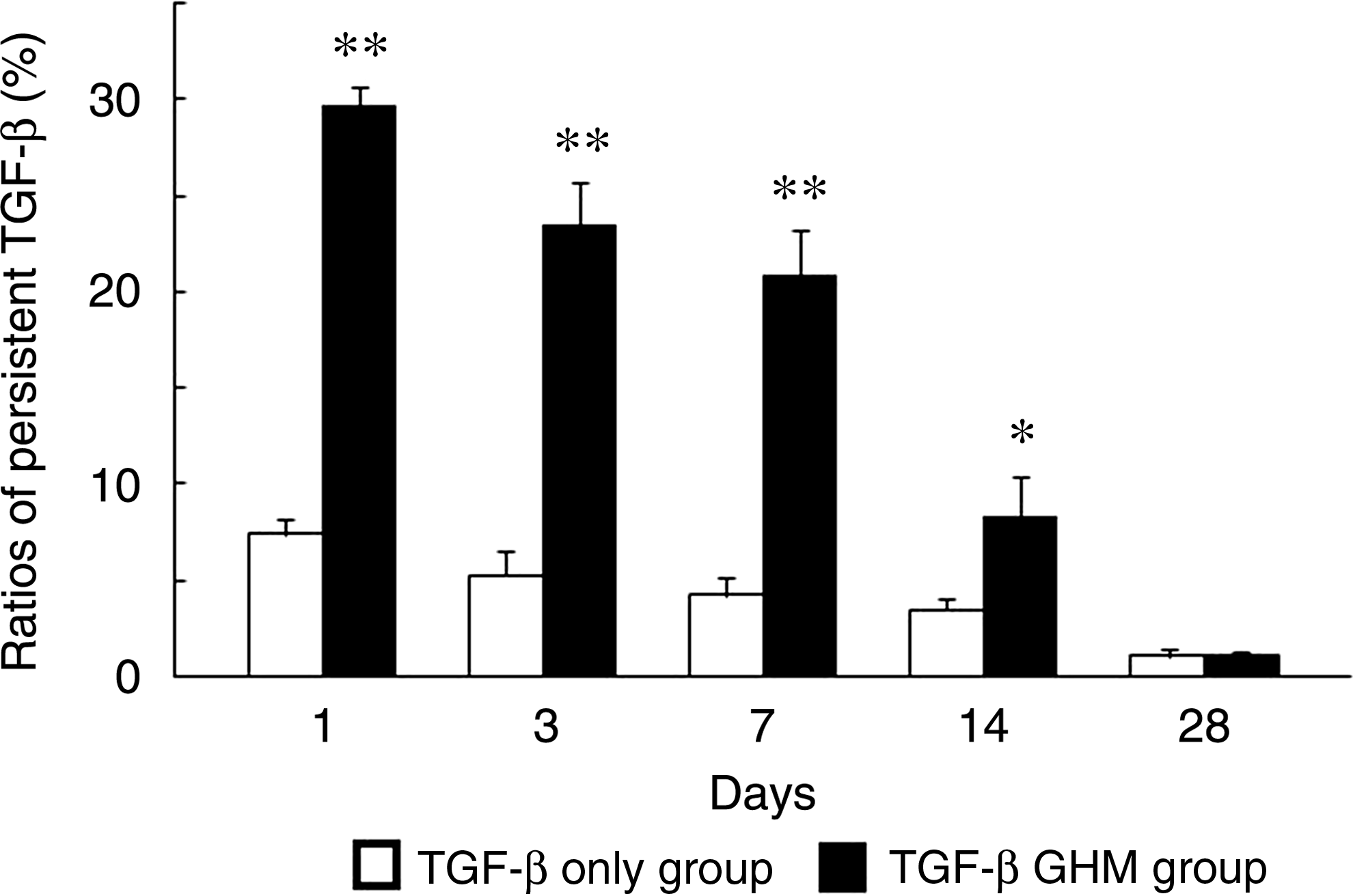
Ratio of remaining radioactivity in IVDs injected with labeled transforming growth factor (TGF-β) GHM and labeled TGF-β only. Each value is expressed as the mean ± standard error. **p < 0.01 versus the TGF-β–only group. *p < 0.05 versus the TGF-β–only group.
Discussion
Our previous histological evaluation showed that the structure and accumulation of PGs in the IVD were maintained 8 weeks after injection of PRP-GHMs. 20 In contrast, the number of NP cells was notably decreased, and proliferated connective tissues were observed at 4 weeks after control injections without PRP-GHM. To test whether PRP-GHMs affect the water content and gene expression of ECM components in NPs in vivo, we investigated the effects of PRP-GHMs using MR imaging and real-time PCR in the 8-week period after injection, and performed PCNA immunohistochemistry and TUNEL staining for detection of cell proliferation and antiapoptotic effects in NP cells through 8 weeks after the injection. Additionally, to confirm sustained release of TGF-β, a major constituent of PRP, an in vivo kinetic analysis of TGF-β release in IVDs was performed.
The results showed that the disc height and water content were preserved, and that mRNA expression levels of PG core protein and type II collagen were significantly higher in PRP-GHM–treated IVDs. There were no PCNA-positive cells in the NP and inner AF, but the percentage of apoptotic cells in the NP was significantly reduced by PRP-GHM treatment compared to controls. Sustained release of TGF-β in the IVDs with GHMs continued for more than 14 days.
PRP is used clinically in dentistry to enhance osteogenesis35,36 and has also been reported to promote wound healing.37–40 PRP is an autologous concentration of platelets in a small volume of plasma, and the α-granules in platelets contain growth factors such as TGF-β, IGF-1, and PDGF.15–19 TGF-β and IGF-1 have strong effects on ECM production and proliferation in IVD cells,8,9,41 and Gruber et al. showed that PDGF and IGF-1 inhibit apoptosis of AF cells. 42 Akeda et al. and Chen et al. have shown promotion of ECM synthesis by PRP in IVD cells in vitro.15,43 In the current study, a significant increase of the gene expression, due to the PRP-GHM injection, was found only at 2 weeks after injection. In other words, ECM synthesis in NP cells was promoted by PRP-GHM treatment for 2 weeks.
We prepared biodegradable GHMs from acidic gelatin for administration of PRP to degenerated IVDs. The characteristics of gelatin are biosafety, electrical nature, and biodegradability. The electrical nature can be changed by the processing method.44,45 Growth factors, such as TGF-β1 and PDGF, with isoelectric points higher than 8.5 are likely to be absorbed into our GHMs, which have an isoelectric point of 5.0, and we have previously shown that most TGF-β1 and PDGF-BB are immobilized in the GHMs using an enzyme-linked immunosorbent assay. 20 These growth factors are continuously released with degradation of the gelatin, and the release rate depends on the rate of gelatin degradation and can be controlled by the degree of crosslinking with glutaraldehyde. 46 Degradation of almost all our GHMs was shown at 8 weeks after injection in a previous histological analysis. PRP-impregnated gelatin hydrogel induced regeneration of bone and meniscal tissue, as previously described,47–49 which suggests that PRP growth factors released upon GHM degradation acted on NP cells and upregulated the synthesis of PG core protein and type II collagen for 2 weeks after injection. However, the gene expression levels did not differ significantly from those with control treatment at 4 and 8 weeks after injection.
In our previous immunohistochemical study of PG, intense immunostaining in the NP and inner AF was observed at 8 weeks after administration of PRP-GHMs, 20 and the disc height and signal intensity on MR images were maintained through 8 weeks after injection of PRP-GHMs relative to control treatment. The disc height decreases with IVD degeneration, 24 and abundant water in IVDs is maintained by interaction of water molecules with PG core protein-attached glycosaminoglycans.50,51 Roughley et al. showed that turnover of the ECM in IVDs is very slow, with turnover of PG requiring approximately 20 years. 52 The kinetic analysis confirmed that TGF-β remained in GHMs administered to the IVD for more than 14 days. Our findings indicate that PRP-GHM promotes ECM synthesis in NP cells for 2 weeks after administration, and that water content is maintained more effectively after PRP-GHM treatment compared to control injections.
The proliferative activity of human NP cells in vitro depends on the concentration of growth factors such as TGF-β1 and IGF-1.41,43 Therefore, PRP growth factors released from the GHM were expected to have a proliferative effect on IVD cells in vivo. However, PCNA immunohistochemistry did not indicate proliferation of NP cells after PRP-GHM treatment, which may be due to a concentration dependence of the proliferative effect of PRP growth factors on NP cells. Peng et al. also found no cells with positive staining for PCNA in IVDs from humans without lower back pain, 53 and NP and AF cells may have little or no proliferative activity in vivo. Proliferative activity of NP and inner AF cells was also low in the IVD degeneration model in our study, and it is unlikely that PRP-GHM administration could change this activity.
TUNEL staining suggested that the percentage of apoptotic cells in the NP was reduced at 2 weeks after administration of PRP-GHM compared to other groups. Gruber et al. and Wei et al. reported antiapoptotic effects using PDGF, IGF-1, and BMP-7 on IVD cells in vitro.42,54 This suggests that PDGF and IGF-1 in the PRP were able to inhibit apoptosis of the NP cells in vivo. This indicates that progression of IVD degeneration may be suppressed by inhibition of apoptosis of NP cells and subsequent induction of ECM synthesis by the continuous action of PRP growth factors on IVD cells in vivo.
Based on the Pfirrmann classification, the degenerated IVDs were of grades 1 to 2 (data not shown) at the time of PRP-GHM injection. This indicates that growth factor therapy using PRP-GHMs may be effective for early IVD degeneration in which the signal intensity and disc height are maintained on MR images. However, the effects of PRP-GHMs in the long term and the need for multiple PRP-GHM injections remain unclear. Several obstacles remain to be overcome for clinical use, but this study suggests that PRP-GHMs offer a new therapeutic modality for degenerated IVDs in which NP cells are present.
Footnotes
Acknowledgment
This work was supported by the Grant-in-Aid for Scientific Research (C 19591729) from Japan Society for the Promotion of Science.
Disclosure Statement
No competing financial interests exist.