




Abstract
In bone tissue engineering, bioglass coating of titanium (Ti) scaffolds has drawn attention as a method to improve osteointegration and implant fixation. In this in vitro study, bioactive glass layers with an approximate thickness of 1 μm were deposited at 200°C onto a three-dimensional Ti-6Al-4V scaffold using a radio frequency (r.f.) magnetron sputtering system. After incubation with SAOS-2 human osteoblasts, in comparison with the uncoated scaffolds, the bioglass-coated scaffolds showed a twofold increase in cell proliferation (p < 0.05) up to 68.4 × 10⁶, and enhanced the deposition of extracellular matrix components such as decorin, fibronectin, osteocalcin, osteonectin, osteopontin, and type-I and -III collagens (p < 0.05). Calcium deposition was twofold greater on the bioglass-coated scaffolds (p < 0.05). The immunofluorescence related to the preceding bone matrix proteins and calcium showed their colocalization to the cell-rich areas. Alkaline phosphatase activity increased twofold (p < 0.001) and its protein content was threefold higher with respect to the uncoated sample. Quantitative reverse transcriptase–polymerase chain reaction analysis revealed upregulated transcription specific for type-I collagen and osteopontin (p < 0.001). All together, these results demonstrate that the bioglass coating of the three-dimensional Ti scaffolds by the r.f. magnetron sputtering technique determines an in vitro increase of the bone matrix elaboration and may potentially have a clinical benefit.
Introduction
Ti and its alloys are generally well integrated into bone, and many studies have highlighted the potential of Ti to form a direct interface with bone cells and tissues.2–4 However, particularly in bony tissue, loosening has recently been indicated as one of the major causes for implant failure because of the lack of tissue adherence with the biomaterial surface.5–8 The host response to Ti-6Al-4V is not always favorable because a fibrous layer may form at the skeletal tissue–device interface, causing aseptic loosening.9–15 Depending on the type of implant, the loosening rate is reported to be in the range of 4–6%. Successful fixation of the biomedical implants can be achieved by encouraging intimate bone apposition (osseointegration) to the implant surface. To obtain primary stabilization of the implant and subsequent osteointegration, macroporous and rough implant surfaces have been developed.16,17 This morphological fixation is a mechanical interlock, which requires a long immobilization period. 17 The early osteointegration of a macroporous or rough metallic surface depends mainly on the ingrowth of the bone tissue onto the material surface. To improve and accelerate the osteointegration during the patient's clinical immobilization period, bioactive coatings (biocoatings) and micro- or nanostructured modifications of the metallic surfaces have been developed.18,19 The most studied biocoatings are based on the apatites and hydroxyapatite (HA)-based materials, because of their chemical similarity with the natural components of bone tissue. 20 However, among the bioactive materials, silica-based bioglasses seem to fit better with the bone cavity and show stronger bone-bonding properties. Bioactive glasses are known to enhance proliferation and downward dedifferentiation of osteoblasts in vitro.21–23 Bioglasses and glass ceramics are composed of multicomponent oxide systems, for example, (Na2O)-CaO-SiO2-P2O5, which easily react with the body fluids to form a calcium phosphate layer.24,25 This layer, which progressively crystallizes into HA, is structurally very similar to the apatite present in the bone and forms a more stable interface with the bone tissue with respect to the synthetic HA.20,26 An optimal biocoating should have three main perquisites: (i) high adhesion to the metal substrate; (ii) adequate thickness and uniformity; and (iii) dense structure, at least at the implant interface, to prevent the corrosive effects from reactions between the body fluids and the metal. In previous years, to address these aims, both chemical and physical deposition techniques have been investigated. 27 In the first case, bioglass-based films have been deposited via the sol–gel techniques, like dip coating, with the aim of reducing processing temperatures, modulating the layer porosity and to design implants with drug delivery properties, in addition to improving their bioactivity and biocompatibility.28,29 The physical preparation of biocoatings has been chiefly based on the plasma spray approaches.18,20 However, problems related to bad adhesiveness and nonuniform thickness have been reported. 30 More recently, other methods like radio frequency magnetron sputtering, 30 pulsed laser, 31 and electrophoretic deposition32,33 have been tested with more success in the preparation of biocoatings for endoprosthesis. Among these, r.f. magnetron is of particular interest because of its capability to deposit, even at low temperatures, films of controlled stoichiometry with homogeneity at the molecular level and high adhesiveness because of the kinetic energy of the impinging atomic, molecular, and clustering species which ranges in the interval of 100–1000 eV, depending on the radiofrequency power used. 34 This can enhance the resistance of coatings against attack by biological fluids, thus favoring the formation of the HA layer that is mandatory for the osteointegration process. Moreover, r.f. magnetron sputtering is a relatively inexpensive and simple technique, which can be used at the industrial level for large-scale production. Finally, because of the nature of plasma deposition, this technique is well suited for the coating of the three-dimensional (3D) structures characterized by complex morphology and microstructure, for example, cavities and macropores. The main advantage with respect to the wet techniques like dip coating is the absence of solvents which renders the final product intrinsically safer.
To demonstrate the potential for osseointegration using an in vitro model, our study evaluated the response of human osteoblast-like cells to a bioglass-coated Ti mesh and determined whether the bioglass coating of 3D Ti could improve the healing and bone formation processes by enhancing cell proliferation and activating the bone matrix deposition. The main goal of this study was to demonstrate that the deposition, performed at low cost and with a simple technique, of a controlled and homogeneous film of bioglass coating onto a 3D Ti scaffold enhances osteointegration and hence potentially accelerates the healing and bone formation processes.
Materials and Methods
3D Ti-alloy scaffolds
Porous 3D Ti scaffolds (diameter 12 mm; height 4 mm) were obtained by sintering a powder (average diameter 20 μm) of Ti6Al4V Ti alloy (ISO 5832-3 standard practice) at 1100°C for 2.5 h (Ufficio Italiano Brevetti e Marchi; Patent Number: UD2007000092 [Italian Patent pending]) (Lima-Lto S.p.A., Lima Group, Villanova di San Daniele del Friuli, Italy).
The result of the sintering process was a multilayered “warp and weft” 3D Ti net with a density of 2.7 g/cm3. This 3D warp and weft net had completely interconnected square pores, the sides of which were ∼800 μm in a scanning electron microscopy (SEM) orthogonal view (Fig. 1A, B).
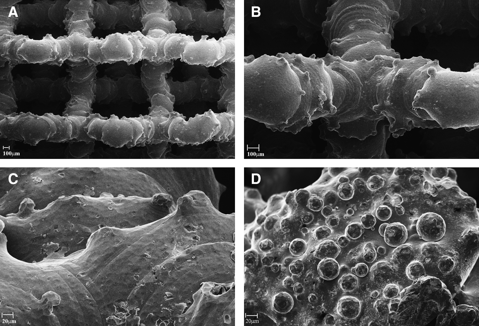
Scanning electron microscopy images of an unseeded 3D Ti scaffold at 20 × (
Synthesis of bioactive glasses
The bioglass SiO2 (58 wt%)–CaO (33 wt%)–P2O5 (9 wt%) (58S), selected as a powder target for the deposition of biocoatings, was synthesized by means of a sol–gel route. 35 Briefly, the synthetic process consisted of the initial hydrolysis of the silicon precursor tetraethoxysilane, followed by the addition of proper amounts of triethylphosphate and subsequently Ca(NO3)2:4H2O. The sol was stored until gel formation, and the resulting gel was dried at 700°C to remove any organic contamination.
Deposition of biocoatings by means of radio frequency magnetron sputtering
Biolayers with a thickness of ∼1 μm were deposited at 200°C in a high-vacuum (10−3 Pa) r.f. magnetron sputtering system, which consists of three confocal cathode guns placed 40–50 mm from the substrate centre. The cathode alignment is offset at 30° offset from the substrate normal with an off-axis geometry, to minimize the remission effect of the atom flow. Ti-based 3D scaffolds were carefully degreased and washed with ethanol and water before use. Pure argon (>99%) was chosen as the process gas at a pressure of 2.2 Pa. The sputtering power was set at 130 W. The deposition time was about 4 h.
Cells
The human osteosarcoma cell line SAOS-2 was obtained from the American Type Culture Collection (Rockville, MD) (HTB85; ATCC). The cells were cultured in McCoy's 5A modified medium with 1.5 mM
Cell seeding and culture
The 3D Ti scaffolds were sterilized with ethylene oxide at 38°C for 8 h at 65% relative humidity. After 24 h of aeration to remove residual ethylene oxide, the scaffolds were placed inside a standard 24-well plate and were washed first with sterile distilled water, then with 0.9% NaCl sterile solution, and finally with culture medium. To ensure a maximum number of attached cells for scaffolds, a cell suspension of 1 × 10⁶ cells was added in two steps onto the top of each scaffold and, after 0.5 h, 1 mL of culture medium was added to cover the scaffolds. The culture medium was changed every 3 days.
SEM analysis
Before and after the sterilization procedure, the scaffolds were observed by SEM and showed similar surface morphology. At the end of the culture incubation, the scaffolds were fixed with 2.5% (v/v) glutaraldehyde solution in 0.1 M Na-cacodylate buffer (pH 7.2) for 1 h at 4°C, washed with Na-cacodylate buffer, and then dehydrated at room temperature in a gradient ethanol series up to 100%. The samples were kept in 100% ethanol for 15 min, and then critical point dried with CO2. The specimens were sputter coated with gold and observed at 20 × , 50 × , and 100 × magnification with a Leica Cambridge Stereoscan 440 microscope (Leica Microsystems, Bensheim, Germany) at 8 kV. 36
3-(4,5-dimethylthiazole-2-yl)-2,5-diphenyl tetrazolium bromide test
To evaluate the mitochondrial activity of the seeded cells, that is, the cell viability on the 3D Ti scaffolds during the culture period, a test with 3-(4,5-dimethylthiazole-2-yl)-2,5-diphenyl tetrazolium bromide (MTT) (Sigma-Aldrich), was performed on days 1, 3, 14, and 27 (end of the culture period). The culture medium was replaced by a 0.5 mg/mL solution of MTT in phosphate-buffered saline (PBS) (137 mM NaCl, 2.7 mM KCl, 4.3 mM Na2HPO4, 1.4 mM KH2PO4, pH 7.4), and the cell cultures were incubated for 4 h. Viable cells are able to reduce MTT into formazan crystals. After removing the MTT solution, to solubilize the formazan products, 500 μL of dimethyl sulphoxide (Sigma-Aldrich) were added, and the well plate containing the cultured 3D scaffolds was agitated for 20 min on a shaker. Aliquots of 200 μL were sampled, and the related absorbance values were measured at 570 nm by a microplate reader (BioRad Laboratories, Hercules, CA). A standard curve of cell viability was used to express the results as percentage.
DNA content
On days 1, 3, and 27 of incubation, the cultured scaffolds were extensively washed with PBS, and the cells were lyzed by a freeze–thaw method in sterile deionized distilled water. The released DNA content was evaluated with a fluorometric DNA quantification kit (PicoGreen; Molecular Probes, Eugene, OR). A DNA standard curve, 36 obtained from a known amount of osteoblasts, was used to express the results as cell number attached per scaffold.
Set of rabbit polyclonal antisera
Dr. Larry W. Fisher (http://csdb.nidcr.nih.gov/csdb/antisera.htm; National Institutes of Health, Bethesda, MD) provided us with the rabbit polyclonal anti type-I and -III collagens, anti-decorin, anti-osteopontin, anti-osteocalcin, anti-osteonectin, and anti-alkaline phosphatase (ALP).
For polyclonal antibody production against human fibronectin (HFN), New Zealand rabbits were injected intraperitoneally five times at 12-day intervals with 100 μg of the purified HFN. 37 The antigen was emulsified with an equal volume of complete Freund's adjuvant for the first immunization followed by four injections with incomplete adjuvant. The rabbit was bled, and the sera were tested for reactivity to the purified HFN using an enzyme-linked immunosorbent assay (ELISA). The specific anti-Fn immunoglobulin (Ig)Gs were purified by affinity chromatography on protein G-Sepharose columns according to the manufacturer's recommendations (Amersham Biosciences, Piscataway, NJ). Antibody titers were assayed by ELISA.
Set of purified proteins
Decorin, type-I collagen, and FN were purified as described previously38–40 ; osteocalcin was acquired from Biomedical Technologies (Stoughton, MA); osteopontin and osteonectin were obtained from Assay Designs (Ann Arbor, MI); type-III collagen and ALP were purchased from Sigma-Aldrich.
Extraction of the extracellular matrix proteins from the cultured scaffolds and ELISA
On days 3 and 27, to evaluate the amount of the extracellular matrix (ECM) constituents throughout the scaffolds surface, the uncoated and bioglass-coated scaffolds were washed extensively with sterile PBS to remove the culture medium, and then incubated for 24 h at 37°C with 1 mL of sterile sample buffer (20 mM Tris–HCl, 4 M GuHCl, 10 mM ethylenediaminetetraacetic acid, 0.066% [w/v] sodium dodecyl sulfate, pH 8.0). The sample buffer aliquots were removed, and then both types of 3D Ti scaffolds (after 3 or 27 days of culture incubation) were centrifuged at 4000 rpm for 15 min to collect the sample buffer entrapped in the pores. The total protein concentration in both culture systems, uncoated and bioglass coated, was evaluated by the BCA Protein Assay Kit (Pierce Biotechnology, Rockford, IL). The total protein concentration was detected after 3 and 27 days of culture: on the uncoated 3D Ti scaffolds, it was reported to be 123 ± 0.5 and 1006 ± 13.5 μg/mL on days 3 and 27, respectively; on the bioglass-coated 3D Ti scaffolds, the protein content was 134 ± 1.1 and 1531 ± 26.1 μg/mL on days 3 and 27, respectively. After matrix extraction, the scaffolds were incubated, once again, for 24 h at 37°C with 1 mL of sterile sample buffer, and no protein content was further detected.
Calibration curves to measure type-I and -III collagens, decorin, osteopontin, osteocalcin, osteonectin, FN, and ALP were performed. Microtiter wells were coated with increasing concentrations of each purified protein, from 10 ng to 2 μg, in coating buffer (50 mM Na2CO3, pH 9.5) overnight at 4°C. Control wells were coated with bovine serum albumin (BSA) as a negative control. To measure the ECM amount of each protein by ELISA, microtiter wells were coated, overnight at 4°C, with 100 μL of the previously extracted ECM (20 μg/mL in coating buffer). After three washes with PBS containing 0.1% (v/v) Tween 20, the wells were blocked by incubating with 200 μL of PBS containing 2% (w/v) BSA for 2 h at 22°C. The wells were subsequently incubated for 1.5 h at 22°C with 100 μL of the anti-type-I and -III collagens, anti-decorin, anti-osteopontin, anti-osteocalcin, anti-osteonectin, and anti-ALP rabbit polyclonal antisera (1:500 dilution in 1% BSA), kindly provided by L. Fisher. The same dilution was used for the anti-FN rabbit polyclonal IgG. After washing, the wells were incubated for 1 h at 22°C with 100 μL of horseradish peroxidase (HRP)-conjugated goat anti-rabbit IgG (1:1000 dilution in 1% BSA). The wells were finally incubated with 100 μL of the development solution (phosphate-citrate buffer with o-phenylenediamine dihydrochloride substrate). The color reaction was stopped with 100 μL of 0.5 M H2SO4, and the absorbance values were measured at 490 nm with a microplate reader (BioRad Laboratories). An underestimation of the absolute protein deposition is possible because the sample buffer, used for matrix extraction, contained sodium dodecyl sulfate, which may interfere with the protein adsorption during ELISA. The amount of ECM constituents throughout the 3D Ti scaffolds was expressed as fg/(cell × scaffolds).
Indirect immunofluorescence staining
At the end of the culture period, the scaffolds were fixed with 4% (w/v) paraformaldehyde solution in 0.1 M phosphate buffer (pH 7.4) for 8 h at room temperature and washed with PBS three times for 15 min. The scaffolds were then blocked by incubating with PAT (PBS containing 1% [w/v] BSA and 0.02% [v/v] Tween 20) for 2 h at room temperature and washed. Anti-type-I and -III collagens, anti-decorin, anti-osteopontin, anti-osteocalcin, anti-osteonectin, and anti-ALP rabbit polyclonal antisera were used as the primary antibody with a dilution equal to 1:500 in PAT. The same dilution was performed with anti-FN rabbit polyclonal IgG. The incubation with the primary antibodies was made overnight at 4°C, whereas the negative controls were incubated overnight at 4°C with PAT instead of the primary antibodies. The scaffolds and the negative controls were washed and incubated with Alexa Fluor 488 goat anti-rabbit IgG (H + L) (Molecular Probes) at a dilution of 1:750 in PAT for 1 h at room temperature. At the end of the incubation, the scaffolds were washed in PBS, counterstained with a solution of propidium iodide (2 μg/mL) to target the cellular nuclei, and then washed. The images were taken by blue excitation (bandpass, 450–480 nm; dichromatic mirror, DM500; barrier filter, BA515) with a fluorescence microscope (BX51; Olympus, Tokyo, Japan) equipped with a digital image capture system (Olympus) at 20 × magnification. The fluorescence background of the negative controls was almost negligible.
Quantification of calcium
To evaluate the calcium deposition over the uncoated and bioglass-coated scaffolds, calcein detection and calcium–cresolphthalein complexone methods were performed as described previously.41,42
Calcein
At the end of cell incubation, scaffolds were rinsed with sterile PBS and stained with a calcein solution (5 μM in PBS; Invitrogen, Carlsbad, CA) for 30 min at 22°C. The scaffolds were counterstained with a solution of propidium iodide (Sigma-Aldrich; 2 μg/mL) to target the cellular nuclei, and then washed with PBS. The images were taken by blue excitation (bandpass, 450–480 nm; dichromatic mirror, DM500; barrier filter, BA515) with a fluorescence microscope at 40 × magnification.
Calcium–cresolphthalein complexone method
The calcium content of each scaffold was assayed to quantify the amount of mineralized matrix present and was measured using a Calcium Fast kit (Mercury SPA, Naples, Italy) according to the manufacturer's instructions. The colorimetric end point assay measures the amount of purple-colored calcium–cresolphthalein complexone complex formed when cresolphthalein complexone binds to free calcium in an alkaline solution. 42 Briefly, an aliquot (1 mL) of 1 N HCl was added to each scaffold and incubated for 24 h at room temperature to release calcium into solution. The sample supernatant was diluted 1/10 with the Assay Working Solution previously prepared by mixing equal parts of calcium-binding reagent and calcium buffer reagent provided by the Kit. Ca2+ standards in concentrations ranging from 0 to 10 μg/mL were prepared from dilutions of a 100 μg/mL stock solution of Ca2+. The absorbance reading was performed at 595 nm with a microplate reader (BioRad Laboratories) using 100 μL of standard or sample placed into individual wells of a 96-well plate. Samples were run in triplicate and compared against the standard solution calibration curve.
ALP activity
ALP activity was determined using a colorimetric end point assay as previously described. 43 The assay measures the conversion of the colorless substrate p-nitrophenol phosphate (PNPP) by the enzyme ALP to the yellow product p-nitrophenol, where the rate of the color change corresponds to the amount of enzyme present in the solution. Briefly, an aliquot (1 mL) of 0.3 M PNPP (dissolved in glycine buffer, pH 10.5) was added to each scaffold at 37°C. After incubation, the reaction was stopped by the addition of 100 μL 5 M NaOH. Standards of PNPP in concentrations ranging from 0 to 50 μM were freshly prepared from dilutions of a 500 μM stock solution and incubated for 10 min with 7 U of ALP (Sigma-Aldrich) previously dissolved in 500 μL of ddH2O. The absorbance reading was performed at 405 nm with a microplate reader (BioRad Laboratories) using 100 μL of standard or sample placed into individual wells of a 96-well plate. Samples were run in triplicate and compared against a calibration curve of p-nitrophenol standards. The enzyme activity was expressed as micromoles of p-nitrophenol produced per minute per milligram of enzyme.
Assay for gene expression
Cells were recovered on days 3 and 27, and the total RNA was extracted from the cultured scaffolds using the Trizol® reagent system according to the manufacturer's protocol (Invitrogen). Reverse transcriptase–polymerase chain reaction (RT-PCR) was performed to evaluate the gene expressions for decorin, FN, osteocalcin, osteopontin, type-I collagen, type-III collagen, human transforming growth factor bone sialoprotein, bone morphogenetic protein 2, and the housekeeping gene expression for glyceraldehyde-3-phosphate dehydrogenase (GAPDH). The RT reaction was performed with 300 ng of the total RNA using the Biorad® kit. The primers (Primm s.r.l., Milan, Italy) were designed according to the published gene sequences, and the PCRs were performed with the GeneAmp PCR System 9700 (Applied Biosystems, Foster City, CA). The primers used are indicated in Table 1. 36
BMP-2, bone morphogenetic protein 2; BOSP, bone sialoprotein; COLI, type-I collagen; COLIII, type-III collagen; DEC, decorin; FN, fibronectin; GAPDH, glyceraldehyde-3-phosphate dehydrogenase; TGF, transforming growth factor-beta; OC, osteocalcin; OP, osteopontin.
The PCRs were performed with 2 μL of the cDNA mixture using the Platinum Taq DNA Polymerase (Invitrogen): 30 cycles were run at 94°C for 45 s (denaturation), 58°C for 45 s (annealing), and 72°C for 1 min (extension). The gene expression of all proteins was normalized to the housekeeping gene expression of GAPDH. To obtain a specificity control, the same PCRs were performed with 2 μL of the total RNA mixture without the RT reaction. The resulting products were fractionized by electrophoresis through an ethidium bromide–stained 1% agarose gel in 1 × Tris–borate ethylenediaminetetraacetic acid buffer. Images were taken with a CANON PowerShot G6 digital camera.
Real-time PCR
Total RNA from the chemically treated samples (treat) and the nontreated samples (control) was extracted with the Trizol reagent (Invitrogen) and retrotranscribed into cDNA with the iScript™ cDNA Synthesis Kit (BioRad Laboratories). Quantitative (q) RT-PCR analysis was performed in a 48-well optical reaction plate using an MiniOpticon Real-Time PCR System (BioRad Laboratories). Oligonucleotide primers were designed with gene sequences published in GeneBank and are indicated in Table 2. Reactions were performed in 20 μL with 2 μL of cDNA, 10 μL Brilliant® SYBER® Green qPCR Master Mix (Stratagene, La Jolla, CA), 0.4 μL of each primer, and 7.2 μL H2O. PCR conditions were as follows: 3 min at 95°C, 30 cycles of 5 s at 95°, and 23 s at 60°C. Gene expression was normalized to the GAPDH gene expression. Each sample was analyzed in triplicate and correlated against a standard curve. The reaction mixture, without the cDNA, was used as a negative control in each run.
Statistics
The 3D Ti scaffold number was 40 for each repeated experiment (20 uncoated scaffolds and 20 bioglass-coated scaffolds). Each experiment was repeated three times. Results are expressed as mean ± standard deviation. To compare the results between the two types of scaffold, one-way analysis of variance with the post hoc Bonferroni test was applied, electing a significance level of 0.05.
Results
Characterization of the 3D Ti scaffolds
The structure of the unseeded scaffolds, uncoated or bioglass coated, were observed by SEM. The uncoated Ti scaffold appeared to be composed of a 3D mesh showing regular squares and interconnected holes (Fig. 1A, B). At higher magnification, because of the sintering process, the scaffold surface appeared nonhomogeneous and somewhat rough (Fig. 1C). The appearance of the bioglass-coated 3D Ti scaffold surface was very similar to that of the surface of the uncoated scaffold. At higher magnification, a distribution of small droplets of bioglass above the scaffold surface was observed (Fig. 1D). The droplets have diameters in the range 5–40 μm, and their thickness is in the order of 1 μm. The droplet distribution was relatively uniform, despite the high bending of the scaffold surface. The actual composition of the bioglass coating was determined by SEM energy dispersive X-ray (EDX) analysing system on a layer deposited on a flat Ti6Al4V surface, and resulted equal to the nominal one with an uncertainty of less than 5%.
Cell morphology and viability
SAOS-2 cell morphology cultured on the uncoated and the bioglass-coated 3D Ti scaffolds was viewed by SEM (Fig. 2). Figure 2 is a representative image of 27 days of cell culture showing adherence of cells to the surface on both types of scaffolds. In particular, the cells more homogeneously covered the surface and spanned to the neighboring fibers on the bioglass-coated scaffold than they did on the uncoated scaffold (Fig. 2A, C). At higher magnification, some differences were observed: on the uncoated scaffold (Fig. 2B) cells showed a rounded shape, whereas on the bioglass-coated scaffolds individual cells were no longer discernable within a dense layer of ECM (Fig. 2D).

Representative scanning electron microscopic images of the cells cultured on the uncoated (
To evaluate the cell viability on the uncoated and bioglass-coated 3D Ti scaffolds during the culture period, a MTT test was performed. On days 1, 3, 14, and at the end of the culture period, the average cell viability was in the 88–93% range with no statistically significant difference in the cell viability (p > 0.05) between both types of scaffold at each culture period.
Cell attachment
To assess whether, in comparison with the uncoated scaffolds, the bioactive coating of the 3D Ti scaffolds could influence the initial cell attachment and thus the ECM deposition, the number of osteoblasts attached to both types of scaffold was detected earlier on days 1 and 3 and later on day 27. The longer incubation time was chosen to allow the in vitro cell production of detectable bone proteins. The percentage of cell attachment was about 25% ± 2.5% (on day 1) and 31% ± 2.2% (on day 3) for both uncoated and bioglass-coated 3D Ti scaffolds, showing no significant difference (p > 0.05). After 27 days of cell culture, a significantly consistent increase in the measurement of DNA content was detected. On the uncoated 3D Ti, the cell number per scaffold rose to 35.2 × 10⁶ ± 12.2 × 10⁴, whereas on the bioglass-coated 3D Ti scaffolds it reached 68.4 × 10⁶ ± 10.4 × 10⁴ (p < 0.05). An underestimation of the culture cellularity is possible because of the trapping of DNA within the thick ECM.
Characterization of the calcified ECM deposition
At the end of cell culture, the immunolocalization of type-I collagen, decorin, osteopontin, and osteonectin showed a more intense green fluorescence on the bioglass-coated cultured scaffolds (Fig. 3B, D, F, H), revealing the stimulatory effects of the biocoating in terms of higher cell proliferation and more intense fluorescent staining of the ECM: cells adhering to the uncoated 3D Ti scaffolds were few and surrounded by a thin and discontinuous ECM (Fig. 3A, C, E, G). The immunolocalization of other proteins such as type-III collagen, osteonectin, or FN showed similar patterns (data not shown).

Immunolocalization of type-I collagen (
To evaluate the amount of the ECM constituents produced throughout both types of 3D Ti scaffolds, an ECM extraction was performed on days 3 and 27 of incubation. Unfortunately, on day 3 even if the total protein content was determined, the levels of the specific bone proteins were too low to be detected in both types of Ti scaffolds. At the end of the culture period, in comparison with the culture on the uncoated 3D Ti scaffolds, the deposition of bone proteins throughout the bioglass-coated 3D Ti scaffolds was considerably enhanced (p < 0.05) (Table 3). These data are in accordance with the immunofluorescence analysis performed on the uncoated and bioglass-coated cultured scaffolds (Fig. 3).
In comparison with the uncoated sample, the coating with bioactive glass promoted a better deposition of bone extracellular proteins throughout the entire scaffold.
3D, three-dimensional; Ti, titanium.
The enhancement of protein deposition was particularly marked for ALP, which was threefold greater when compared with the uncoated sample (Table 3). Figure 4 shows the ALP activity measured on both types of scaffold at the end of the culture period: the level of the ALP activity was consistently higher on the bioglass-coated 3D Ti than on the uncoated scaffolds (p < 0.001).

The ALP activity of SAOS-2 cells seeded onto the uncoated and the bioglass-coated 3D Ti scaffolds. The ALP activity was determined colorimetrically, corrected for the protein content measured according to the BCA Protein Assay Kit, and expressed as nanomoles of p-nitrophenol produced per minute per milligram of protein. Bars express mean values ± standard error of the mean of results from three experiments (*p < 0.05). ALP, alkaline phosphatase.
The relative amount of calcium contained in both types of scaffold was evaluated by the calcein method (Fig. 5A, B) and quantified by the calcium–cresolphthalein complexone method (Fig. 5C) to evaluate the matrix calcification. Figure 5 shows a qualitative assessment of cell and calcium deposition onto the scaffolds: the mineralization of the ECM produced by SAOS-2 cells was shown by an intense green fluorescence colocalized within the cells on the bioglass-coated scaffolds over the uncoated sample. The qualitative evaluation of the calcium deposition was confirmed by the quantification method: calcification of the deposited ECM was considerably greater on the bioglass-coated 3D Ti scaffolds than on the uncoated scaffolds (p < 0.05) (Fig. 5C). It is possible that the larger amount of Ca2+ found in the bioglass-coated scaffold may be because of leaching of the calcium present in the coating itself. However, an order-of-magnitude estimation rules out this hypothesis. In fact, an estimated layer thickness of ∼1 μm gives, at best, a coating weight of the order of 1 mg, which accounts for ∼200 μg of Ca2+ in the layer. Because we can expect leaching of 0.2% Ca2+ under physiological conditions, 44 the overall expressed calcium should be in the order of 0.4 μg per sample, whereas the difference reported in Figure 5C gives an estimate of ∼140 μg for a cell population of ∼7 × 10⁶ (see above).

Representative fluorescence images at 40 × magnification (the scale bar shown represents 50 μm) of calcium deposits from SAOS-2 cells cultured onto uncoated (Panel
Characterization of gene expression in bone
To characterize the gene expression in bone, RT-PCR and qRT-PCR analyses were performed after 3 and 27 days of culture. On day 3, the qualitative RT-PCR and qRT-PCR did not show a significant difference in the gene expression levels for ECM proteins on the bioglass-coated scaffold when compared with the uncoated scaffold (data not shown). The main difference in the gene expression levels between both types of scaffold was observed after 27 days of culture.
At the end of the cell culture period, the qualitative RT-PCR performed on the cells grown on the bioglass-coated 3D Ti scaffolds revealed a bright band on the agarose gel for all the indicated genes and particularly for the transcripts specific for bone morphogenetic protein 2, type-I and -III collagens, and osteopontin (Fig. 6). To further examine these data, a qRT-PCR for the gene expression profiles of bone-specific proteins was performed after 27 days. The results showed a significantly enhanced fold difference for osteopontin (p < 0.001) (Fig. 7). No evident fold difference was detected for the gene expression profiles of the other indicated proteins (p > 0.05).
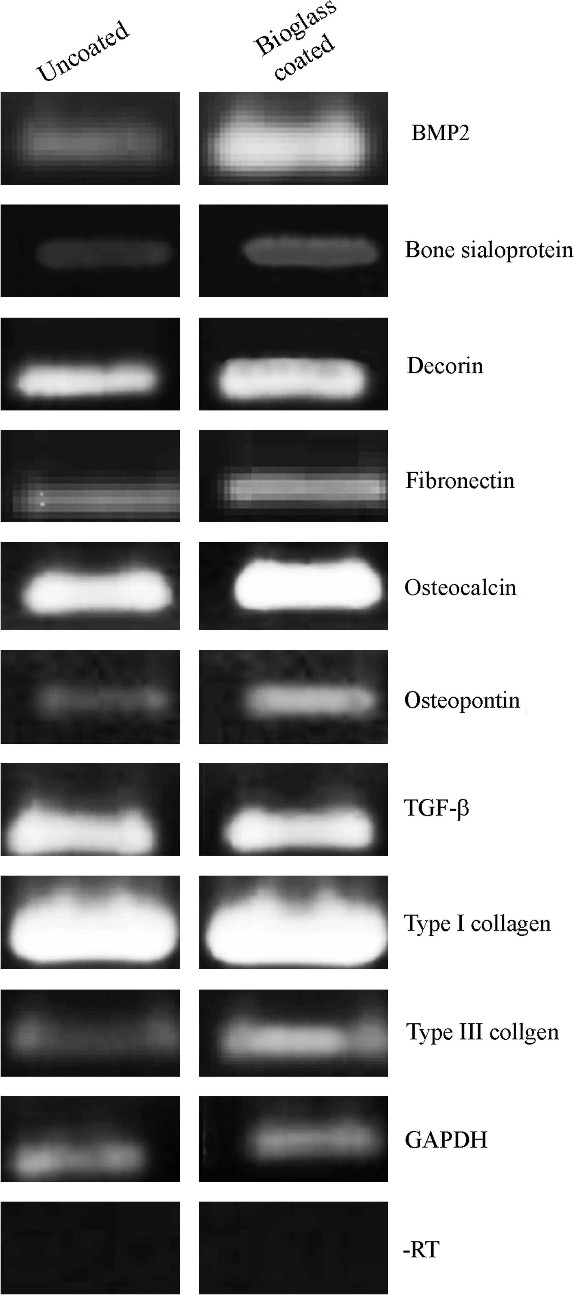
Assay for gene transcription of the uncoated and the bioglass-coated 3D Ti scaffold. Reverse transcriptase–polymerase chain reaction of the indicated products were subjected to electrophoresis on 2% agarose gel and viewed by UV exposure. The level of specific bands was normalized for GAPDH cDNA. Similar results have been obtained by normalization with respect to the β-actin cDNA content (data not shown). GAPDH, glyceraldehyde-3-phosphate dehydrogenase.

Gene expression of the indicated bone-specific markers as determined by quantitative reverse transcriptase–polymerase chain reaction. The data represent fold difference in expression of three samples normalized to the uncoated 3D Ti scaffold (*p < 0.05, #p > 0.05).
Discussion
The predominant tissue found at the implant interface is affected by the implant stability, material biocompatibility, implant design, and implant placement at the surgical site. Biomaterials currently available for clinical use are known for their good biocompatibility, and most of these have the mechanical properties required for a defined implantation site. However, biomaterial properties such as good tissue integration and regeneration could be improved. In this study, we evaluated the deposition by r.f. magnetron sputtering of a thin film of bioactive glass onto a 3D Ti scaffold, by means of analysis of osteoblast morphology, proliferation, and deposition of a calcified ECM. In particular, the Ti6Al4V scaffold used in this study is especially appropriate for implants under high stress, such as hip implants. 45
To test the bioactivity of bioglass-coated 3D Ti scaffolds, the SAOS-2 cell line was selected as it exhibits several fundamental osteoblast characteristics 46 and represents a widely used model for in vitro osteoblast study. In contrast to the other cell lines, these osteoblasts can be grown indefinitely, and they exhibit unique osteoinductive activity.47–49 At the end of incubation, SEM examination of the 3D Ti scaffolds incubated with SAOS-2 cells showed a layer of cells and matrix throughout the scaffold. However, the appearance of the cell layer differed between the uncoated and the glass-coated surfaces of the scaffolds: the latter showed a more uniform distribution and much higher infiltration of cells and matrix throughout the entire scaffold. The round shape of the cells seen on the uncoated scaffold was probably not affected by ion release: in fact the metal ions liberated in the physiologic solutions was less than 1 ppm. Confirming the SEM observation, the cell proliferation onto 3D Ti scaffolds coated with bioactive glass was almost twofold higher in comparison to the uncoated scaffold.
A temporal and functional pattern of gene expression characterizes the osteoblast maturation process, which can be divided into proliferation, differentiation, and mineralization stages. 50 Initially, actively proliferating cells, expressing cell-cycle- and cell-growth-regulated genes, produce a FN/type I collagen ECM. Later, the decline in the proliferative activity and the subsequent induction of genes associated with matrix maturation and mineralization is supported by two key aspects: (1) enhanced expression of ALP immediately following the proliferative period, and, subsequently, an increased expression of osteopontin at the onset of mineralization; and (2) enhanced levels of expression of the osteoblast markers when collagen deposition is promoted. Therefore, an increase in the bone formation depends on the enhancement of ECM synthesis. 51 The thin film of bioactive glass deposited by r.f. magnetron sputtering onto the metallic scaffolds caused a significant increase in the ECM components produced by mature SAOS-2 osteoblasts: in comparison with the uncoated scaffolds, the scaffold deposition of type-I collagen showed ∼1.5-fold difference. Bone type-I collagen, designated [alfa1(I)2alfa2], comprises 85–90% of the total organic bone matrix, and its synthesis is upregulated at the proliferation stage and downregulated during the subsequent stages50,52; type-III collagen, a fibrous scleroprotein in bone, is frequently observed in association with type-I collagen; and decorin, a member of a small leucine-rich repeat family of proteoglycans, colocalizes with collagen, aids the assembly of collagen fibers and regulates HA crystal growth. 53 In comparison to the uncoated scaffolds, a slight increase in type-III collagen and decorin was also shown. Other fundamental bone matrix constituents like osteopontin, osteocalcin, osteonectin, and FN were also investigated. In comparison with the uncoated scaffolds, the deposition of osteopontin and osteocalcin was ∼2.40- and 1.31-fold, respectively greater on the bioglass-coated 3D Ti scaffolds. The data reported for osteonectin were not significantly increased. All of these ECM proteins are organic components of bone and are implicated in bone formation and remodeling: osteopontin, a glycosylated phosphoprotein, plays an important role in cell attachment 54 and calcification of mineralized tissue 55 ; osteocalcin, a member of the bone Gla protein family, 56 is the latest of secreted ECM protein and constitutes 1–2% of the total bone protein; osteonectin, a noncollagenous phosporylated glycoprotein, is a calcium and collagen-binding ECM glycoprotein and also acts as a modulator of cell–matrix interaction. 57 The FN value (Table 3) is not completely reliable because contamination due to the presence of serum in the culture medium is possible. However, these data are quite important: FN has been reported to promote both cell adhesion and proliferation in many cell types.58–61 FN expression matches with the condensation of preosteoblasts before calcification and decreases once bone mineralization has commenced. 62 Because this molecule is involved in the differentiation process of osteoblasts, the inductive effect of the bioactive glass on FN deposition may facilitate both adhesion and differentiation of osteoblasts. In view of this, the important increases in in vitro protein levels of ALP (makes phosphate available for calcification), osteopontin (anchors the bone cells via their αVβ3 integrin to the mineralized bone surface), type-I collagen (the major organic component of bone matrix produced by osteoblasts), and FN are quite important (Table 3). All together these results suggest that the main effect of the bioactive glass coating scaffolds by r.f. magnetron sputtering favors osteoblast proliferation and differentiation, and promotes bone ECM deposition. An higher number of differentiated osteoblasts permits stimulation of specific bone protein production and more rapid regeneration of the damaged bone tissue. Further, one may argue that the observed changes in matrix production are simply because of the differences in the initial cell attachment levels versus any real effect of the coating substrates: on days 1 and 3, comparable values of cell attachment on both uncoated and bioglass-coated 3D Ti scaffolds were observed, suggesting that the variation in matrix production may be because of the bioglass coating effects and not because of the diverse early cell attachment.
Interestingly, qRT-PCR analysis showed an increase in type-I collagen and osteopontin gene expression levels on the bioglass-coated 3D Ti scaffolds. As type-I collagen and osteopontin are critical in mediating the signal cascade for the expression of the mature osteoblastic phenotype and the mineralization of the ECM,63–65 the higher COLI and OP gene expressions in SAOS-2 cells could be related to the ability of the cells to better differentiate toward mature osteoblasts and to deposit a mineralized bone matrix onto a bioglass-coated 3D scaffold. The increase in the transcript levels of COLI and OP genes was supported by both the immunolocalization analysis and the mineralization data. Quantitative analysis of calcium mineral content showed that SAOS-2 cells were able to deposit significantly higher amounts of newly mineralized matrix onto the bioglass-coated scaffolds than onto the uncoated scaffolds: with an approximate difference of almost twofold in matrix calcification in comparison to the uncoated scaffolds. Moreover, the calcification was localized to the cell-rich areas, like the above bone matrix protein constituents: this may suggest that calcium and bone matrix proteins are colocalized.
In this study, the increase in calcium deposition was consistent with the rise in the ALP expression: the protein content was threefold greater than on the uncoated scaffolds. An increase of almost twofold in the ALP activity was shown on the bioglass-coated surface. It is believed that the mineralization of the matrix is initiated by the expression of the membrane-bound glycoprotein ALP on the osteoblasts. Previous authors have reported that ALP is expressed in large amounts in osteoblasts in vivo, 66 but it has also been found in in vitro differentiation studies with osteoblast-like cell lines. 67 The elevated expression of ALP, which is produced at the end of the cell proliferative state, and of osteopontin may suggest that the osteoblasts on the bioactive scaffolds are more differentiated than on the uncoated scaffolds and have already started to promote bone ECM deposition.
Our results are in agreement with data reported by other authors regarding the mechanism of gene expression activation and protein secretion by the bioactive glass materials.63,64 Cell surface receptors such as integrins interact with the ECM causing a cascade of intracellular cell signaling molecules that ultimately, via the transcription factors Cbfa1 in osteoblasts, activate or deactivate gene expression: bioactive glass stimulated genes are unwound from DNA, transcribed into mRNA, and translated to proteins, which determine the cell phenotype and thereby the response to the initial stimuli.68,69 The bioactive glass materials influence gene expression in the local environment through surface chemistry, topography, rate, and type of dissolution ions release, and shear stress at the implant surface. In particular, the rapid reactions occurring on the bioactive glass surface involve ionic dissolution of critical concentrations of soluble Si, Ca, P, and Na ions that cause the upregulation of numerous genes expressing growth factors, cytokines, and ECM components.70–72 Further, a stable bioactive glass made by sol–gel processing should favor the rapid formation of HA.44,73 Bioactive glass deposition by the magnetron sputtering technique allows the formation of a film of controlled stoichiometry, showing good homogeneity and adhesive properties that should promote the formation of a HA layer which is, ultimately, functional for the osteointegration processes.
In conclusion, this study supports the hypothesis that a thin bioglass coating onto a 3D Ti scaffold is sufficient to provide a favorable environment for osteoblast proliferation and function by enhancing the deposition and mineralization of the ECM. For the first time, bioactive coating of a Ti 3D scaffold by r.f. magnetron sputtering has shown its potential as a favorable interface for osteoblast interaction. Further advantages of this technique include the following: (i) it can be scaled up at the industrial level, (ii) it is relatively cost-effective, and (iii) last but not least, the final coated products are intrinsically safer.
Prospectively, the bioglass-coated scaffold by r.f. magnetron sputtering may be successfully used, in clinical applications, to coat biomedical implants to improve implant osteointegration and fixation and, consequently, reduce patient healing time. Further in vivo studies to evaluate its potential clinical applications are warranted.
Footnotes
Acknowledgments
This work was supported by Fondazione Cariplo Grants (2006.0581/10.8485), by a PRIN Grant (2006) from the Italian Ministry of Education, University and Research to L. V., and by a FIRB Grant (RBIP06FH7J) from Italian Ministry of Education, University and Research to M.G. C. De A. We are grateful to G. Mazzini and D. Picenoni for their technical assistance in the immunofluorescent and scanning electron microscopic studies. A special thanks to G. Magenes, F. Benazzo, and D.Galli for reviewing the manuscript and Laureen Kelly for correcting the English.
Disclosure Statement
All the authors state that no competing financial interests exist.