
Abstract
Autologous bone graft, the standard of bone grafting in achieving spinal fusion, is associated with several limitations and complications. The use of bone marrow cells (BMCs) as a potential cell source for spinal fusion, combined with a suitable scaffold to promote bone formation, may be a better choice. The aims of this study were to evaluate the efficacy of natural bone collagen scaffold (NBCS) combined with autologous-enriched BMCs for induction of osteogenesis in vitro and in vivo. Ovine-enriched BMCs were co-cultured with NBCS for 1, 2, 3, and 4 weeks to investigate whether NBCS would support the population expansion and differentiation of enriched BMCs. Using an ovine interbody fusion model, NBCS seeded with autologous enriched BMCs was implanted into the lumbar disc space. Fusion outcomes were compared with the use of the autograft, NBCS without BMCs, and BMCs without NBCS. In vitro results demonstrated that NBCS facilitated the population expansion and differentiation of ovine-enriched BMCs, promoting the expression of collagen type I and the formation of a mineralized matrix. The use of NBCS combined with enriched BMCs in vivo enhanced the spinal fusion rate (6 of 6 at 10 week) (p < 0.05), the biomechanical stiffness of fusion masses, and bone volume at the fusion site (p < 0.05). Histological findings also revealed that a combination of NBCS and BMCs induced new bone formation that integrated well with host bone tissue. In conclusion, NBCS is an effective scaffold that supports ovine-enriched BMCs. The combination of NBCS and BMCs may be a useful alternative for autograft in induction of spinal fusion.
Introduction
Osteoconductive scaffolds are an essential element in bone tissue engineering. In recent years, a number of materials, such as collagen type I, 12 hydroxyapatite, 13 and tricalcium phosphate, 14 have received attention as potential osteoconductive scaffolds and have demonstrated varied efficacy in preclinical studies and clinical trials. However, the use of osteoconductive materials alone has generally not been considered to be an effective alternative to the autograft, because they do not induce efficient bone formation in the absence of osteoinductive factors or osteogenic progenitor cells.15,16 Alternative options include the use of osteoinductive growth factors, such as recombinant human bone morphogenetic protein-2 and osteogenic protein-1, which are potent inducers of osteoblast differentiation, but their high costs and the risk of bone overgrowth and spinal stenosis limit their clinical applications. 17 Focus is now on mesenchymal stem cells (MSCs), which have pluripotent potential to differentiate into multiple mesenchymal lineages, including osteoblasts, chondrocytes, and adipose cells. 18 The use of osteogenic cells has led to success in bone tissue engineering applications, including spinal fusion.14,19–24 Thus, a combination of osteogenic cells and an osteoconductive scaffold may be a better alternative for achieving osteogenesis in procedures such as spinal fusion.
Natural bone collagen scaffold (NBCS) is a novel scaffold acid extracted from human organic bone particles. The osteoconductivity of NBCS has been evaluated previously in a rabbit spinal fusion model,25,26 which showed that NBCS enhanced bone formation induced by osteogenic protein-1. However, the potential use of NBCS as a scaffold for bone marrow cells (BMCs) in bone tissue engineering applications, in particular spinal fusion, has not been evaluated. The aim of this study was to evaluate the efficacy of NBCS as a scaffold to support the population expansion and differentiation of ovine-enriched BMCs in vitro and in vivo, using an ovine interbody spinal fusion model.
Materials and Methods
Preparation of NBCS
NBCS was prepared as previously described. 25 In brief, bone particles were pre-washed with saline at 40°C–45°C for 5 min and then treated with 0.6 N hydrochloric acid and 2.0 M calcium chloride for up to 12 h at 4°C. Bone was then washed with 0.5 M ethylenediaminetetraacetic acid (EDTA) for 4 h at 4°C followed by sterilized milli-Q water at 55°C for 4 h. Lastly, the NBCS was sterilized using gamma radiation before use.
Harvesting BMCs
Approximately 15 mL of bone marrow per animal was harvested from the iliac crest using bone marrow aspiration needles (Angiotech, DBMNI 1501, Gainesville, FL) connected with 20 mL heparinized syringes. To prevent peripheral blood contamination, multiple aspirations were performed using varying depths and punch points, and no more than 2 mL of bone marrow was harvested from each point. 27 To remove red blood cells, bone marrow was centrifuged at 1500 rpm for 10 min, and the buffy coat was removed. Buffy coat was then centrifuged with Ficoll-Paque Olus (Amersham Pharmacia Biotech, Uppsala, Sweden) at 1500 rpm for 10 min. Enriched BMCs were then used for in vitro population expansion and in vivo implantation.
Co-culture of enriched BMCs with NBCS in vitro
Enriched BMCs were cultured with medium composed of alpha minimum essential medium (α-MEM) supplemented with 10% heat-inactivated fetal bovine serum (FBS), 100 U/mL penicillin, 100 μg/mL streptomycin, and 2 mM L-glutamine (all from ThermoTrace, Melbourne, Australia) using methods described by Chen et al. 28 Cells at a density of 1 × 10⁵ cells/mL were cultured in 75-cm2 culture flasks and incubated at 37°C, 90% humidity, and 5% carbon dioxide. After 48 h of incubation, nonadherent cells were discarded, and adherent cells were washed twice with serum-free medium and replaced with fresh complete medium. The medium was changed every 3 days. Once the cell population reached 80% confluence, cells were passaged into two 75-cm2 flasks. The cells were loaded onto the NBCS at a density of 1.5 × 10⁴ cells per scaffold (approximately 0.01 g) and spun down at 500 rpm for 5 min to enhance the attachment of the cells to the scaffold. To induce osteoblastic differentiation and mineralization, cells were cultured on the scaffold in a 48-well plate with complete medium supplemented with 2.0 × 10⁻⁴M ascorbic acid, 7 × 10⁻³M β-glycerophosphate, and 1.0 × 10⁻⁸M dexamethasone. The medium was changed every 2 days. Scaffolds were sampled at 1, 2, 3, or 4 weeks after commencement of co-culture. Histological examination, scanning electrical microscopy (SEM), and semi-quantitative reverse transcriptase polymerase chain reaction (RT-PCR) were performed to evaluate the integration of enriched BMCs with NBCS.
Histological examination
Scaffolds used in in vitro studies were fixed with 4% paraformaldehyde for 48 h. Samples were embedded in paraffin and sectioned at a thickness of 5 μm. All sections were evaluated using hematoxylin and eosin (H&E). Mineralization of the extracellular matrix was also detected using von Kossa staining. Dewaxed sections were incubated with a 5% silver nitrate solution under ultraviolet light for 60 min. Un-reacted silver was removed with 5% sodium thiosulfate for 5 min and sections counterstained with H&E.
Ovine lumbar samples from in vivo studies were fixed with 4% paraformaldehyde and then decalcified with 10% EDTA. Decalcified samples were embedded in paraffin, sectioned at a thickness of 5 μm, and stained with H&E.
All stained slides were scanned and converted to digital images using Aperio's 120-slide ScanScope XT system (Aperio Technologies, Vista, CA).
Immunohistochemistry
Type I collagen immunohistochemistry was performed on NBCS samples from in vitro studies. Dewaxed slides were digested with 0.1% trypsin for 20 min and blocked with 3% hydrogen peroxide for 5 min followed by 20% FBS for 30 min. Sections were incubated with a primary antibody for collagen type I (MP Biomedicals, New South Wales, Australia; Clone I – 8 H5, 1:1000) for 3 h at room temperature in a moist chamber. Slides were then incubated with immunoglobulin G antibody (Dako Pty Ltd., Botany, NSW, Australia) for 1 h followed by three 5-min washes in tris-buffered saline. Signals were developed using the liquid 3,3′-diaminobenzidine substrate-chromogen solution kit (Dako Pty Ltd.) according to the manufacturer's instruction. Slides were counterstained in hematoxylin and mounted in Depex mounting medium.
Scanning electron micrography
SEM was performed to characterize cell growth within the NBCS. Specimens were fixed with 1% glutaraldehyde for 5 min and dehydrated using a series of ethanol solutions at increasing concentrations followed by critical point drying. The samples were then coated with 5-nm-thick platinum (SEM coating unit, E 5100, Polaron Equipment LTD, Watford, United Kingdom) and viewed under a scanning electron microscope (XL-30, Philips, Portland, OR) at a low voltage (15 kV).
RNA extraction and semi-quantitative RT-PCR
The gene expression of osteogenic markers, type I collagen, osteopontin, and osteocalcin in enriched BMCs cultured on NBCS was determined using semi-quantitative RT-PCR. Total RNA was extracted using the RNeasy Mini kit (Qiagen, Valencia, CA) according to the manufacturer's instructions. Single-strand complementary DNA (cDNA) was prepared from 2 μL of total RNA using reverse transcriptase with an oligo-dT primer. Two μL of cDNA was then subjected to PCR amplification using Taq-polymerase (Promega, Alexandria, NSW, Australia) with specific primers (Table 1). The reaction was carried out to 30 cycles (94°C, 40 sec; 60°C, 40 sec; and 72°C, 40 sec) in a thermal cycler (Perkins-Elmer 2400, Walthan, MA). The housekeeping gene glyceraldehyde 3-phosphate dehydrogenase was amplified as an internal control. PCR products were then quantified using gel electrophoresis using a 2% agarose gel. Complementary DNA from ovine organic bone was used as a positive control. Complementary DNA prepared from NBCS without BMCs was used as a negative control.
Ovine interbody lumbar fusion model
An ovine interbody lumbar fusion model was used to investigate the efficacy of NBCS combined with enriched BMCs to induce new bone formation in vivo. The Animal Ethics Committee of the University of Western Australia, Australia, approved the experimental protocol. Twenty-eight female sheep, approximately 2 years old with an average weight of 58.4 kg (range 54.5–64.2 kg), underwent interbody lumbar fusion surgery. A two-segment spinal fusion (L3/4 and L4/5) was performed on each animal. Animals were divided into four different experimental groups, with one of the following materials implanted into each segment in each group.
Autograft: approximately 2 g of autologous corticocancellous bone chips harvested from the iliac crest (8 sheep). NBCS: approximately 1 g of NBCS (8 sheep). BMCs: 1.5 × 10⁶ enriched BMCs in a volume of 0.2 mL (6 sheep). BMCs-NBCS: approximately 1 g of NBCS combined with 1.5 × 10⁶ enriched BMCs in a volume of 0.2 mL (6 sheep). Using a 1-mL syringe with a 26 G needle, cells were gently seeded onto the scaffold after its implantation. The depth and direction of the injection needle within the scaffold was varied during the injection to ensure even distribution of cells over the scaffold.
Fusion outcomes were assessed at two time points, 6 weeks and 10 weeks postoperatively, based on assessment from previous literature.29–31 One segment lumbar fusion was performed in one animal in the NBCS group (6 weeks) and one animal in the BMCs-NBCS group (6 weeks) group because of unexpected bleeding during the procedure.
Surgical procedure
Under intubated anesthesia with isoflurane (2% isoflurane, O2 1 L/min), the animal was positioned on its right side, its skin was prepared, and it was draped and sterilized in a standard surgical fashion. The anterior aspect of the lumbar spine was approached retroperitoneally. After ligation of the segmental vessels, the intervertebral discs of L3/4 and L4/5 were removed, together with the cartilage layers of the vertebral body to the layer where subchondral bone was exposed. Fusion materials were then implanted into the empty disc space with appropriate impact. The surgical segments were fixed with bone staples (2 staples per segment) to enhance fusion.32–34 The abdominal cavity was carefully checked for any iatrogenic injury before closing. Wounds were closed to the fascial and subcutaneous layers. All animals received prophylactic amoxicillin (20 mg/kg intramuscularly (IM)) daily for 5 days. Analgesia was achieved with IM analgesic injections of buprenorphine (0.005 ∼ 0.01 mg/kg IM every 4 ∼ 6 h) for 5 days. No restrictions were made to animal mobility and diet, and no orthotic devices were applied. Animals were euthanized 6 and 10 weeks postoperatively, and lumbar specimens were harvested and stored at −20°C for further assessment.
Manual palpation
To assess structural integrity of fusion masses, harvested lumbar segments were manually palpated. Assessments were made blindly. Each segment was graded as solid or not solid. Only segments graded solid were considered fused.
Radiography
Lumbar spines were examined using anterior-posterior (AP) (66 kV, 6.4 mA, 17 ms) and lateral (58 kV, 6.4 mAs, 17 ms) plain radiographs (Philips, Maximus M80). Two independent orthopedic surgeons blindly scored spinal fusion status using a 4-point grading scale. 35 Absence of new bone formation was graded 0, presence of new bone that attempted to bridge from one side (or both sides) was graded 1 or 2, and grade 3 was assigned when a continuous bone mass connecting both vertebral bodies was observed.
Biomechanics
To assess the stiffness of surgical lumbar segments, nondestructive biomechanical testing was performed using a 4-point flexion-bending test model. 36 All posterior structures and surrounding muscles and ligaments were removed from intact lumbar vertebral bodies L3 to L5, keeping the annulus fibrosus intact. End vertebrae were potted in stainless steel tubes and loaded in a 4-point bending setup, with an outer span of 138 mm and an inner span of 100 mm. Four planes of motion: flexion, extension, and right and left lateral bending were applied using Instron Materials Testing Machine System (Instron, 5566, Norwood, MA). The loading protocol was designed in a stepwise fashion using an initial loading of 0.0095 Nm and sequentially increased to 0.162, 0.323, and 0.475 Nm using a loading time of 10 s, followed by release for 5 s. To monitor the motions of segments, two reflecting balls were fixed on each vertebral body and tracked using a video camera. The range of motion (ROM) and neutral zone (NZ) were then analyzed using the software of SIMI Motion (Simi Reality Motion Systems GmbH, Unterschleissheim, Germany). ROM was defined as the segmental displacement at the end of each loading, and NZ was defined as the segmental displacement after all the load was released. 37
Micro-computed tomography
Lumbar samples were scanned using micro-computed tomography (CT) using the SkyScan-1076 in vivo high-resolution micro-CT system (Skyscan N.V., Aartselaar, Belgium). New bone volume was assessed from a cylindrical region of interest (ROI), 5 mm high and 5 mm in diameter, within the middle line of the surgical site. Bone volume in the ROI was calculated using CT-Analyser (Skyscan, 1.5.0.0). Three-dimensional (3-D) micro-CT images were generated using Cone-Beam Reconstruction and 3-D realistic visualization software (Skyscan).
Statistical analysis
Fusion rates determined using manual palpation were compared using the Pearson chi-square test. Statistical analysis of biomechanical testing, radiographic score, and bone volume was made using one-way analysis of variance using the Tukey post hoc test. Significance for all tests was defined as p < 0.05.
Results
Characterization of BMC cultures seeded on the NBCS constructs in vitro
Histological examination of ovine-enriched BMCs cultured on NBCS demonstrated that NBCS supported the population expansion of BMCs (Fig. 1). After 1 week in culture, BMCs had formed a monolayer over the superficial face of the NBCS (Fig. 1A). At this time, no cellular growth was noted within the porous structure of the NBCS, but with increasing time in culture, the BMC population expanded into the porous 3-D network of the NBCS (Fig. 1B, C). At the fourth week, cells continued to infiltrate the porous structure of the NBCS and began to form new matrix (Fig. 1D).

Population expansion of ovine bone marrow cells (BMCs) seeded onto natural bone collagen scaffold (NBCS). Histological assessment (
SEM also confirmed that NBCS supported the population expansion of ovine BMCs. After 1 week in culture, a monolayer of BMCs displayed good adhesion to the surface of NBCS (Fig. 1E). With increasing time in co-culture, multiple layers of BMCs covered the surface of the NBCS, as showed in Figures 1F and G. Cells were clearly embedded within the matrix after 4 weeks (Fig. 1H).
The capacity of NBCS to support osteogenic differentiation of BMCs was demonstrated using immunohistochemistry, von Kossa staining, and semi-quantitative RT-PCR. New matrix generated by BMCs stained strongly for collagen type I, whereas NBCS stained weakly (Fig. 2A–D). At an early time point, collagen type I was detected only at the surface of the NBCS (Fig. 2A), but after longer culture time, collagen type I expression was evident within the entire porous structure of the NBCS (Fig. 2D), suggesting greater bone matrix generation by BMCs. Additionally, mineralized nodules, detected using von Kossa staining, were localized along the surface of the NBCS after 3 weeks of culture (Fig. 2E). At 4 weeks, the number and size of mineralized nodules had increased (Fig. 2F). Moreover, the expression of osteogenic genes, collagen type I, osteopontin, and osteocalcin was observed in cells during co-culture within NBCS at all time points. The expression of these three genes was absent in NBCS without cells (Fig. 2G).
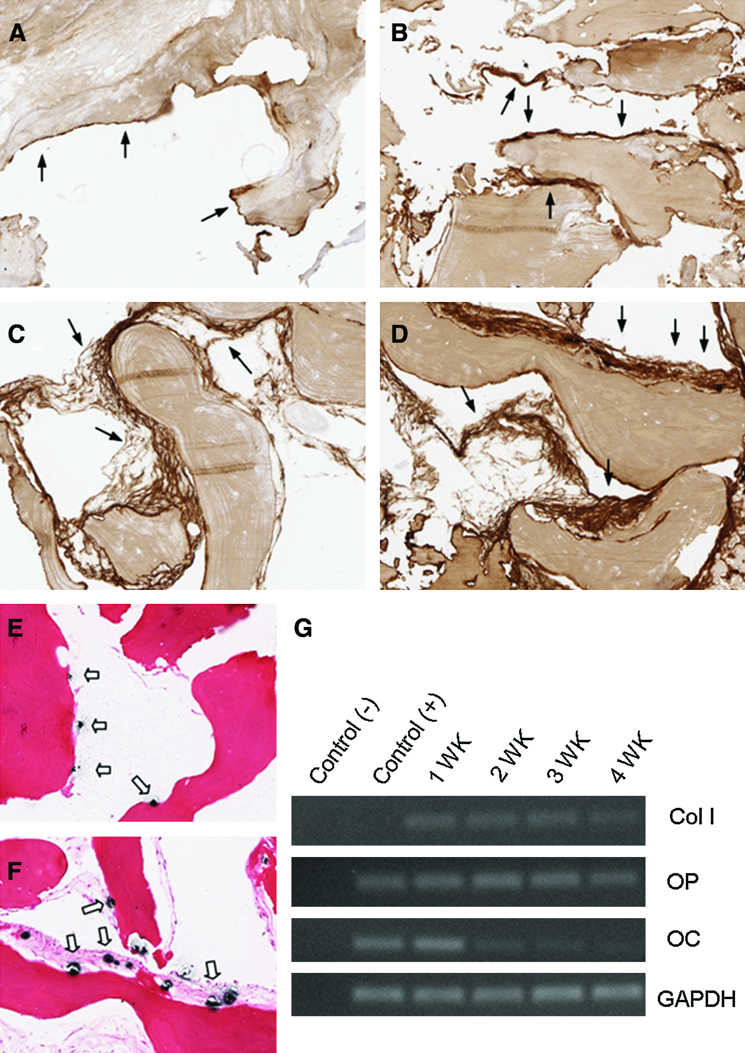
New bone matrix generation and mineralization in vitro. Newly generated matrix stained strongly for collagen type I (black arrows) (
Efficient induction of bone formation by NBCS seeded with enriched BMCs in an ovine spinal fusion model
New bone formation induced in vivo by NBCS seeded with enriched BMCs in an ovine spinal fusion model was assessed at two time points (6 and 10 weeks). Interbody fusion masses were most prominent in the BMC-NBCS group than in controls. At 6 weeks, fusion rate according to manual palpation was 40% in the BMC-NBCS group, compared with 12.5% in the autograft group, 0% in the NBCS group, and 33.3% in the BMCs group. Solid spinal fusion was achieved in all six segments in the BMC-NBCS group at 10 weeks, compared with four of eight segments in the autograft group, two of eight segments in the NBCS group, and three of six segments the BMCs group (Table 2). Similarly, 6 weeks postoperatively, the radiographic score in the BMC-NBCS group was 1.60 ± 0.52, with no significant differences from the control groups. At 10 weeks, the radiographic score reached 2.33 ± 0.49 in the BMC-NBCS group, which was significantly higher than in the autograft, NBCS, or BMC group (p < 0.05, Table 2).
p < 0.05, **p < 0.01, statistically significant difference between the bone marrow cell–natural bone collagen scaffold (BMC-NBCS♂) construct group and the control groups.
Biomechanical testing demonstrated that fusion segments in the BMC-NBCS group had significantly greater mechanical strength than in the other groups. The ROM and NZ of surgical segments were not significantly different between the groups at 6 weeks (Fig. 3A, B). However, lumbar segments obtained from the BMC-NBCS group were stiffer in all directions (flexion, extension, left and right bending) than the control groups (Fig. 3C, D). This was emphasized at the maximum loading moment of 0.475 Nm, where the difference between the BMCs-NBCS group and the control groups reached statistical significance, with the exception of the ROM of the BMCs group during flexion and left bending (Table 3).
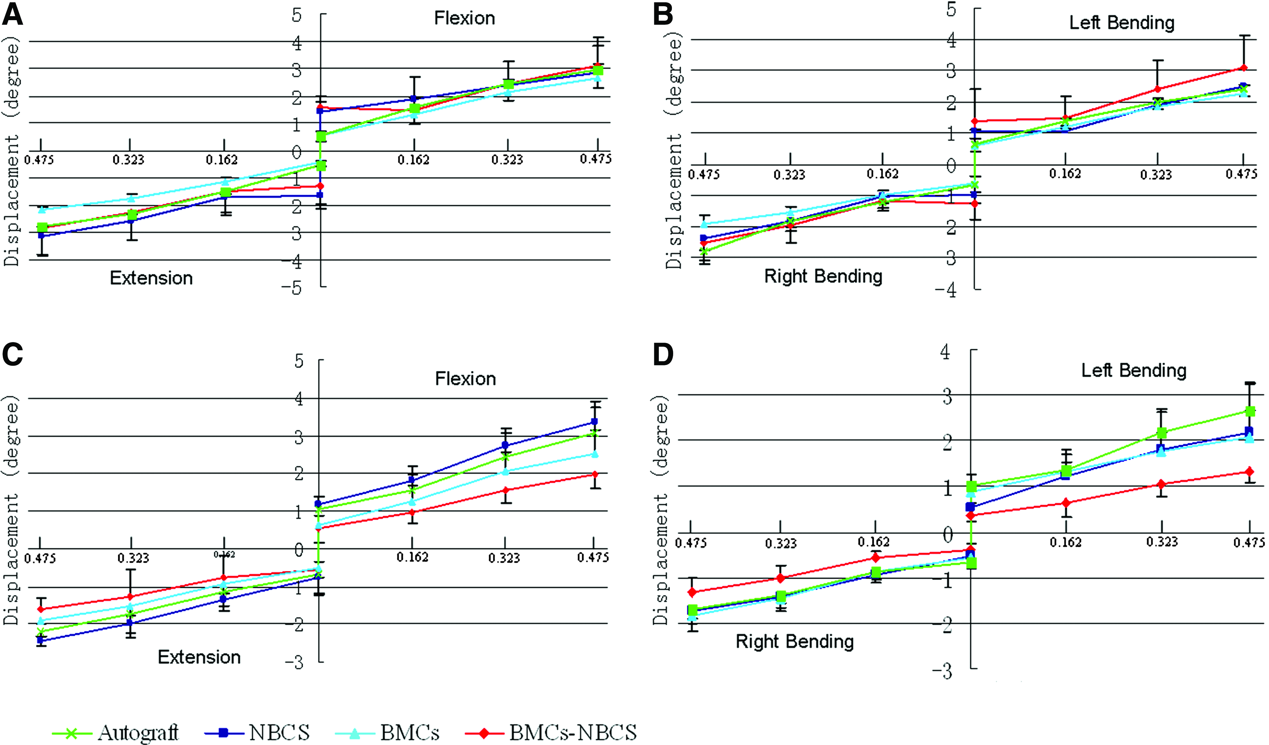
Biomechanical properties of lumbar segments at 6 and 10 weeks postoperatively. There was no difference in range of motion (ROM) and neutral zone (NZ) between groups in flexion, extension (
p < 0.05, **p < 0.01, statistically significant difference between the bone marrow cell–natural bone collagen scaffold (BMC-NBCS) construct group and the control groups.
Micro-CT scanning was performed to investigate the morphology and volume of new bone formation at surgical fusion sites. Three-dimensional reconstructions demonstrated that there was little new mineralized bone in the surgical disc space between two adjacent vertebral bodies in the NBCS, BMC, or BMC-NBCS group at 6 weeks (Fig. 4B–D). However, the surgical disc space of the autograft group was filled with uneven bone chips that were originally implanted into the segment (Fig. 4A). Ten weeks postoperatively, continuous new bone formation was observed in surgical sites connecting two adjacent vertebral bodies in the BMC-NBCS group (Fig. 4H). In the other three groups, new bone had formed from subchondral bone of adjacent vertebral bodies, and gaps remained within the disc spaces (Fig. 4E–G). New bone volume in the BMC-NBCS group reached 18.17 ± 0.64 mm3 10 weeks postoperatively, which was significantly higher than in the autograft, NBCS, or BMC groups (p < 0.01).
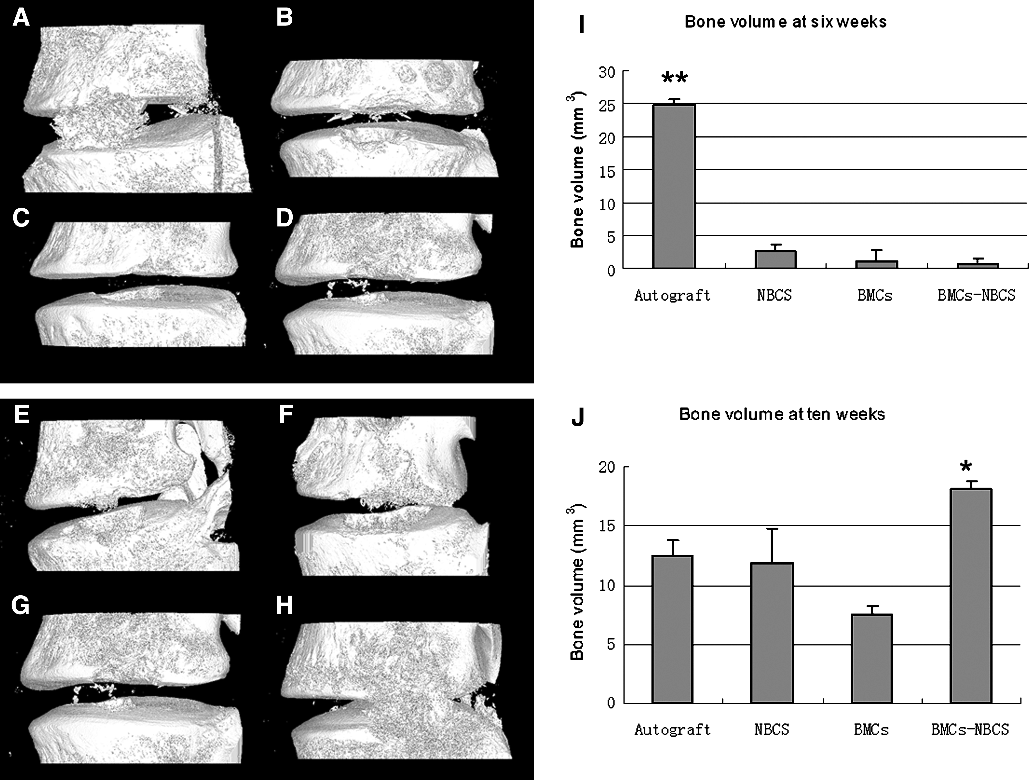
Micro-computed tomography of spinal fusion segments. At 6 weeks, minimal bone formed in the surgical disc space and in subchondral bone defects in vertebral bodies of the NBCS (
Histological assessment demonstrated that new bone was identified within the implanted NBCS in the BMC-NBCS group at 6 weeks (Fig. 5). However, minimal new bone formation was observed in the autograft, NBCS, and BMC groups. A small number of chondroblasts were observed lining subchondral bone. Inflammatory cells were noted surrounding the implanted NBCS. More significantly, new bone tissue that formed in the BMC-NBCS group bridged the disc space and integrated well with host bone tissue from the vertebral bodies at 10 weeks. Endochondral bone formation was noted lining subchondral bone, but new endochondral bone formation mixed with fibrous tissue was observed within the disc space at the implantation site in the autograft, NBCS, and BMCs groups.

Photomicrographs of histology sections 6 and 10 weeks after spinal fusion. After 6 weeks, new bone formation (black arrows) was noted surrounding the NBCS implantation (IP) seeded with BMCs. The autograft also induced new bone formation but to a lesser extent than the BMC-NBCS group. NBCS alone and BMCs alone induced minimal new bone formation at 6 weeks. At 10 weeks, new bone formation induced by BMC-NBCS integrated well with host bone tissue (HB) from vertebral bodies. In the other groups, new bone formation was observed along vertebral bodies, whereas fibrous tissue (FT) filled the disc space. Color images available online at www.liebertonline.com/ten.
Discussion
The goal of spinal fusion is to achieve a bony union between the involved vertebrae. Successful spinal fusion relies on sufficient new bone formation induced using an autologous bone graft or alternative tissue engineering methods. Effective osteogenic cytotherapy for spinal fusion requires an osteoconductive scaffold and cells that are capable of inducing bone formation. A suitable osteoconductive scaffold enhances the support of osteoblastic differentiation and guides bone matrix generation 38 while cells from an appropriate source possess osteogenic differentiation potential and generate new bone matrix, which integrates with host bone tissue. The combination of NBCS with autologous enriched BMCs was considered as a potential substitute to the autograft in bone tissue engineering applications. Our study demonstrated that NBCS supported the population expansion and osteogenic differentiation of ovine-enriched BMCs, facilitating the production of a mineralized collagen matrix within the scaffold. In an in vivo ovine model, NBCS seeded with BMCs enhanced spinal fusion, promoting bone formation and yielding fusion masses of a higher stiffness. These results suggest that NBCS is an efficient scaffold for enriched BMCs and that NBCS combined with autologous enriched BMCs can be used as an alternative to autograft in bone regeneration.
Porous structure and bioactivity are two important elements in the design and use of osteoconductive materials.39–41 Based on its bioactivity, collagen type I has been popular as a scaffold material for bone tissue engineering. Collagen can be made into a porous sponge,42–44 or in a gel form, used in combination with porous hydroxyapatite, 12 hydroxyapatite–tricalcium phosphate, 45 or sintered bovine bone 46 to improve its osteoconductive propertities. Additional materials have also been used as a scaffold, including hydroxyapatite,30,47 β-tricalcium phosphate,48,49 and polylactic glycolic acid.8,50 However, although some of these scaffolds have produced encouraging results in preclinical studies, they do not always display ideal properties in terms of physical structure, chemistry component, and biological interaction with cells and host tissue. In this instance, NBCS is unique, given that it is a bioactive material with a favorable natural bone microstructure, which may be optimal for bone formation. NBCS retains the original porous structure of organic bone and therefore exhibits suitable pore size, porosity, and interconnecting architecture to provide a optimal environment to support the population expansion and differentiation of BMCs. 51 Moreover, NBCS retains the main collagen type I component from organic bone, which has been shown to promote osteoblastic differentiation via the BMP signaling pathway.52–54
This present study used centrifugation enrichment methods55–57 to harvest enriched BMCs. BMCs contain a mixed cell population that includes MSCs and other mononuclear cells that have been shown to positively influence osteogenesis. 58 Unfractionated BMCs and cultured MSCs have also been used as cell sources in bone tissue engineering,57,59,60 but these cells are associated with limitations that hinder their wider use. The major concern regarding using unfractionated bone marrow is its low concentration of MSCs. The bone marrow of healthy adults contains only one MSC for every 50,000 nucleated cells,27,61,62 and this population is further diminished if bone marrow is diluted by peripheral blood during aspiration. 27 In a clinical trial of nonunion fracture repair conducted by Connolly, 57 only 80% of patients treated with unfractionated BMCs obtained a favorable clinical outcome, with the remaining 20% demonstrating inadequate bone formation in response to this therapy. Using specific purification and subcultivation techniques, MSCs can be separated from bone marrow and expanded in vitro while retaining their multilineage potential.10,63,64 Culture-expanded MSCs have been successfully used to repair critical-sized long-bone defects in animal models, 65 but several problems associated with in vitro culture need to be resolved before cultured BMCs can be used widely in clinical applications, including contamination, carcinogenicity, and immunological rejection. 66 For example, when human embryonic stem cells are cultured with medium containing animal serum or serum replacement, the binding of nonhuman sialic acid Neu5Gc with human sera antibodies can result in cell death in vivo. 67 Our present study indicated that enriched BMCs are a potential cell source for bone tissue engineering, with positive effectiveness and safety.
We selected an ovine interbody fusion model to evaluate new bone formation based on the anatomic and biomechanical similarities between sheep and human lumbar vertebrae.68–70 Our results indicated that a 6-week time point was insufficient to assess spinal fusion. Intra-group differences were detected only after 10 weeks. From histological assessment, it is apparent that the complete spinal fusion process requires longer than 10 weeks, given that incomplete ossification is still observed at this time point, however, it is difficult to decide on an ultimate time point for assessing spinal fusion, given that multiple factors, including animal species, fusion approach, materials used, and surgical technique, influence the ossification process. 71 Previous studies using an ovine model of interbody fusion have assessed end points that vary from 6 to 12 weeks.29–31 Because different devices and materials were used in these studies, the outcomes widely varied. The fusion outcomes in this study at 10 weeks were sufficient to show the differences between the BMC-NBCS group and controls.
A limitation of this study is that no other commonly used scaffolds such as collagen type I or tricalcium phosphate were included as a control.17,72 The lack of comparison with one or more of these scaffolds weakens the conclusions of this study. In addition, no specific markers were used to identify the origin of the cells that were involved in facilitating new bone formation, whether from implanted enriched BMCs or from fusion beds on the vertebral body.
Conclusion
In summary, this study showed that NBCS is effective as a scaffold for ovine-enriched BMCs in vitro and in vivo. The in vitro study demonstrated that NBCS facilitated adhesion, population expansion, and osteoblast differentiation of ovine BMCs and guided bone matrix generation and mineralization. In vivo, the combination of NBCS and enriched BMCs in an ovine interbody fusion model enhanced fusion outcomes, promoting bone volume and fusion stiffness above that of controls. Together, these results strongly suggest that NBCS is a potent osteoconductive scaffold that facilitates osteogenesis of enriched BMCs. The combination of NBCS and BMCs has potential in clinical applications using osteogenic cytotherapy.
Footnotes
Acknowledgments
The authors would like to thank Dr. Peter Self from Adelaide Microscopy, University of Adelaide for performing micro-CT scans, Ms Anne Cowie from the Perth Bone and Tissue Bank for the preparation of NBCS, and Mr. Andy Wilson and Miss Astrid Armitage for their assistance with animal care.
Disclosure Statement
There are no known conflicts of interest associated with this publication, and there has been no significant financial support for this work that could have influenced its outcome. All named authors have read and approved the manuscript, and no other persons satisfied the criteria for authorship but are not listed. We have given due consideration to the protection of intellectual property associated with this work, and there are no impediments to publication, including the timing of publication, with respect to intellectual property. In so doing, we confirm that we have followed the regulations of our institutions concerning intellectual property.
Any aspect of the work covered in this manuscript that has involved experimental animals or human patients has been conducted with the ethical approval of all relevant bodies, and such approvals are acknowledged within the manuscript.