




Abstract
The three-dimensional scaffolds play a very important role in regulating cell adhesion and the production of extracellular matrix molecules in in vitro regeneration of cartilage. This study evaluates how the three-dimensional structure and physicochemical properties of the polymeric scaffolds influence in vitro regeneration of cartilage tissue. A synthetic poly(vinyl alcohol)–poly(caprolactone) semi-interpenetrating polymer network (IPN) scaffold and gelatin–albumin, made of natural polymers, are used for the study. The polymers in the semi-IPN synthetic scaffold mimic the properties of collagen and glycosaminoglycans present in native cartilage. Its appropriate swelling and pore structure enabled cell–cell and cell–matrix interactions. This helped the chondrocytes to retain its spherical morphology and resulted in enhanced secretion of extracellular matrix components. In contrast, the biomimetic structure in gelatin–albumin scaffold induced chondrocytes to loose its phenotype by spreading and becoming fibroblastic in morphology. Its high swelling and the large pore size failed to recreate an appropriate microenvironment for chondrogenesis that resulted in less secretion of cartilage-specific molecules. Mesenchymal stem cell differentiation to chondrocytes in the presence of growth factors is also enhanced in the synthetic semi-IPN scaffold. The study thus indicates that the chemical composition and the physicochemical properties of the scaffolds play a very important role in providing appropriate niche in in vitro tissue regeneration.
Introduction
Scaffolds are 3D structures that serve as an analog of native ECM and create a microenvironment similar to that of the native tissue. It provides appropriate physical, biochemical, and molecular signals for tissue regeneration. The two-dimensional cultures are inadequate to model the complex cellular interactions compared to the 3D culture that promotes high cell–cell and cell–matrix interactions, which is a better representation of actual tissue. Tissue engineering of cartilage utilizes natural or synthetic polymers to serve as a 3D template for growing cells. 3 The overall morphology of the 3D scaffolds, inbuilt properties of the materials, and surface chemistry may directly or indirectly influence the behavior of cells cultured on scaffolds.
Mesenchymal stem cell (MSC) differentiation to chondrocytes is also influenced by appropriate signaling received from the surrounding microenvironment called niche. The polymeric scaffolds along with the growth factors play an important role in creating this microenvironment for the differentiation process. In this study, we evaluate the hypothesis that physicochemical properties and 3D structure of the scaffold play a significant role in maintaining the chondrogenic phenotype, stem cell differentiation to chondrocytes, and the secretion of ECM that plays a key role in the function of articular cartilage. To evaluate this hypothesis, two scaffolds are fabricated that mimic the ECM of cartilage in its properties. A 3D semi-interpenetrating polymer network (IPN) scaffold, PVA–PCL, made of synthetic polymers poly(vinyl alcohol) (PVA) and poly(caprolactone) (PCL) and another made from a blend of natural polymers gelatin–albumin (GA) are used for the study. Both the scaffolds have been characterized and are found to be favorable for chondrocyte culture in our previous studies.4,5 This study evaluates how physicochemical properties of these 3D scaffolds influence chondrogenesis in in vitro engineering of cartilage tissue. The results of our study indicate that the synthetic scaffold is more appropriate for chondrogenesis.
Materials and Methods
Fabrication of 3D scaffolds
A semi-IPN of hydrophilic PVA (Mw 12,000–23,000, Sigma-Aldrich, St. Louis, MO) and hydrophobic PCL (Mw 65,000; Sigma-Aldrich), with differing polar constituents, was prepared to impart synergistic properties of both the polymers. Briefly, the synthesis of PVA–PCL semi-IPN (50:50) involves high-speed mixing (3000 rpm) of an aqueous solution of PVA and solutions of PCL in chloroform (CHCl3) at concentrations favoring miscibility to form homogeneous foam. The foam was lyophilized, and PVA in the scaffold was crosslinked with 1% gluteraldehyde solution to form the semi-IPN. The scaffolds were washed to remove traces of gluteraldehyde and lyophilized to get 3D scaffolds with an interconnected porous structure.
Three-dimensional scaffold of GA was fabricated in a similar manner by high-speed mixing of aqueous solutions of gelatin (Porcine skin, Type A, ∼300 blooms; Sigma, St. Louis, MO) and albumin (Egg albumin; Spectrochem, Mumbai, India) in percentage weight ratio (90:10) to get a uniform foam. The foam was freeze-dried and crosslinked using 1-ethyl-3(3-dimethylaminopropyl) carbodiimide hydrochloride (Sigma). The crosslinked scaffolds were washed and freeze-dried to obtain a stable 3D porous GA scaffold.
Pore structure, swelling, and chemical composition of 3D scaffolds
The 3D scaffolds were characterized for their physicochemical properties using various techniques. The overall porous structure of the scaffold was imaged using a high-resolution microcomputed tomography (μCT 40 Scan CoMedical, Bassersdorf, Switzerland) and scanning electron microscope (Model-S2400; Hitachi Inc., Brisbane, CA). The presence of open continuous pores in the dry and wet state of the scaffold was evaluated using liquid extrusion porosimeter (LEP–Model 1100A; PMI Porous Materials Inc., Ithaca, NY). The medium uptake ability was determined by the swelling ratio, and the in vitro degradation was studied in phosphate-buffered saline for a period of 3 months. Nicolet 5700 FTIR spectrophotometer with deutrated triglycine sulfate detector (Thermo Electron Corporation, Madison, WI) using diamond attenuated total reflectance (ATR) accessory in reflectance mode was used to determine the chemical composition.
Chondrocyte culture and MSC differentiation in 3D scaffolds
Cartilage tissue was obtained from porcine articular joints. The chondrocytes were isolated in a sterile condition as per the accepted protocol. 6 The cells were resuspended in chondrogenic media containing Dulbecco's modified Eagle medium (DMEM), 10% fetal bovine serum, penicillin (100 U/mL), streptomycin (100 μg/mL), sodium pyruvate (1 mM/mL), glutamine (1.4 mM/mL), non-essential amino acids (NEAA) (0.1 mM/mL), ascorbic acid (50 μg/mL), proline (40 μg/mL), and 10−7 M dexamethasone (Sigma). For culture on 3D scaffolds (10 mm diameter and 2–3 mm thickness), chondrocytes were seeded on both the scaffolds at a density of 2 × 10⁶ cells per scaffold and cultured in chondrogenic medium at 5% CO2 and 37°C for a period of 2 months. The medium was replaced every 3 days. The constructs were evaluated for chondrogenesis at the end of 2 months.
MSCs were isolated from bone marrow of 2-week-old Wistar rats as per the accepted protocol. 7 The cells were cultured in alpha modified Eagle medium (αMEM) containing 15% fetal bovine serum, and cells from passage 1 were seeded onto 3D scaffolds at a density of 1 × 10⁶ cells per scaffold. They were cultured in chondrogenic medium supplemented with three different growth factors. The three growth factor–supplemented groups were transforming growth factor beta3 (TGFβ3) (10 ng/mL) (Sigma, St. Louis, MO), bone morphogenetic protein 2 (BMP2) (25 ng/mL) (R&D Systems, Minneapolis, MN), and combination of TGFβ3 (10 ng/mL) and BMP2 (25 ng/mL). The medium was replaced every 3 days. The constructs were evaluated for chondrogenesis at the end of 28 days.
The morphology and viability of cells on scaffolds after seeding were evaluated using scanning electron microscopy (SEM) and live dead staining. The constructs were evaluated for matrix deposition by SEM and for cartilage-specific molecules by histological staining and biochemical assays.
Evaluation of constructs for chondrogenic markers
The cell morphology, overall cell distribution, and deposition of ECM were evaluated using SEM. The total amount of glycosaminoglycans (GAGs) was quantified using dimethyl methylene blue (DMMB) assay. 8 The secretion of GAGs within the interior of constructs was detected using Safranin O staining. 9 The deposition of two major chondrogenic markers such as aggrecan and collagen type II was detected by immunostaining in paraffin sections using specific antibodies and visualized using Ultra Tech HRP (DAB) Streptavidin–Biotin Detection System kit (Beckman Coulter, Marseille, France).
Statistical analysis
Three samples were used for each of the quantitative experiments. Again from each material, all measurements were done in duplicate to confirm the reproducibility. Each parameter was expressed as mean of all values with standard deviations. Statistical analysis was performed using one-way analysis of variance test. Values of p < 0.05 were considered significant (*) and p < 0.0001 were considered very significant (**).
Results and Discussion
Several cell-friendly natural polymers like alginate, agarose, fibrin, and hyaluronic acid have been used as carriers for chondrocyte culture.10–14 It was found that chondrocytes maintain differentiated phenotype and produce GAGs in these hydrogels, but most of them are mechanically weak for long-term in vivo applications and have been observed to degrade before proper formation of cartilaginous tissue. Collagen type II scaffolds have shown promising results. 15 Many of these scaffolds fail to recreate the optimal properties of native cartilage, like high water holding ability of GAGs entangled in a mechanically stable network of collagen fibers that provide the cushioning effect and helps to resist compression. Concerns about the feasibility of obtaining large amounts of naturally derived polymers for clinical applications with assurance of pathogen removal have prompted researchers to investigate the use of synthetic polymers.
Synthetic biodegradable polymers poly(glycolic acid), poly(L-lactic acid), and their copolymers have been widely applied in bone and cartilage repair.16,17 The physicochemical properties of synthetic polymers are finely tunable according to our requirements. Despite the extensive use of these synthetic polymers, one of the limitations is the accumulation of the degradation products leading to drastic decrease in pH that may affect the viability of the cells at the implant site. This is a major concern in orthopedic applications where implants with considerable size would be required. Greater rigidity of these polymers also limits its application to cartilage tissue engineering. PCL and their blends have shown promising results. 18 Recently, researchers are more focused onto the fabrication of hybrid polymers combining the advantages of both natural and synthetic polymers.19,20
Selection of the polymers and fabrication of 3D scaffolds
The selection of polymers in this study was made with a view to mimic the properties of native components of the cartilage. The ECM of cartilage consists of collagen that provides mechanical strength to cartilage, while the GAGs with a larger amount of water retention capacity help to resist compression. A single polymer is not expected to provide all the requirements to mimic the ECM of native cartilage. So each scaffold was a blend of two polymers, so that they exhibit synergistic properties of both the polymers in its composition.
PCL is an Food and Drug Administration–approved biodegradable polymer and is widely used to fabricate 3D scaffolds, particularly for load-bearing applications.21,22 PCL is extremely hydrophobic, and this limitation of PCL was overcome with the addition of PVA, an Food and Drug Administration–approved hydrophilic polymer. Hydrogels of PVA have been used as an artificial cartilage, 23 artificial meniscus, 24 and biocompatible viscoelastic material with mechanical property comparable to that of native cartilage. 25 PVA of low-molecular weight has been shown to be completely excreted from body through urine and with no accumulation of the harmful degradation products in any internal organs.26,27 The PCL in the semi-IPN PVA–PCL scaffold is expected to provide the mechanical strength while the PVA has high water holding capacity similar to the proteoglycans. In the semi-IPN, PCL is retained as the linear polymer, while the PVA chains are partially crosslinked with gluteraldehyde. The crosslinking resulted in interchain and intrachain bonding by covalent linkage of PVA, within which the PCL chains are entangled resulting in a semi-interpenetrating polymer network. This entangled network of both the polymers resembles the ECM of cartilage and enhances mechanical stability of the system. The crosslinking also prevented the phase separation of the two polymers.
The GA scaffold is made from natural polymers and is a stable hydrogel that retains many of the biomimetic sequences of native collagen that helps in cell attachment and ECM synthesis. The biomimetic structure and biocompatibility of gelatin has led to its use in wide range of biomedical applications.28–30 Hydrogels of gelatin have been used widely to serve as cell carriers but they lack the mechanical stability required for long-term load-bearing applications.31,32 In this work, a 3D scaffold was fabricated using a blend of gelatin and egg white albumin. Albumin was added in trace amounts as a nontoxic foaming agent, to attain a hydrophilic–hydrophobic balance and is expected to improve the overall stability of the scaffold by reducing the hydrophilicity of native gelatin. The hybrid scaffold is expected to retain the biomimetic nature of gelatin, and albumin in the blend was added to overcome the limitations of gelatin. The choice of crosslinking agent was based on the reactive groups present on the amino acid side chain and appropriate ambient conditions that do not negatively affect the scaffold and its application. The interchain and intrachain covalent peptide linkage formed is similar to that found in collagen fibers of cartilage. The schematic representation of the two scaffolds is represented in Figure 1.
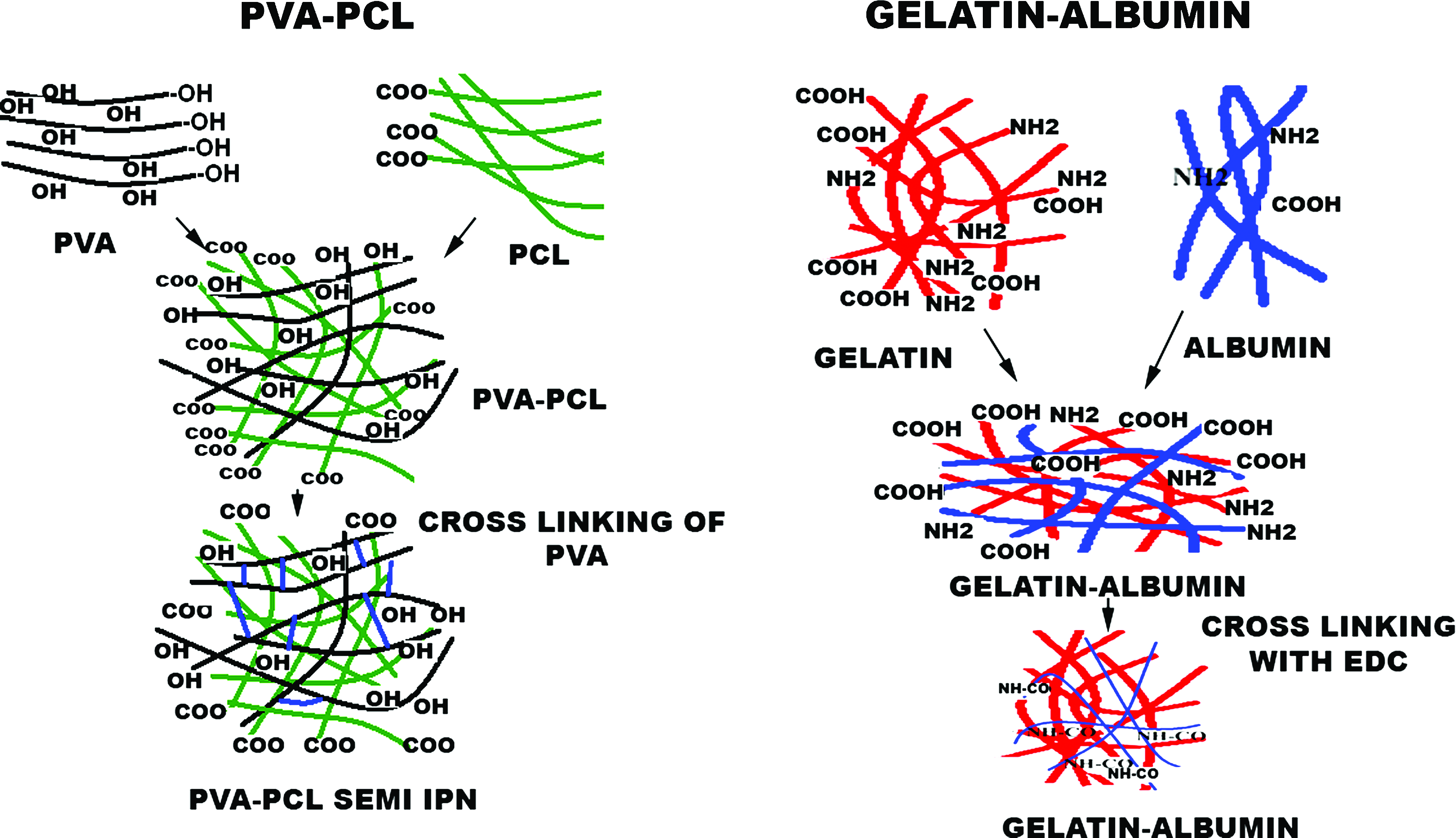
Schematic representation of three-dimensional (3D) poly(vinyl alcohol)–poly(caprolactone) (PVA–PCL) semi-interpenetrating polymer network (IPN) and gelatin–albumin (GA) scaffolds. Color images available online at www.liebertonline.com/ten.
Characterization of 3D scaffolds
Pore structure
Both the scaffolds were highly porous with open interconnected pores. This helps in uniform distribution of cells during seeding and in the free diffusion of the nutrients and waste materials to keep the cells viable. The porous structure also helps in the deposition of ECM secreted by the cells in a 3D pattern.
The microcomputed tomography images of the 3D porous scaffolds in the dry state indicate that both the samples were porous throughout the entire length. The SEM images indicate the presence of open interconnected pores (Fig. 2). The pore distribution of continuous interconnected pores in dry and wet state of the scaffold was estimated by liquid extrusion porosimeter. The pore size distribution was slightly different in the two scaffolds (Fig. 3). In dry PVA–PCL scaffold, the pore size distribution was within the range of 7–192 μm and wet was of 100–500 μm, respectively. In aqueous solution there was a fivefold increase in pore size from the dry scaffold due to the swelling of PVA but excessive swelling was prevented by the semi-interpenetrating structure. About 95% of the open continuous pores in dry GA scaffold were found to be in narrow range of 50–70 μm. Swelling produced a very large increase in the pore size of GA scaffold. Chondrocyte cultures have been conducted previously in scaffolds in different pore size ranges such as 0.2–8, 5, 10–20, 40–60, 70–120, 100, 200, 400, and 380–405 μm.33–35 Most of these studies identified that chondrocytes proliferated and secreted matrix components in the largest pore size used for each study. In general chondrocytes prefer an optimum pore size, promoting free diffusion of nutrients, which allows sufficient space for the secretion and localization of large aggregating proteoglycans and collagen molecules, at the same time enabling cell–cell and cell–matrix interactions.

Microcomputed tomography images of the 3D porous scaffolds in the dry state (

The pore distribution of 3D scaffolds in dry and wet state. (
Chemical composition, swelling, contact angle, and degradation profile of scaffolds
Figure 4 represents the chemical composition, swelling, and degradation profile of both the 3D scaffolds. Fourier transform infrared spectrum gives information on the chemical structure of the 3D scaffolds. The PVA–PCL shows all the characteristic peaks of PCL and PVA such as carbonyl stretching of PCL at 1724 cm−1 and free hydroxyl (−OH) groups of PVA at 3330 cm−1. The GA blend retained the carbonyl (−C = O) stretching peak (amide I) and N-H bending peak (amide II) of peptide bond at 1630 cm−1 and 1539 cm−1 respectively. Both the scaffolds retained all the characteristic peaks of the parent material indicating that the hybrid compositions showed synergistic properties of both the polymers. The spectra of the hybrid composition also proved that neither the fabrication methodology nor the crosslinking procedures produced any change in the chemical structure of the polymers.

(
The swelling percentage of the PVA–PCL was 500% and that of GA is 1500%. Both the scaffolds had the potential to absorb and supply nutrients to all the cells that are seeded within the porous structure of the scaffold. This is an essential requirement for the scaffolding material particularly for chondrocyte culture, as the tissue is avascular and the cells depend entirely on the nutrients that are diffused into the matrix from the surrounding area. Moreover, it is a measure of the inherent capacity of the scaffolding material to hold large amount of water similar to the GAGs in the native ECM of the cartilage. The excessive swelling of PVA in the semi-IPN structure was prevented by the interpenetrating structure. The higher uptake of medium in GA scaffold is also attributed to the wicking action of solution through the pores of the scaffold as well as by the hydration of free (−OH) groups in the protein structure. This swelling resulted in large increase in pore size. However, the high swelling nature did not result in collapse of the scaffold as observed in many other hydrogels. The scaffold remained stable in medium for long period with a good handling property that was attained due to its adequate crosslinking.
The wettability or the surface property of the scaffolds is known to regulate the cell attachment and spreading. The PVA–PCL and GA had a hydrophilic–hydrophobic balance with a water contact angle value of 55.2° and 65°, respectively. These data confirm the above explanation that high medium uptake of the GA scaffold was by a wicking action of the porous scaffold and not due to higher hydrophilicity. The addition of albumin might have helped to attain the hydrophilic–hydrophobic balance and might have a role in the stability of the scaffold. Previous reports have shown that hydrophilic–hydrophobic balance in contact angle values is optimal for cell adhesion.36,37 Both the scaffolds had comparable degradation profile of 20% weight loss in a period of 3 months.
Morphology and viability of chondrocytes in 3D scaffolds
The retention of spherical morphology is an indication of maintaining chondrogenic cell phenotype. The chondrocytes retained the typical spherical shape in PVA–PCL scaffold while they were spread and showed a fibroblastic morphology in the GA scaffold as shown by SEM images (Fig. 5). The cells were viable in both the scaffolds as shown by the live dead staining (Fig. 6a, b). The spherical shape of the cells in PVA–PCL scaffold was also very well evident in the confocal images.

The chondrocytes retained the typical spherical shape in PVA–PCL scaffold (
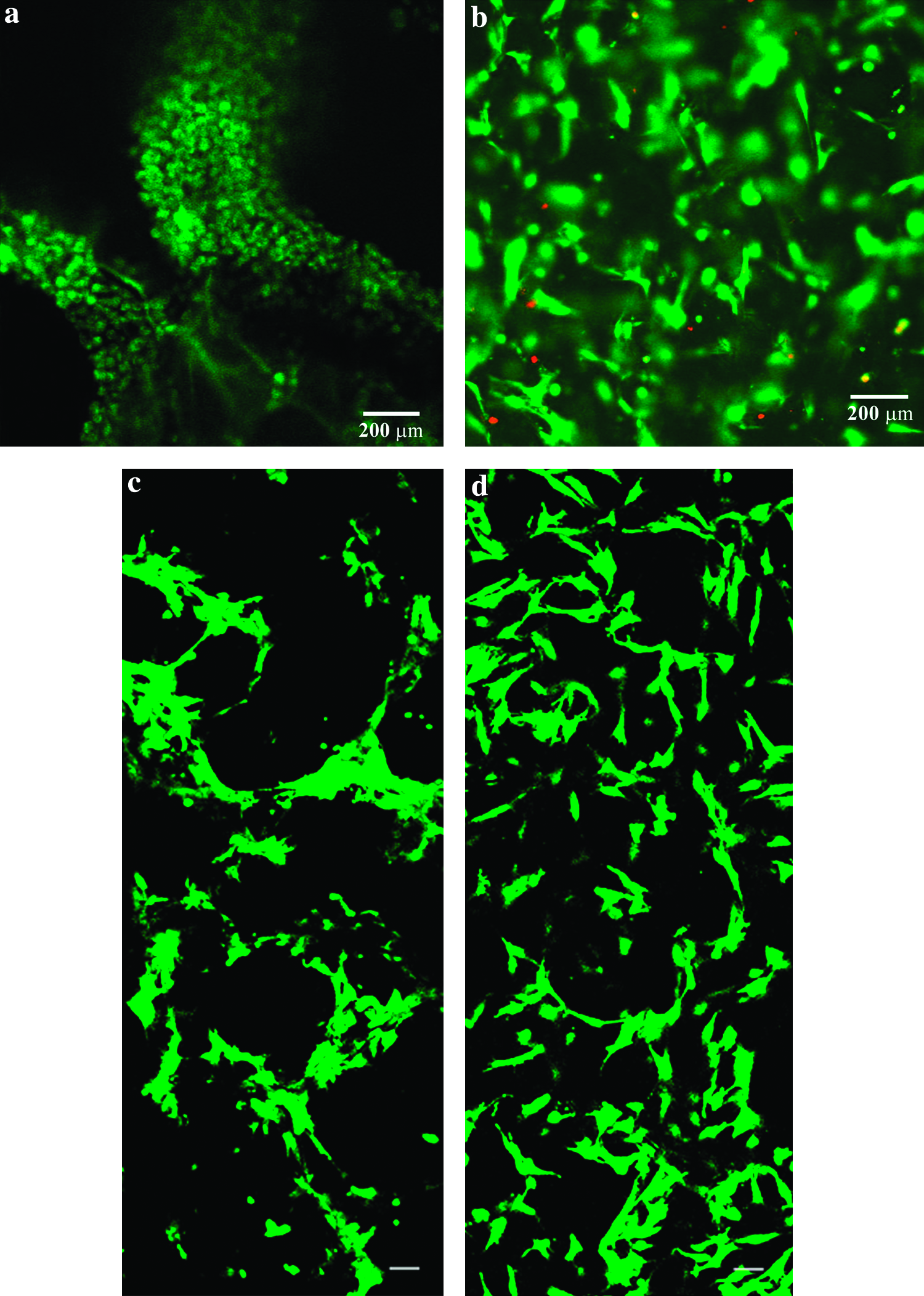
Live dead stain showing viable cells in scaffolds. Chondrocytes are spherical in (
The peaks in Fourier transform infrared spectrum indicate that GA scaffold retains all the typical bands of native gelatin. The biomimetic sequences in this protein might have promoted the spreading of cells in GA scaffold. The wettability of the two scaffolds was comparable with a hydrophobic–hydrophilic balance. So it should be inferred that the chemical composition had a significant influence on cell shape and spreading. The MSCs remain viable and retain their typical spreaded morphology in both the scaffolds (Fig. 6c, d). The pore characteristics have influenced the distribution of cells in these scaffolds.
Evaluation of 2-month constructs
The environmental scanning electron microscope (ESEM) images of PVA–PCL construct showed uniform ECM deposition with chondrocytes retaining its characteristic spherical morphology (Fig. 7a, b). The surface of the GA scaffold showed high ECM deposition or formation of a fibroblastic layer. Cells with spherical morphology could not be observed on the surface (Fig. 7c, d). The deposition of GAGs within the interior was evaluated by Safranin O staining of sections of the scaffolds. The PVA–PCL scaffolds showed a high and uniform deposition of GAG, while the GAG deposition was confined to the periphery in GA (Fig. 8). A significant decrease was also observed in the total GAG content in GA scaffold when compared to PVA–PCL (Fig. 9). Immunostaining for aggrecan also showed a similar pattern of deposition (Fig. 10). The results on the overall ECM deposition indicate that PVA–PCL matrix have favored the cells to maintain a chondrogenic phenotype influencing cell shape and ECM synthesis. The formation of a fibroblastic skin layer on the surface and dilution of cell density due to excessive swelling might have prevented a uniform ECM deposition in the interior of GA. This might have resulted in reduced cell–cell and cell–matrix interactions within each pore and failed to create an appropriate microenvironment for chondrogenesis.

The environmental scanning electron microscope (ESEM) images of PVA–PCL construct showing uniform deposition of extracellular matrix with chondrocytes retaining its characteristic spherical morphology (

Safranin O staining for deposition of glycosaminoglycans (GAGs) in the interior of the construct. (
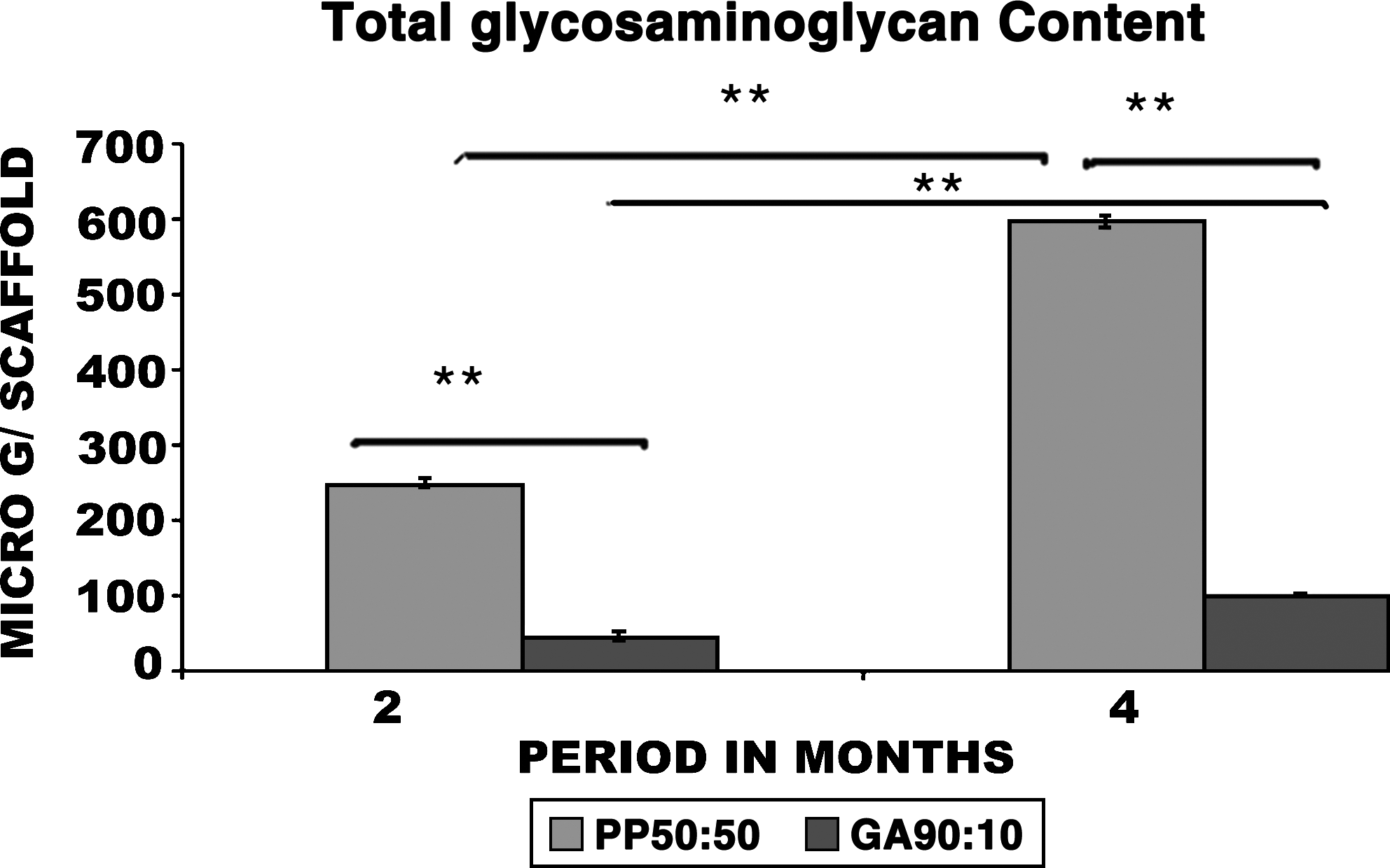
Total GAG content in PVA–PCL semi-IPN and GA constructs. Statistical analysis was performed using one-way analysis of variance test. The values were expressed as the mean of three replicates. Values of p < 0.05 were considered significant (*) and p < 0.0001 were considered very significant (**).

Immunostaining for deposition of aggrecan in the interior of the construct. (
Differentiation of MSCs to chondrocytes in 3D scaffolds
The chondrogenic differentiation of MSCs was found to be better in PVA–PCL scaffold in TGFβ3, BMP2, and in group supplemented with combination of growth factors (TGFβ3 + BMP2). The SEM results reveal that there was a high deposition of ECM and cells that covered the entire surface of both PVA–PCL and GA constructs supplemented with combination of growth factors (TGFβ3 + BMP2). In PVA–PCL constructs, the differentiated cells in both TGFβ3 and BMP2 supplemented constructs had spherical shape like the chondrocytes. However, the spherical shape was more evident in BMP2-supplemented PVA–PCL constructs (Fig. 11). The total GAG content (Fig. 12) was higher and its deposition was uniform (Fig. 13a–c) in PVA–PCL scaffolds than GA. The deposition of collagen type II was also higher in PVA–PCL constructs (Fig. 14a–c). In GA, the cells were flattened and had a fibrous appearance, and the ECM deposition was confined to the margins of the pores and in the exterior of scaffold. This was evident from the SEM results, Safranin O (Fig. 13d–f), and immunostaining for collagen type II (Fig. 14d–f). Induction of chondrogenesis in MSCs depends on the coordinated activities of many factors like cell adhesion, cell density, and appropriate signaling. The general response of cells to different growth factors was more or less similar in both the scaffolds. Chondrogenesis was initiated when densely packed precursor cells form appropriate cell–cell contact in PVA–PCL. In GA scaffold, excessive swelling reduced cell density. The differentiation of MSCs was favored in PVA–PCL scaffold than the GA not only by its 3D structure but also by its semi-IPN composition. The amount and pattern of ECM deposition indicate that the properties of biomaterial scaffolds may have a substantial effect on the action of growth factors on cellular performance.
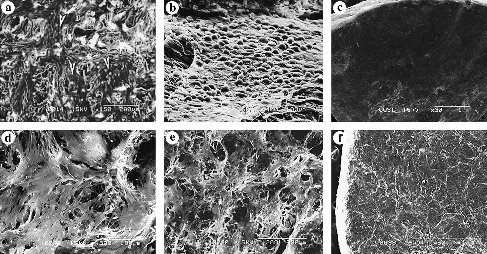
The SEM of 28-day differentiated constructs cultured in different growth factor supplements in PVA–PCL (
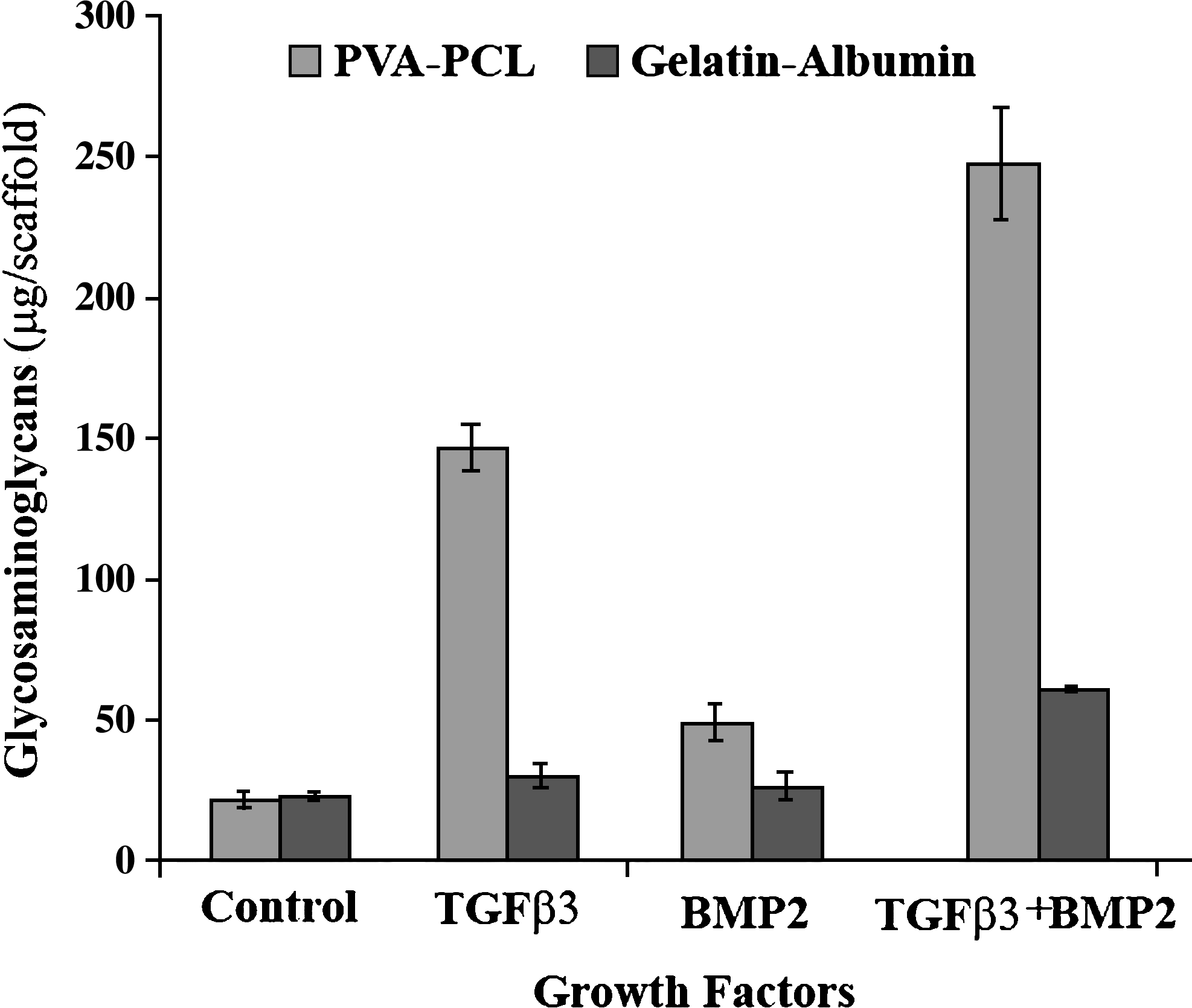
The total GAG content in differentiated PVA–PCL and GA constructs supplemented with different growth factors.

Safranin O staining for deposition of GAGs in the differentiation of mesenchymal stem cells to chondrocytes. (

Immunostaining for collagen type II deposition in the differentiated constructs. (
Summary and Conclusions
The 3D structure, chemical composition, and physicochemical properties of scaffolds influenced the chondrogenic responses like cell morphology, ECM secretion, and distribution. In this study, chondrogenic response was found to be better in synthetic PVA–PCL scaffold than GA scaffolds. This was analyzed in terms of cell morphology, GAG, and collagen type II deposition. Cell response varied from initial process of cell adhesion that regulated subsequent events of tissue growth and maturation. PVA–PCL is a synthetic scaffold that had no inbuilt nature to favor cell response. The chondrocytes retained the spherical morphology and maintained its phenotype in PVA–PCL scaffold, while the MSCs maintained their typical spread morphology. The cell attachment in it might be nonreceptor-mediated cell adhesion or by adsorption of serum proteins. The basic structure of the PVA–PCL semi-IPN was that of mechanically stable PCL chains, forming interpenetrating network, with hydrophilic PVA. This was similar to the network of collagen and GAGs in native cartilage. This structure might have provided a better microenvironment for the chondrocytes to maintain its morphology and to retain the chondrocyte phenotype. In GA scaffold, the biomimetic sequences of gelatin and albumin stimulated the cell to attach and spread on to the material to form a fibroblastic morphology. The adhesion and growth of cells on the surface are considered to be strongly influenced by the hydrophilicity and hydrophobicity. There was no significant difference in water contact angle values between these scaffolds. Both the scaffolds had wettability with a hydrophobic–hydrophilic balance. So it should be inferred that the chemical composition had a significant influence on the initial process of cell adhesion and spreading.
The factors like 3D architecture of the scaffold and the swelling characteristics also regulate the chondrogenic response. The porosity was within the general accepted range for cartilage regeneration for both the scaffolds. Pore structure in the semi-IPN was in the wide range of pore sizes that were similar to anisotropy in native cartilage, while GA had a uniform pore size. The native cartilage is composed of different zones with variation in the structure of matrix assembly. Studies have shown that the chondrocytes favor anisotropy than a uniform ordered structure. In culture, the swelling behavior was different in both the scaffolds that widely influenced the quality of in vitro–generated construct. The semi-interpenetrating polymer network structure with PCL optimized the swelling nature of the scaffold. This situation was favorable for cells in creating a suitable 3D environment, favoring cell–cell and cell–ECM interactions that resulted in uniform distribution of ECM. GA scaffold had a very high swelling that reduced the optimum cell density within each pore. It resulted in uneven distribution of ECM, which was confined to the outer surface and edges. There are reports that if the scaffold is incompletely filled with cells, it may result in fibrous ingrowth.38,39 The initial events and further the microenvironment during the course of culture stimulated the cells to produce a skin layer of cells and ECM on the surface of the scaffold. The formation of a skin layer on the surface with spreading of cells and deposition of ECM on the surface might have prevented the supply of nutrients to the interior of the scaffold. This might have resulted in cell death in the interior of GA scaffold. All these factors might have contributed to reduced secretion of chondrogenic markers in the GA scaffold. The results also point out how the physicochemical properties of the scaffold affect the initial cell response that regulated the whole sequence of events, in the further growth and maturation of the construct. It is essential that the scaffolds need to provide a suitable microenvironment for the cells throughout the course of culture period. The variation in the chondrogenic response between the two scaffolds used in this study is as a result of the combined effects of various physicochemical properties of the scaffold. It is necessary to identify the key parameters of the scaffold in regulating the chondrogenic response and optimize these parameters to improve the quality of in vitro regeneration of cartilage. Unlike scaffolds fabricated from a single polymer, the hydrophilic PVA and the mechanically stable PCL in the semi-IPN recreate the optimal properties of native cartilage like high water holding GAGs on a mechanically strong collagen network. This structure will help in the cushioning effect of cartilage and would be useful for long-term in vivo applications.
Footnotes
Acknowledgments
The authors would like to thank the Director SCTIMST and Head BMT WING for providing the facilities for carrying out this work. Neethu Mohan gratefully acknowledges the grant of SCTIMST Institute fellowship. Dr. Anilkumar is acknowledged for confocal images.
Disclosure statement
No competing financial interests exist