




Abstract
Although tissue-engineered scaffolds made from collagen sponge are suitable for cell infiltrating, easily supplying oxygen and nutrients to cells, and removing the waste products, their mechanical properties are not satisfactory to be used as scaffold materials for tissue engineering applications. To improve mechanical properties of collagen, a novel porous scaffold for bone tissue engineering was prepared with collagen sponge reinforced by polypropylene/polyethylene terephthalate (PP/PET) fibers. Collagen solution (6.33 mg/mL) with PP/PET fibers (collagen/fiber ratio [w/w]: 1.27, 0.63, 0.42, 0.25) was freeze-dried, followed by cross-linking of combined dehydrothermal and glutaraldehyde. A scanning electron microscopy-based analysis of surface of the sponges demonstrated that the sponge with collagen/fiber <0.25 exhibited homogenous and interconnected pore structure with an average pore size of 200 μm. Incorporation of PP/PET fibers significantly enhanced the compressive strength of the collagen sponge. Proliferation and osteogenic differentiation of mesenchymal stem cell in collagen sponges reinforced with PP/PET fibers incorporation were significantly enhanced compared with collagen sponge without PP/PET incorporation. We conclude that incorporation of PP/PET fibers not only improves the mechanical properties of collagen sponge, but also enables mesenchymal stem cells to positively improve their proliferation and differentiation.
Introduction
Tissue engineering is a multidisciplinary approach that use three-dimensional (3D) biodegradable scaffolds together with stem cells to promote tissue regeneration. The scaffold should mimic the structure and biological function of native extracellular matrix (ECM) as much as possible, in terms of both chemical composition and physical structure.13–15
Material design of scaffold is one of the key technologies for tissue engineering.15–18 The materials used for scaffold fabrication not only determine physical properties such as biocompatibility, biodegradability, and mechanical stability, but also provide the appropriate signals for directing the cellular process that lead to tissue formation. 19 Collagen as the main component of ECM has become one of the most attractive materials to fabricate an artificial ECM for tissue engineering applications. Collagen sponge that exhibits highly porous, interconnected pores is efficient for cell infiltration, oxygen and nutrient in-diffusion, and waste material out-diffusion. However, the mechanical properties of collagen are not satisfactory for the application in hard tissue engineering. Several biodegradable synthetic polymers such as poly (glycolic acid) (PGA) and poly (lactic acid) indicated sufficient mechanical strength due to their hydrophobic nature.20,21 However, the lower cell attachment properties of biodegradable synthetic polymers in comparison with collagen and degradation products such as glycolic acid that reduce the pH-balanced environment and greatly affect cell growth are disadvantages of using such biodegradable synthetic polymers. Some materials such as polypropylene (PP),22–24 polyethylene terephthalate (PET), 25 modified polystyrene, and PE 26 have been widely reported to use for in vitro cell culture as nondegradable materials that do not change the cell culture environment during tissue regeneration.
The objective of this study was to fabricate a feasible scaffold with the properties of collagen and PP/PET to improve the poor mechanical properties of collagen. The effect of fiber and incorporation amount on the physical and mechanical properties, the mesenchymal stem cell (MSC) proliferation, and osteogenic differentiation were also evaluated.
Materials and Methods
An aqueous solution of type I collagen, prepared from porcine tendon by pepsin treatment (6.33 mg/mL, pH 3.0) in HCL, was purchased from Nitta Gelatin Inc. Dulbecco's modified Eagle's minimal essential medium (DMEM) and fetal bovine serum (FBS) were purchased from Gibco. All water used was double-distilled water (DDW).
Isolation and culture of MSCs
Rats were narcotized by ketamine 10% (40–90 mg/kg; Alfasan) and xylazine 2% (5 mg/kg; Alfasan) and then sacrificed by cervical dislocation. Under sterile conditions, both femurs from each rat were excised. Muscles and the entire connective tissue were detached; additionally, the epiphyses were removed. Femoral bone marrow tissue was extracted from the bones by flushing the medullar cavities of the bones with washing solution using an 18-gauge needle. Washing solution contained high-glucose DMEM (Gibco), supplemented with 15% FBS, 100 U/mL penicillin, and 100 U/mL streptomycin (all from Gibco). Marrow plug suspension was dispersed by passing through two different needles (21 and 23 gauge), respectively, and centrifuged at 500g for 5 min. Supernatant containing thrombocytes was discarded, and the cell pellet was treated for RBC lysing procedures using hypotonic shock. 27 Then, the cells were suspended in a growth medium containing DMEM-high glucose supplemented with 15% FBS, 100 U/mL penicillin, and 100 U/mL streptomycin. Cells in the growth medium were plated on 25 cm2 plastic flasks (Orange) and incubated at 37°C in a 5% CO2/95% air atmosphere. Nonadherent cells were removed after 4 days by medium exchange and adherent cells were allowed to grow for up to 20 days, with medium replacement every 3 days. After 10 days in culture, isolated colonies of MSC became apparent. When the cell cultures reached 80% confluence, they were harvested by trypsinization with aqueous solution of 0.05 wt% trypsin (Gibco) and 1 mM ethylenediaminetetraacetic acid (Merck) in 0.1 M phosphate-buffered saline (PBS, pH 7.4) for 3 min at 37°C and reseeded in new flasks at a density of 10–15 × 103 cells/cm2.
Fabrication of collagen sponge
Collagen sponges with different amounts of PP/PET fibers were fabricated by conventional freeze-drying method, followed by cross-linking of combined dehydrothermal and glutaraldehyde. PP/PET fibers with 20 μm diameter were fabricated in Tarbiat Modares University (95 PP: 5 PET) without any compatibilizer. Briefly, PP/PET nonwoven fabric was immersed in acetone for 1 h to remove oils and fats, and rinsed three times with DDW at 25°C for 10 min. The PP/PET fibers with different collagen/fiber ratio (w/w)—1.27, 0.63, 0.42, and 0.25 (5, 10, 15, and 25 mg)—were homogeneously placed into 24-well polystyrene plates (Orange), and then 1 mL of collagen solution was poured into each mold. The resulting collagen solution was frozen at −20°C and freeze-dried for 24 h. The freeze-dried sponge was dehydrothermally cross-linked at 140°C for 12 h under vacuum conditions and, in addition, underwent chemical crosslinking with 0.3 vol% glutaraldehyde solution (25%; Sigma) in mixture of acetone/0.01M HCl (Merck) (7/3). After stirring cross-linked collagen with 0.1M glycine (Sigma) solution at room temperature, samples were washed with DDW, and freeze-dried. A similar preparation procedure was performed with collagen solution to obtain a sponge without PP/PET fiber incorporation. Then, samples sterilized with ethylene oxide gas at 50°C.
Morphological observation
Appearance and intra-structure of sponges were characterized by scanning electron microscopy (SEM) using a Philips, model XL 30 microscope.
The pore size of collagen sponges was calculated as the geometric mean of the major and minor diameters of 20 pores from the picture of cross section of the sponges.28–30
Sponge characterization
The porosity was measured by liquid substitution method. Isopropanol with density ρi was used as the displacement liquid at 4°C. 31 A density bottle filled with isopropanol was weighed (W1). A sponge sample of weight Ws was immersed into the density bottle, and weighed (W2). The sponge saturated with isopropanol was taken out and then density bottle was weighed (W3).
The following parameters of a scaffold, the volume of the sponge pore (Vp), the density of the sponge skeleton (Vs), and the porosity (ɛ), were calculated.
32
Maximum water absorption of sponges was checked after centrifuging samples in PBS at 300 rpm on an orbital shaker (Heidolph ROTAMAX 120) at 37°C for 8 and 24 h. The formula for the maximum water uptake was as follows
33
:
The diameter of sponge was checked in MSC culture after 14 days. Formula for the shrinkage was as follows:
Mechanical measurement
A DMA instrument (DMA 242C, NETZSCH) was used to measure mechanical properties of the 3D sponges. Cylindrical fixture was chosen to test the specimens and evaluate their behavior as a whole structure along their compression axis in the Z direction. Sponge was loaded with the dynamic force ranging from 0.5 to 5 N in a dynamic stress experiment. More specially, a starting force of 0.5 N was applied and then continuously increased to 5 N with a step function ramp of 0.5 N/min at a constant frequency of 1 Hz. Compression modulus was calculated from the slope of the initial linear portion of the strain–stress curve. The wet samples were prepared by centrifuging PBS at 300 rpm on an orbital shaker for 8 h at 37°C. For each configuration two cycles were done. The temperature was 37°C during all experiments.
Cell seeding into collagen sponge and cell culture
MSCs were seeded into sponges, prewetted in DMEM for 2 h, at a density of 15 × 104 cells per 24.87 mm3 unit volume in 24-well plates. Briefly, 24 μL of cell suspension (65 × 105 cells/mL) was added on each scaffold and incubated for 2 h at 37°C in a 5% CO2/95% air atmosphere. After that, 1 mL of DMEM supplemented with 15% FBS and 1% of 100 U/mL penicillin and 100 U/mL streptomycin (Gibco) was added to each sponge. Every sponge was incubated in DMEM for 4 weeks at 37°C in a 5% CO2/95% air atmosphere.
Osteogenic differentiation of MSCs in collagen sponge
To determine osteogenic differentiation, the MSC-seeded sponge was incubated in DMEM containing 50 μg/mL ascorbic acid (Sigma) and 10 mM β-glycerophosphate with 100 nM dexamethasone (Sigma) at 37°C in a 5% CO2/95% air atmosphere. The medium was changed every other day.
MTT assay
Proliferation of MSCs on sponges was determined using the MTT (3-{4, 5-dimethylthiazol-2yl}-2, 5-diphenyl-2H-tetrazoliumbromide) (Sigma) assay. The sponges were transferred into a new 24-wells plate, and 1 mL of MTT solution (0.5 mg/mL) was added into each well. After incubation at 37°C for 4 h in a 5% CO2/95% air atmosphere, MTT was taken up by active cells and reduced in mitochondria to insoluble purple formazan granules. Subsequently, the medium was discarded and the precipitated formazan was dissolved in isopropanol containing 0.1 N HCl (150 μL/well). Optical density of the solution was evaluated using a microplate spectrophotometer after subtraction of OD690nm from OD570nm. Viable cell number was determined using a linear calibration curve between OD and predetermined cell concentration.
Cell attachment
SEM observation was applied to evaluate the morphology of MSCs attached on collagen sponges in normal and differentiation media. In this method, the culture medium was removed, and sponges were washed with PBS for three times. Then, adhered cells on sponges were fixed with 4 mL of 2.5% glutaraldehyde for 90 min at 4°C. After three times washing with PBS, adhered cells were dehydrated in an ethanol-graded series (30%, 40%, 50%, 60%, 70%, 80%, 90%, and 100%) for 1 h each and allowed to dry in a clean bench at room temperature.
Alkaline phosphate and Alizarin red assay
To evaluate the osteogenic differentiation of MSCs, calcium deposits were qualified by using alizarin red (Sigma), and the intracellular alkaline phosphatase (ALP) activity was measured using ALP assay kit (Parsazmun Co. Ltd.) at Noor Pathobiology lab (Tehran, Iran).
To determine calcium deposition, cultured sponges were washed three times with PBS. Then, MSCs were detached from the scaffold by treatment with an aqueous solution of 0.05 wt% trypsin and 1 mM ethylenediaminetetraacetic acids in 0.1 M phosphate-buffered saline (PBS, pH 7.4) for 3 min at 37°C. After 24 h, MSCs attached on the plate were fixed with 3% glutaraldehyde for 10 min at room temperature. To stain the differentiated cells with alizarin red, the samples were immersed in alizarin red with pH 4 for 10 min at room temperature, and washed with PBS. Then, the alizarin red was removed and replaced by PBS, and pictures were taken.
To assess ALP activity, cultured sponges were washed three times with PBS, minced with scissor, and finally homogenized in lysis buffer solution (0.2% Triton X-100, 10 mM Tris-HCl, and 1 mM MgCl2 [pH 7.5]). Sample lysate (1 mL) was centrifuged at 11,000 rpm for 10 min at 4°C and ALP activity of the supernatant was assayed by use of p-nitrophenyl phosphate as a substrate. Briefly, 20 μL cell lysate was mixed with 800 μL of 1M Diethanolamine (pH 9.8), 0.5 mM magnesium chloride, and 200 μL of 10 mM p-nitrophenyl phosphate aqueous solution and incubated at 37°C for 1 min. Then, the absorbance of the solution mixture was measured after 1, 2, and 3 min at 405 nm by a spectrophotometer (Beckman). The subtraction of optical density at each minute from the previous one was calculated and the mean of them was multiplied by 2757 to obtain the average of ALP activity.
Statistical analysis
All the results were statistically analyzed by one-way analysis of variance with SPSS version 13, and p < 0.05 was considered statistically significant. Data were expressed as means ± the standard deviation of the mean.
Results
Morphology and physical properties of collagen sponges
Figure 1 shows SEM photographs of cross sections of collagen sponges incorporating various amounts of PP/PET fibers. Irrespective of the fiber amount incorporated, every collagen sponge possessed an interconnected porous structure with similar intrastructural appearance. PP/PET fibers were exposed in the pores of PP/PET-incorporated sponges, and the internal structure was similar to that of sponges without PP/PET fibers fiber incorporation. The homogeneity of PP/PET-incorporated sponge at collagen/fiber ratio of 0.25 (Fig. 1E) was decreased, whereas interconnection structure and other collagen/fiber ratio were not decreased. Morphological characteristics are summarized in Table 1.

Cross-sectional scanning electron microscopy photographs of the collagen sponge without PP/PET fiber incorporation (collage/fiber ratio [w/w]: 6.33)
Figures 2 and 3 show decreasing water absorption and shrinkage of collagen sponge with and without PP/PET fiber incorporation. As expected, the hydrophobic property of PP/PET significantly influenced these phenomena.
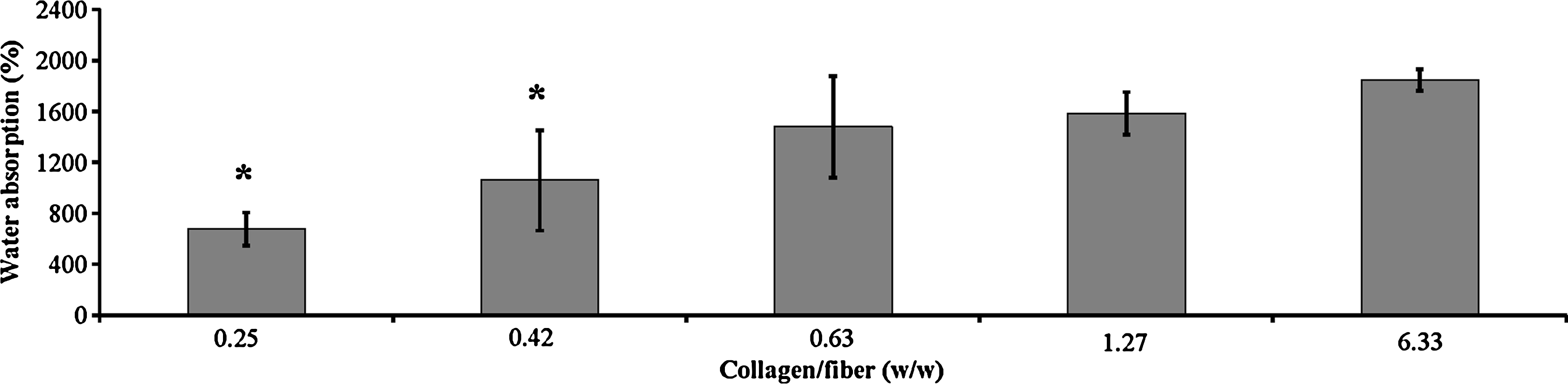
Water absorption of collagen sponges. Sponges were immersed in phosphate-buffered saline at 300 rpm for 8 h at 37°C. Sample number: 3. *p < 0.05; significant against the water absorption of collagen sponge without PP/PET fiber incorporation (collagen/fiber ratio [w/w]: 6.33).
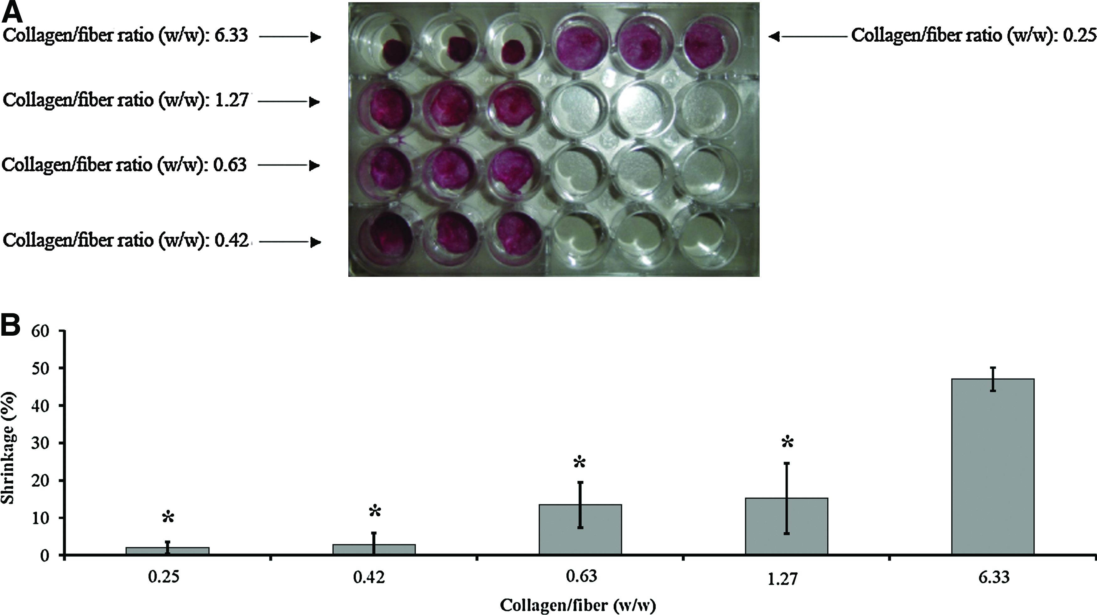
Shrinkage photograph
Mechanical properties of collagen sponges
Figure 4 shows the compression modulus of sponges under wet and dry conditions. Incorporation of fibers enabled sponges to increase the compression modulus. The compression modulus of collagen sponge reinforced with PP/PET fiber incorporation at collagen/fiber ratio (w/w) of 0.25 was decreased as the higher amount of fibers caused deformation of collagen sponge and disarrayed the interconnected pores. As expected, compression modulus decreased under wet condition, but it was not less than dry collagen sponge. Wet modulus of collagen sponge without fibers incorporation was not detectable by DMA.
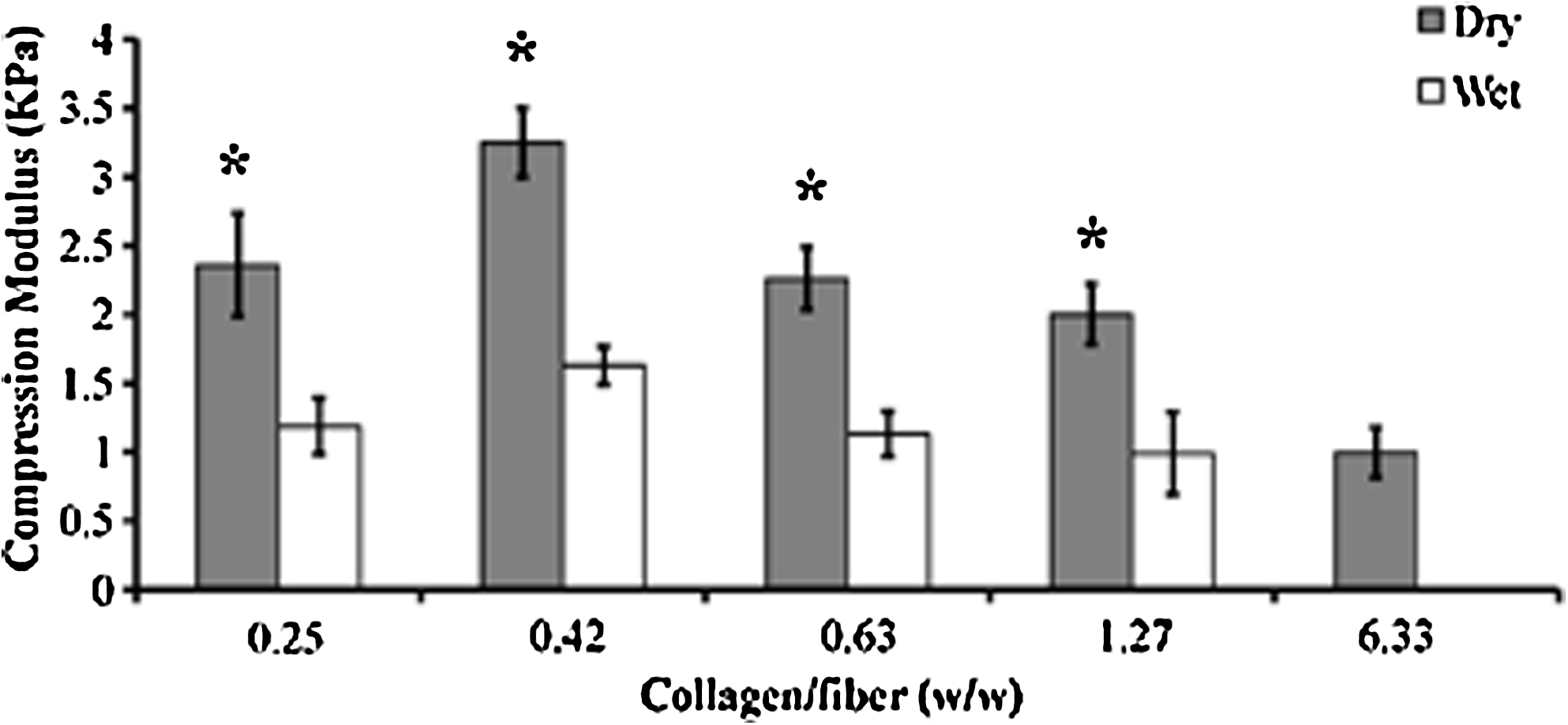
Compression modulus of collagen sponges. Sample number: 3. *p < 0.05; significant against the compression modulus of collagen sponge without PP/PET fiber incorporation (collagen/fiber ratio [w/w]: 6.33).
Cell proliferation of collagen sponges
Figure 5 shows the number of cells attached and proliferated in sponges during 3 weeks. There was a significant difference in cell number between the original collagen sponge and the collagen sponge reinforced with PP/PET fiber incorporation at collagen/fiber ratio (w/w) 0.63.

The number of mesenchymal stem cells attached to collagen sponges in normal media at 37°C in a 5% CO2/95% air atmosphere. *p < 0.05; significant against the cell proliferation of collagen sponge without PP/PET fiber incorporation (collagen/fiber ratio [w/w]: 6.33).
Cell attachment
Figure 6 shows cell attachment and differentiation on surface of fiber and collagen layer in the normal and differentiation media. As it is shown in Figure 6, cells have the tendency to flatten on the surface of fibers.

Scanning electron microscopy photographs of cell adhesion on collagen layer and surface of fibers in normal media
Osteogenic differentiation
Figure 7 shows MSCs differentiated to osteocytes. Calcium deposits of differentiated cells were qualified by alizarin red after 28 days. Figure 8 shows the time course ALP activity of MSCs cultured on collagen sponge and collagen sponge reinforced with PP/PET fibers incorporation at collagen/fiber ratio (w/w) 0.63. ALP activity increased with time for the initial 21 days, and thereafter leveled off. ALP activity of MSC cultured in the bone differentiation medium was always high for the collagen sponge reinforced with PP/PET fiber incorporation compared with collagen sponge without PP/PET fiber incorporation.
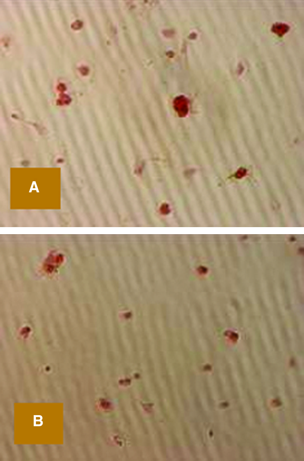
Calcium deposition photographs of collagen sponge with collagen/fiber (w/w): 0.63 in bone differentiation media after 28 days at 37°C in a 5% CO2/95% air atmosphere

Time course of alkaline phosphatase activity in bone differentiation media at 37°C in a 5% CO2/95% air atmosphere. *p < 0.05; significant against the alkaline phosphatase activity of collagen sponge without PP/PET fiber incorporation (collagen/fiber ratio [w/w]: 6.33).
Discussion
It has been recognized that induction of tissue regeneration based on tissue engineering can be achieved by the following three key steps: proliferation of cells, seeding of cells, and proliferation in a suitable scaffold and maintenance of differentiation phenotype of the engineered tissues.34–38 Tissue engineering is an interdisciplinary field that applied principles and methods of engineering toward the development of substitutes to improve the function of damaged tissue and organs. One of the most important factors to enhance regeneration by using tissue engineering is the scaffolding material that should be suitable for cell proliferation and differentiation. The biocompatibility and mechanical properties of the scaffold materials are very important to provide suitable condition for cell seeding. Among many materials currently used as cell scaffolds, collagen has been widely used and in vivo safety has been proven through long-term applications on clinical trials, cosmetics, and food industries. Collagen sponge is a highly porous material with an interconnected pore structure, which is effective in infiltration of cells, supplying oxygen and nutrients to cells, and excluding cell wastes, while shape and bioresorbability can be readily regulated by changing the formulation conditions. However, the drawback of collagen sponge as a scaffold for cell proliferation and differentiation is its poor mechanical strength and high dimension changes. To overcome these inherent materials problems of the sponge, its combination with other materials has been attempted. Several nonbiodegradable synthetic polymers such as PP,22–24 PET, 25 and PE 26 have been used to fabricate scaffolding materials. The mechanical resistance of these scaffolds to compression is acceptable for tissue engineering applications. However, nonbiodegradable materials are relatively or completely hydrophobic and hinder cell seeding. Therefore, hybrid scaffolds that use biodegradable and nonbiodegradable polymers to improve the mechanical property of the scaffolding materials are preferable to overcome the hindrance effect of nonbiodegradable materials on cell attachment. In the present study, we used PP fibers and improved its mechanical strength with 5% PET. 39 PP is a nonbiodegradable polymer that has been shown good biocompatibility under in vivo and in vitro conditions.22–24 Low cost, easy fabrication and characterization, and stability of chemical and mechanical properties of synthesized nonbiodegradable fibers compare with biodegradable ones and are important key factors to develop a new type of scaffolding materials for tissue engineering applications.
Pore size has been observed to influence adhesion, growth, and differentiation of a wide variety of cell types. Studies have shown that a cell does exhibit selectively on pore size according to specific cell type. It is generally reported that pore sizes in excess of 100 μm are required in bone regeneration.40–44
Morphological observation showed that sponges fabricated at −20°C had 3D micro porous structure because of using ice crystals as progenies. This structure plays important roles in mass and flow transport and direction of cell signals. Increasing the amount of PP/PET fibers up to collagen/fiber ratio of (w/w) 0.42 did not have negative effect on interconnected structure and pores of sponge (Fig. 1).
Water absorption behavior is shown in Figure 2. As it is shown, the hydrophobic property of fibers has a major influence on decreasing the tendency of sponge to an aqueous medium. By decreasing water absorption and providing a physical network between fibers and collagen, the shrinkage of collagen sponge decreased (Fig. 3). Shrinkage is one of the most important deficiencies of collagen sponge. Reduction of dimension causes fewer spaces for cell attachment and changes in the sponge structure under wet condition. Glutaraldehyde is a common material to decrease the amount of shrinkage, but it is toxic for clinical application. It is preferable to use the least amount of toxic materials and make strong network without negative effects on cell attachment and proliferation by substituting nontoxic materials. PP and PET, known as hydrophobic polymers, are good candidates to overcome this problem. They are biocompatible and decrease water absorption without producing toxic byproducts.
The compression test indicated that the amount of fibers and interconnected structure of sponge had parallel roles in increasing mechanical properties. Even when fibers added, the hybrid sponge responded efficiently to an applied stress by absorbing energy and dissipating stress. High flexibility of the collagen sponge is beneficial for surgical operation to keep the scaffold safely at the defect site. More importantly, the rigidity and stiffness of fibers made the system resist higher stress levels and exhibit enhanced elastic modulus. The sponge with collagen/fiber ratio of (w/w) 0.42 showed the highest modulus, and decreased by increasing the percentage of fibers (Fig. 4). In fact, by increasing the amount of fibers up to collagen/fiber ratio (w/w) 0.25, the interconnected structure of scaffold destroyed and the pore size distribution was not uniform in sponge. Moreover, as it is shown in Figure 4, although the modulus of sponges under wet condition was lower than the dry one, it was still higher than the dry collagen sponge. In fact, PP fibers that not have any functional group to react with water act as a barrier to prevent water penetration. However, the mechanical strength is lower than that required for bone regeneration, and it is necessary to consider a modified method to improve mechanical properties.
Cells grown in the sponge with collagen/fiber ratio of (w/w) 0.63 showed the highest growth rate during the period of 14–21 days (Fig. 5). Accordingly, Li et al. reported a higher proliferation rate of osteoblasts on collagen scaffolds reinforced by chitosan fibers compared to unreinforced scaffold. 45 These results indicated that the growth rate of the cells on the reinforced scaffold was not abnormally higher than that of the control, although the growth rate on the reinforced scaffold was obviously higher than that of the unreinforced scaffold. The main reason for this is that the chitosan material, which is alkaline in nature, can neutralize the acidity caused by the PLLA degradation, which may provide an advantageous microenvironment for cells living on the fibers and, in addition, may not stimulate the cells to proliferate malignantly. This indicated that the addition of chitosan fibers might increase the biocompatibility of these scaffolds. Also, higher cell attachment on PGA fiber-reinforced collagen sponges compared to collagen scaffold has been previously reported. 20 Herein, although a suitable cell attachment was observed, quantification seems to be needed for a more exact comparison between reinforced collagen sponges in future studies. Surface characteristics of materials, whether their topography, chemistry, or surface energy, play an essential part in cell adhesion on biomaterials. Thus, attachment, adhesion, and spreading belong to the first phase of cell–material interactions, and the quality of the first phase will influence the cell capacity to proliferate and differentiate itself in contact with the implant. 46 Regardless of cell type, it was observed that cells are adhered, spread, and grown more onto the surface of materials with moderate hydrophilic properties than hydrophobic.39,47–50 This maybe closely related to the serum protein adsorption on the surfaces; the preferential adsorption of some serum proteins like fibronectin and vitronectin from culture medium into the moderately hydrophilic surfaces may be a reason for better cell adhesion, spreading, and growth. In our study, SEM results confirmed cell attachment for different scaffolds (Fig. 6) and revealed that the attached cells had flattened adhesion on both collagen and fibers. Our observation manifested that the growth rate on the reinforced scaffold is obviously higher than that of the unreinforced scaffold. The main reason for these phenomena is the moderate hydrophobic and hydrophilic properties of the scaffolds and more space for cell attachment and proliferation due to less shrinkage and cells tendency to adhere on fiber surfaces. Moreover, using nondegradable fibers such as PP/PET can control the negative effect of releasing degradation products in comparison with poly (lactic acid) and PGA in decreasing the pH-balance of environment,20,21,51 and increase the biocompatibility of the scaffold. In fact, the wettability, free spaces, homogenized structure, and finally biocompatible scaffolds helped cells to appropriate signals to direct the cellular process. Besides, they made better conditions for oxygen and nutrient circulation and extraction waste products.
Osteogenic differentiation of cells also revealed the role of fibers. ALP is an ectoenzyme, produced by osteoblasts, that is likely to be involved in the degradation of inorganic pyrophosphate to provide a sufficient local concentration of phosphate or inorganic pyrophosphate for mineralization to precede.52–54 Therefore, ALP is a useful marker for osteoblast activity. Alizarine red is a marker to determine calcium deposits by cells. The ALP activity increased rapidly and saturated at third and fourth weeks (Fig. 8). According to the chemical structure of PP and PET, incorporation fibers do not have any interaction with cells under differentiation media. Also, several studies showed that the initial cell attachment directly affects such other cellular responses as movement, proliferation, and phenotype expression of cells through the internal signal transduction. 46 Therefore, the improvement in cellular activity on the sponge with collagen/fiber ratio (w/w) of 0.63 may be originated from the stability of sponge in differentiation media and initial higher level of cell attachment and proliferation.
Conclusion
Fabrication of collagen sponges reinforced with nonbiodegradable PP/PET fibers as bone tissue-engineered scaffold was studied in this work. The investigation of in vitro study has shown that incorporation of fibers had an impressive effect on mechanical and physical properties as well as increasing cell proliferation and differentiation. These results suggest that PP/PET fiber incorporation is a suitable method to enhance mechanical and physical stability without using toxic materials. Further in vivo investigation is necessary to find the reactions of nonbiodegradable fibers under real conditions.
Footnotes
Acknowledgments
The authors would like to express their gratitude to Dr. Roozbeh Tanhaeivash and Ms. Shahnaz Halimi for their contribution to MSC preparation from rat femoral bone, and Dr. Babak Arjmand for his supervising of sterilization of the scaffolds at Iran Bank of Transplanted Organs.
Disclosure Statement
No competing financial interests exist.