




Abstract
Tissue-engineered blood vessel is one of the most promising living substitutes for coronary and peripheral artery bypass graft surgery. However, one of the main limitations in tissue engineering is vascularization of the construct before implantation. Such a vascularization could play an important role in graft perfusion and host integration of tissue-engineered vascular adventitia. Using our self-assembly approach, we developed a method to vascularize tissue-engineered blood vessel constructs by coculturing endothelial cells in a fibroblast-laden tissue sheet. After subcutaneous implantation, enhancement of graft integration within the surrounding environment was noted after 48 h and an important improvement in blood circulation of the grafted tissue at 1 week postimplantation. The distinctive branching structure of end arteries characterizing the in vivo adventitial vasa vasorum has also been observed in long-term postimplantation follow-up. After a 90-day implantation period, hybrid vessels containing human and mouse endothelial cells were still perfused. Characterization of the mechanical properties of both control and vascularized adventitia demonstrated that the ultimate tensile strength, modulus, and failure strain were in the same order of magnitude of a pig coronary artery. The addition of a vasa vasorum to the tissue-engineered adventitia did not influence the burst pressure of these constructs. Hence, the present results indicate a promising answer to the many challenges associated with the in vitro vascularization and in vivo integration of many different tissue-engineered substitutes.
Introduction
One of the main challenges in tissue engineering is to produce thick tissues while ensuring proper nutrition of all cells within such constructs. Many strategies have been previously devised to enhance in vitro angiogenesis in tissue engineering.13–15 Our group succeeded in creating capillary-like structure in skin tissue-engineered constructs using human umbilical vein endothelial cells (HUVEC).16–18 A prior study has also shown that those cells create long-lasting blood vessels in vivo. 19 The incorporation of a microvasculature in tissue-engineered construct should lead to better tissue integration after implantation by preventing early graft failure due to improper nutrition of the engineered-tissue.20,21
Inosculation is a phenomenon enabling the perfusion of a tissue-engineered capillary network by the vasculature of the host after implantation. We have shown such an event in an unique skin substitute. 22 Inosculation of preexisting capillaries incorporated into a scaffold with mesenchymal or skeletal muscle has previously been described, but no study implicating such a phenomenon with an endothelial capillary network contained within the wall of a vascular structure has been reported.19,23
Tissue-engineered blood vessels (TEBV) would benefit from such an adventitial capillary network, akin to vasa vasorum, created in vitro within the substitutes. The vasa vasorum are capillaries lining the blood vessel walls when passive diffusion is insufficient for appropriate nourishment. Thus, one of the main properties of vasa vasorum is to allow nutrition and oxygenation of the arterial wall.24,25 They have a tree-like structure that allows them to be functional endarteries. 26 The many interesting properties of vasa vasorum should translate into an important improvement in the adventitial wall of TEBV allowing blood perfusion, drainage, and rapid graft integration within the host environment. 27
Using the self-assembly method, our group has developed an approach to produce TEBV with mechanical resistance and burst pressure allowing their implantation. 9 In the present study, it was demonstrated that a capillary system can be added in vitro to the adventitial layer of TEBV and that it allowed for an improve perfusion of the engineered tissue in vivo. Stable capillary structures similar to a physiological vasa vasorum were also observed in long-term in vivo analysis in an athymic mouse model.
Materials and Methods
Tissue culture
The study was approved by the CHA Saint-Sacrement Hospital ethics committee and all tissues were obtained after informed consent was given. Human skin fibroblasts were obtained from the dermis of adult breast skin and cultured as described previously. 28 Briefly, fibroblasts were isolated from skin biopsies using collagenase H (Roche Diagnostics, Laval, Canada) and grown in Dulbecco–Vogt modification of Eagle's medium (DMEM; Invitrogen, Burlington, Canada) supplemented with 10% fetal calf serum (HyClone, Logan, UT) and antibiotics. HUVEC were obtained from healthy newborns umbilical cords as described previously. 16 HUVEC were cultured in M199 medium supplemented with 20% fetal calf serum (HyClone), 2.28 mM glutamine, 40 IU/mL heparin (Leo Laboratories, Pickering, Canada), 20 μg/mL endothelial cell growth supplement (Calbiochem/EMD Biosciences, Gibbstown, NJ), and antibiotics.
To create the tissue-engineered vascular adventitia with vasa vasorum (TEVAwVV), human fibroblasts were cultured for 21 days in DMEM (Invitrogen) with 10% fetal calf serum (HyClone), 100 U/mL of penicillin, 25 μg/mL of gentamicin, and 50 μg/mL of ascorbic acid (Sigma, Oakville, Canada). HUVEC were then seeded on top of the fibroblasts and cultured together in a 1:1 mixture of DMEM with 10% fetal calf serum (HyClone) and M199 culture medium supplemented with antibiotics and 50 μg/mL of ascorbic acid for 7 days.
Tissue-engineered vascular adventitia (TEVA) were cultured in the same conditions using fibroblasts without the addition of endothelial cells for control purposes. Both control TEVA and TEVAwVV were rolled onto a mandrel after 28 days and matured in vitro for 14 days in a 3:1 mixture of DMEM and Ham's F12 (Invitrogen) modified medium with 10% fetal calf serum (HyClone) supplemented with 100 U/mL of penicillin, 25 μg/mL of gentamicin, and 50 μg/mL of ascorbic acid.
Subcutaneous implantation
Athymic mice were chosen to avoid rejection of human cells. They were anesthetized using Isoflurane (Baxter, Mississauga, Canada). TEVAwVV and TEVA onto the mandrel were stitched with Vicryl 5-0 suture (Ethicon/J&J, Montreal, Canada) subcutaneously, without any surgical anastomose, on the dorsal muscle of athymic mice after removal of the fascia. Vessels were explanted at 48 h (n = 4 mice), 7 days (n = 6 mice), 14 days (n = 6 mice), and 90 days (n = 3 mice) for postimplantation analysis.
Mechanical testing
Uniaxial tensile testing was performed on 5 mm sections of vascular constructs, mounted between two hooks adapted to a tensile apparatus (Electropuls E3000; Instron Corp., Norwood, MA). The rings were loaded to failure at a displacement rate of 0.2 mm/s. Ultimate tensile strength (UTS) and failure strain were defined by the peak stress and maximum deformation withstood by the samples before failure. The modulus was defined as the slope of the linear portion of the stress–strain curve comprised between 25% and 80% of the UTS of the sample. Engineering stress and strain were used to determine the mechanical properties of the samples. Stress–strain curves were plotted and analyzed using a Matlab script developed in house (The Mathworks, Natick, MA) for the calculation of the tensile testing parameters. Experimental data of the mechanical properties are expressed as mean ± SD. Uniaxial tensile testing was performed on a minimum of three distinct sections of the same construct for three different specimens for each type of construct.
Statistical analysis
Unpaired two-sample t-tests were performed to assess the differences in perfusion of the capillaries between the conditions at 48 h, 1 week, and 2 weeks. Statistical significance was established using a standard p < 0.05.
Burst pressure testing
Burst pressure measurements were performed by inflating the tissue-engineered constructs up to failure using a custom-built experimental setup (Levesque et al., in preparation). Tissues were mounted and secured with o-rings on in-house-designed canulas and were loaded in a chamber containing phosphate-buffered saline (PBS) at 37°C. Vascular constructs were pressurized with PBS using a syringe pump activated by a stepper motor (Excitron, Boulder, CO) controlled by a LabView virtual instrument (VI; National Instruments, Austin, TX) at a constant 4 mL/min flow rate. Pressure data were recorded by a pressure transducer (68846-series; Cole Parmer, Montreal, Canada) connected to an acquisition card (NI PCI-6221; National Instruments) and acquired using the previously described VI. Burst pressure was considered to be the highest pressure value recorded before failure of the construct. Burst pressure testing was performed on three different specimens for each type of construct.
Histology and immunochemistry
For histological staining, tissue samples were fixed in Histochoice's solution (Amresco, Solon, OH), dehydrated, and embedded in paraffin. Five-micron sections were cut and stained with Masson's trichrome. For hematoxylin and eosin staining after optimal cutting temperature (OCT) (Tissue-Tek/Somagen, Edmonton, Canada) embedded and immunofluorescence stained sections, microscope coverslips were removed after incubation in warm water, and sections were stained for hematoxylin and eosin.
Immunofluorescence assays were performed to label human and mouse endothelial cells, nuclei, and red blood cells. Tissues were embedded in OCT and cut in 5 μm consecutive sections for fluorescence microscopy and 30 μm sections for confocal microscopy. Tissue sections were fixed in acetone for 10 min at 4°C and washed three times for 10 min after the fixation in PBS. Tissues were incubated with antibodies, diluted in PBS containing 1% bovine serum albumin (Sigma) for 30 min (primary antibody), and washed three times for 10 min in PBS before 30 min of incubation in the dark (conjugated antibody). Cell nuclei were labeled with Hoechst reagent 33258 (Sigma) after immunofluorescence staining. For 5 μm consecutive sections, the first microscope slide was stained with tetramethyl rhodamine isothiocyanate (TRITC)-conjugated lectin from Ulex europaeus (Sigma), rabbit anti-mouse red blood cells (Cederlane, Burlington, Canada), and fluorescein isothiocyanate (FITC)-conjugated sheep anti-rabbit IgG (Chemicon, Temecula, CA); the following microscope slide containing the section 5 μm deeper was stained with rat anti-mouse CD-31 (Platelet endothelial cell adhesion molecule [PECAM-1]) IgG2a (Pharmingen/BD Biosciences, Mississauga, Canada) and Alexa 594–conjugated goat anti-rat IgG (Molecular Probes, Eugene, OR) followed with rabbit anti-mouse red blood cells (Cederlane) and FITC-conjugated sheep anti-rabbit IgG (Chemicon). For confocal microscopy imaging, 30-μm-thick tissue sections were stained with TRITC-conjugated lectin from Ulex europaeus (Sigma), rat anti-mouse CD-31 (PECAM-1) IgG2a (Pharmingen), and Alexa 488–conjugated goat anti-rat IgG (Molecular Probes). For 3D confocal imaging, 30-μm-thick tissue sections were stained with TRITC-conjugated lectin from Ulex europaeus (Sigma) and rabbit anti-mouse red blood cells (Cederlane) and FITC-conjugated sheep anti-rabbit IgG (Chemicon) or rat anti-mouse CD-31 (PECAM-1) IgG2a (Pharmingen) and Alexa 488–conjugated goat anti-rat IgG (Molecular Probes). Immunofluorescence was measured using a Nikon Eclipse E800 confocal microscope or epifluorescence microscope for histological analysis.
Results
Culture and characterization of TEVAwVV in vitro
In previous experiments, the self-assembly method was used to reconstruct TEBV via the addition of ascorbic acid in the culture medium to hasten and optimize extracellular matrix formation. 9 Using a modified self-assembly approach, a vasa vasorum in a tissue-engineered adventitia (Fig. 1A) was elaborated. Fibroblasts were cultured for 21 days in the presence of ascorbic acid; HUVEC were then added and cultured for another 7 days. At this point, endothelial cells had started to form capillary-like structures inside the fibroblast sheet. The tissue sheet was then rolled onto a mandrel and matured for another 14 days. The tissue sheet has been detached without the use of any proteolytic enzymes. This is explained by the fact that the cohesive forces inside the tissue sheet were greater than the molecular adhesion forces bounding the tissue sheet to the culture plastic dish. After that maturation period, both TEVA and TEVAwVV were ready for implantation (Fig. 1B). TEVAwVV constructs had an average thickness of 280 ± 30 μm and TEVA constructs were 300 ± 30 μm thick. Capillary-like structures similar to a vasa vasorum were created in vitro, as observed by histological (Fig. 1C, D), immunofluorescence (Fig. 1E), and 3D confocal (Fig. 1F) analysis. The number of capillaries in TEVAwVV is 3.9 ± 1.4 capillaries/mm2.
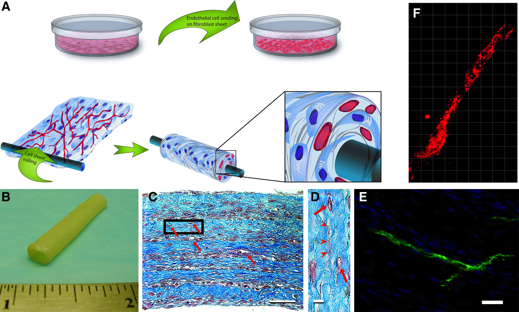
Fabrication method and characterization of TEVAwVV. (
Characterization of the mechanical properties of TEVA and TEVAwVV was assessed by uniaxial ring testing and burst pressure testing to determine the effect of vascularization on the resistance of the tissue. Tensile testing results displayed constant mechanical properties for every vascular construct in presence or absence of a vasa vasorum within the vessel wall. UTS, modulus, and failure strain measured for both TEVA and TEVAwVV were similar to those of pig coronary (Fig. 2A–C). Moreover, burst pressure values recorded showed that resistance of the tissue remain the same even with the addition of a vasa vasorum to the tissue-engineered adventitia (Fig. 2D). Results from explanted pig coronaries in burst pressure are not presented since we were not able to properly rupture the left coronary artery for comparative study purpose due to the many collateral vessels of such coronaries. Results from our tissue-engineered constructs suggest that both TEVA and TEVAwVV displayed mechanical properties suitable for implantation.
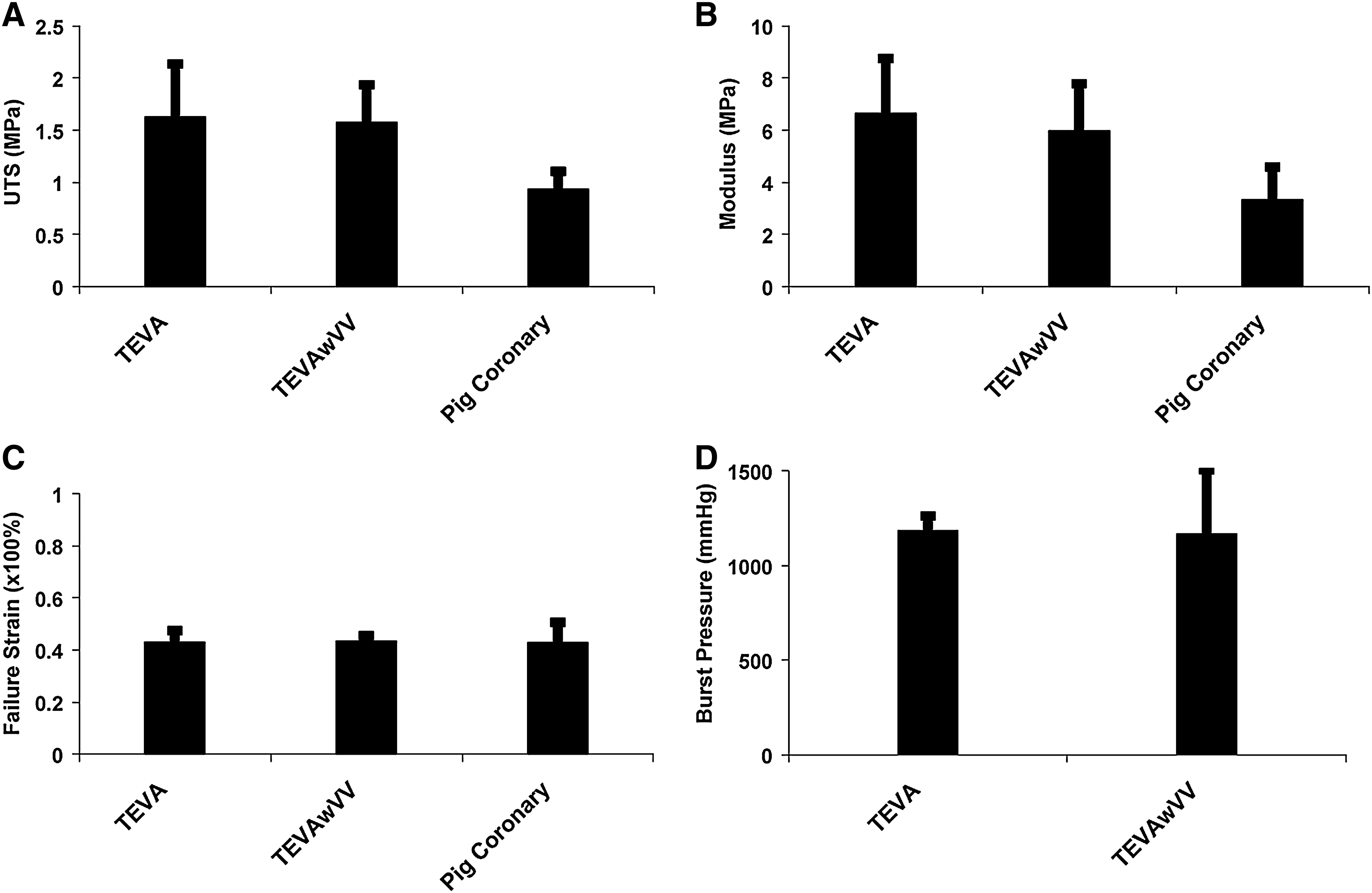
Impact of a vasa vasorum on the mechanical properties of tissue-engineered vascular constructs. Tensile properties of TEVA, TEVAwVV, and pig coronary were measured by uniaxial ring testing. Ultimate tensile strength (
Implantation of the human tissue-engineered adventitia with vasa vasorum
To ensure that the adventitial vasa vasorum engineered in vitro inosculates with the recipient vascular bed, 20-mm-long TEVAwVV and TEVA were subcutaneously implanted in athymic mice without surgical anastomoses. After removal of the fascia, a TEVA and a TEVAwVV on their respective mandrel were sutured on each dorsal muscle of the same athymic mouse. Macroscopic observations (Fig. 3) revealed no vascular connection during the first 48 h postimplantation, but graft embedding of TEVAwVV was already improved compared to TEVA, which was not integrated into the host tissue. At 1 week, TEVA started to adhere onto the dorsal muscle, but no signs of vascularization were observed. In contrast, TEVAwVV were well integrated and some blood perfusion was noticed within the vessel wall. At 2 weeks, control vessels were attached to the wound bed but did not seem to be vascularized according to macroscopic observations. In contrast, TEVAwVV were well integrated and perfused throughout the construct with mouse blood.
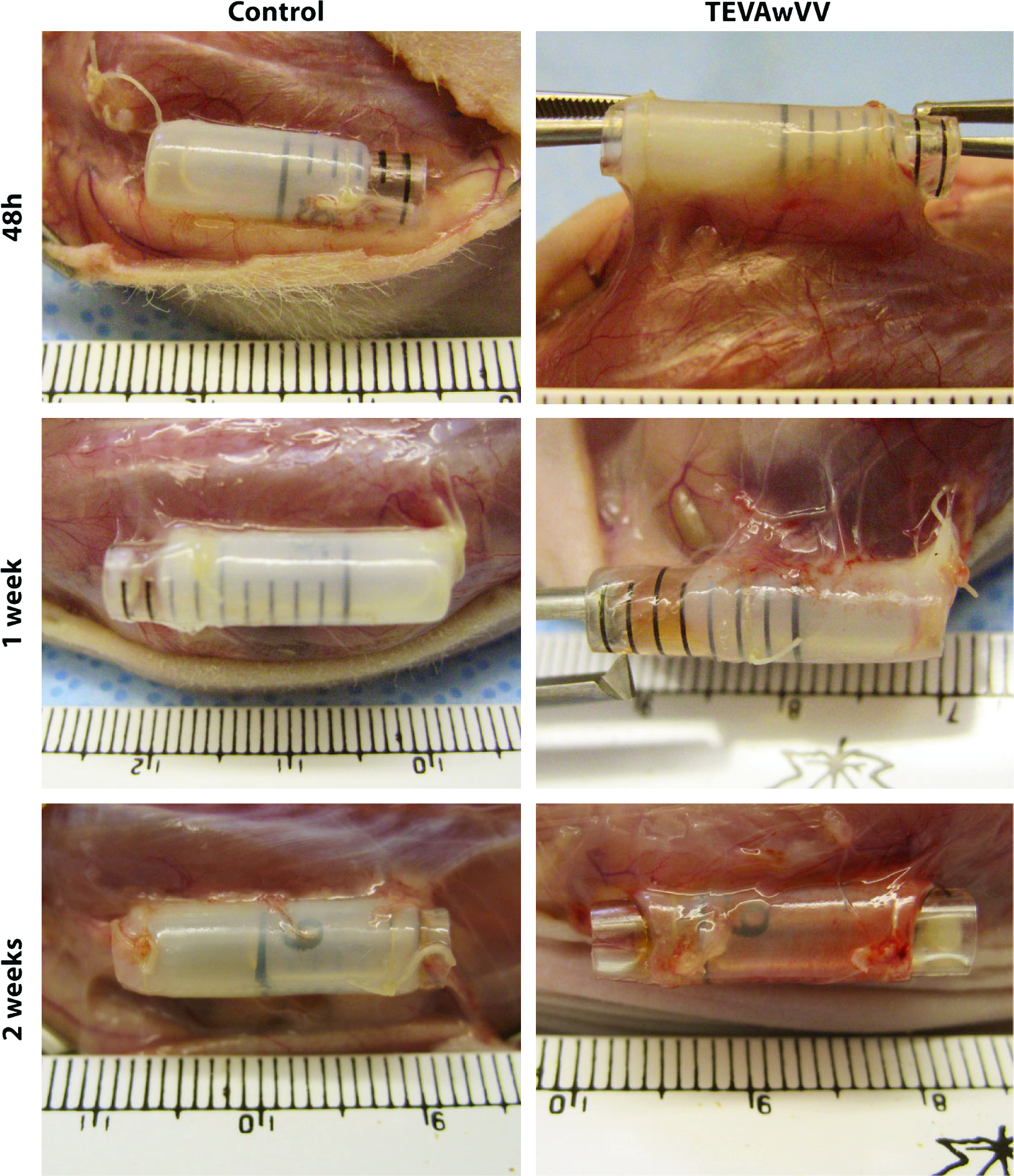
Subcutaneous implantation of TEVAwVV. Macroscopic images of TEVAwVV and control TEVA at 48 h, 1 week, and 2 weeks postimplantation. At 48 h (n = 4 for each condition), controls TEVA were held in place through sutures. Once the sutures are removed, the control TEVA can be lifted with no adherence on the dorsal muscle. TEVAwVV were embedded to the host since once the sutures were removed, a cut into the dorsal muscle was needed to lift the vessel. At 1 week (n = 6 for each condition), control vessels started to make adherences onto the dorsal muscle, but no signs of vascularization were observed. TEVAwVV are well integrated and some blood perfusion can be observed within the vessel wall. At 2 weeks (n = 6 for each condition), control vessels are integrated but do not seem to be vascularized from macroscopic observations. TEVAwVV are well integrated and perfused throughout the construct with mouse blood. Color images available online at www.liebertonline.com/ten.
Postimplantation analysis
Histological stainings were performed to verify the inosculation of the vasa vasorum with the mouse vascular bed. TEVAwVV presented mouse erythrocytes within the vessel wall 1 week postimplantation (Fig. 4A). In contrast, almost no erythrocytes were found in the control TEVA after 1 week. Further, after 2 weeks, a very dense capillary network filled with red blood cells was observed within the adventitial wall. Histological analysis revealed how the human TEVAwVV construct had nicely integrated into the mouse connective tissue (Fig. 4A). In a similar time frame, very little capillaries originating from the mouse vascular bed were observed within the vessel wall of the TEVA without vasa vasorum. Consecutive immunofluorescence sections highlighting human and mouse endothelial cells, as well as mouse red blood cells and nuclei, confirmed the inosculation process between the mouse vascular bed and the human capillary networks in the TEVAwVV at 1 and 2 weeks postimplantation (Fig. 4A).
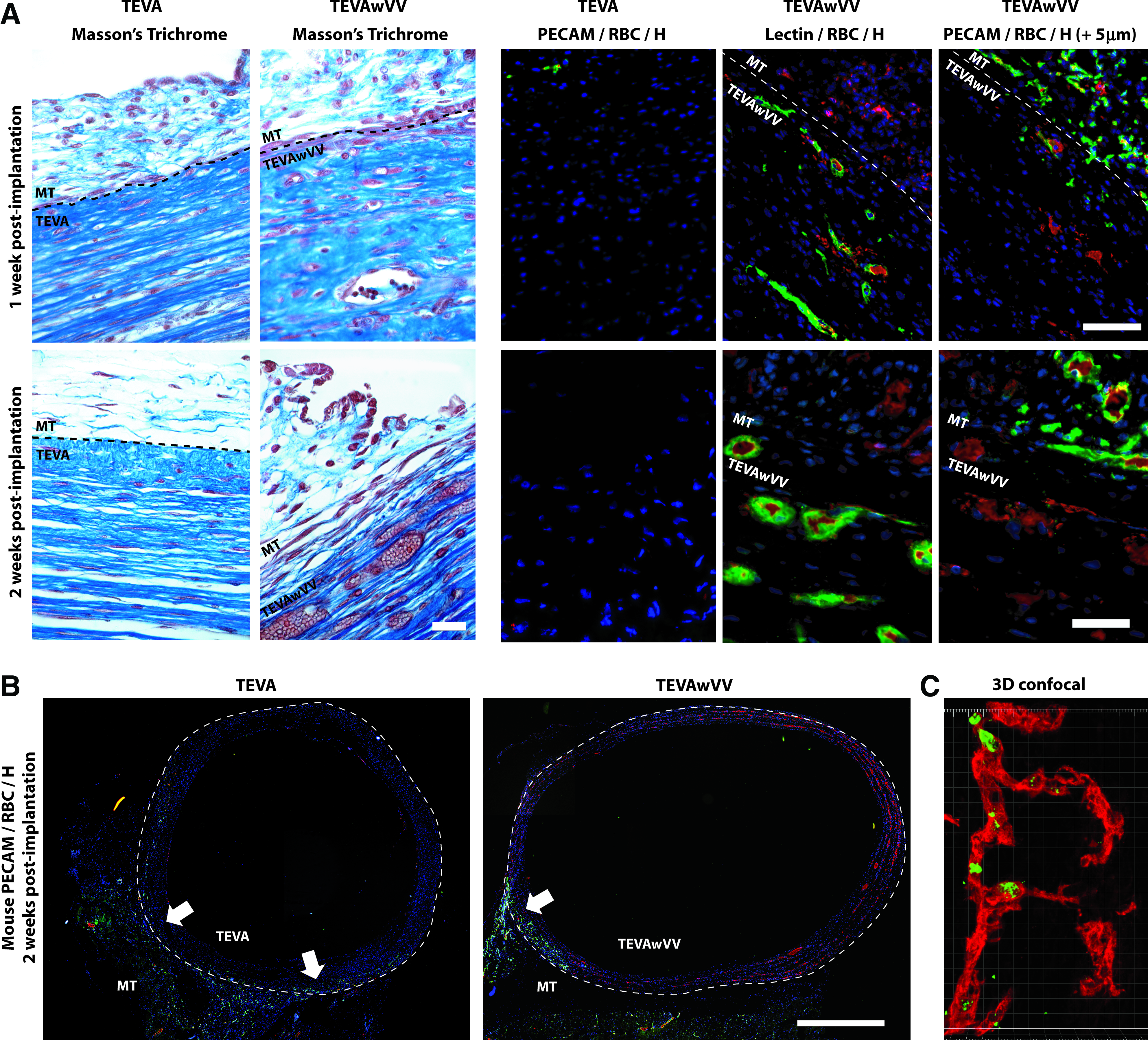
Postimplantation analysis of erythrocytes' presence in TEVAwVV. (
To locate specific zone of inosculation as well as to study the extent of vasa vasorum perfusion with mouse erythrocytes, immunostaining of mouse endothelial cells, red blood cells, and nuclei were performed. In TEVAwVV, the mouse capillaries invaded the tissue-engineered construct and inosculation occurred between the two capillary networks, thus allowing the blood flow through this connection to perfuse the in vitro created vasa vasorum (Fig. 4B). The invasion of mouse capillaries within the wall of the TEVA was observed after 2 weeks of implantation, but the majority of the vessel wall did not contain red blood cells. Mouse erythrocytes in TEVAwVV were also observed using 3D confocal microscopy (Fig. 4C).
Quantification of perfused capillaries was assessed by calculating erythrocyte-positive capillaries on randomly selected histological section of every explanted TEVA and TEVAwVV (Fig. 5). Analysis revealed a 4.6-fold increase in perfused capillaries density at 1 week postimplantation and a 12-fold increase after 2 weeks.
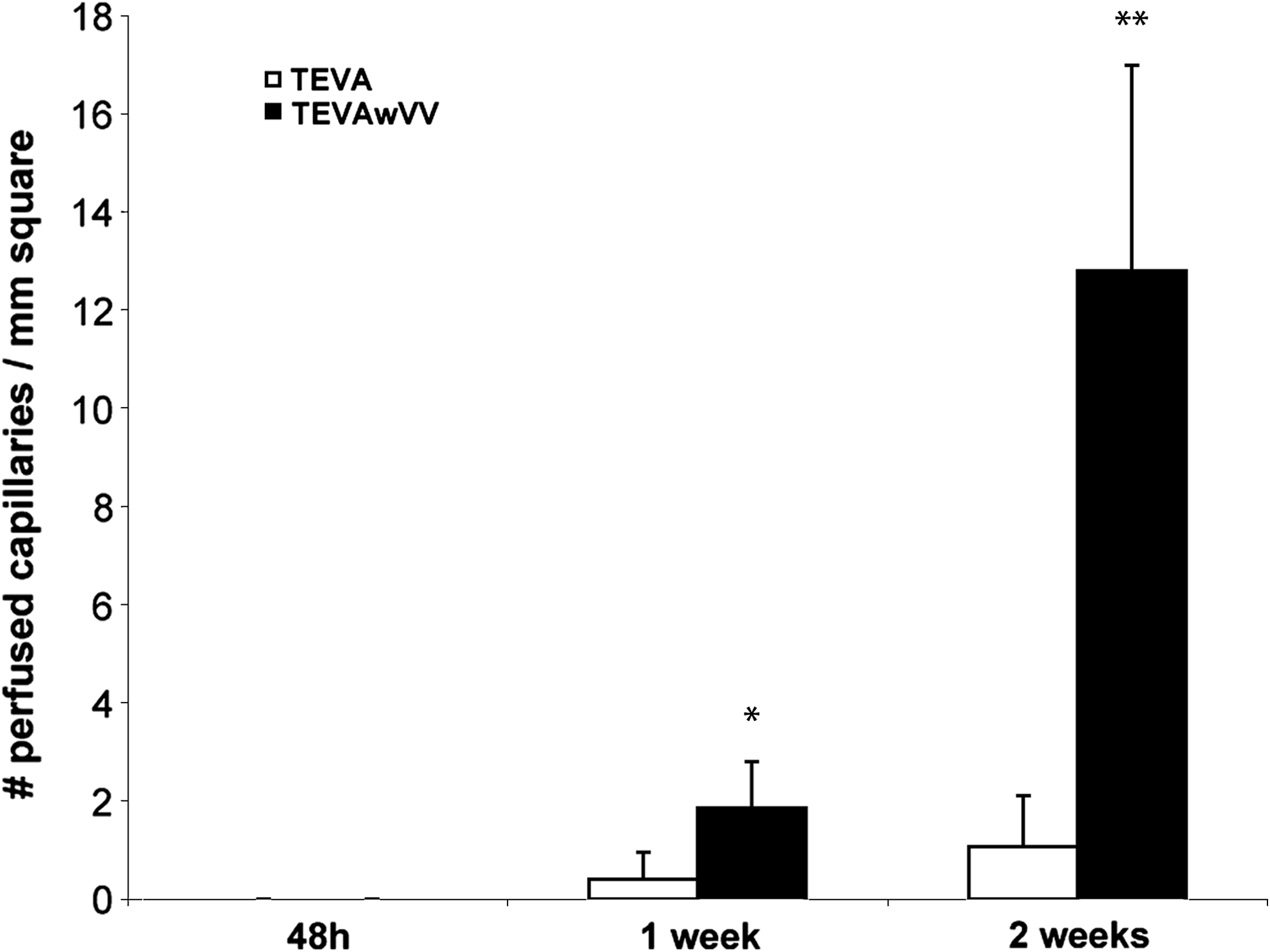
Statistical analysis of the number of perfused capillaries per millimeter square. Presence of erythrocytes was not detected at 48 h. The number of erythrocytes at 1 and 2 weeks was significantly higher for TEVAwVV when compared to TEVA. *p < 0.05, **p < 0.01. n = 6 vessels for every time point and every condition.
Long-term study of in vivo remodeling
To evaluate morphology, stability, and functionality of human TEVAwVV, a 90-day-long in vivo study was performed using an athymic mouse model. Macroscopic observations (Fig. 6A) revealed a normal vasculature within the vessel wall of TEVAwVV, as opposed to the irregular distribution of vasculature within the control TEVA. Immunofluorescence staining of consecutive sections of mouse erythrocytes and either human or mouse endothelial cells (Fig. 6B) demonstrated the presence of mouse erythrocytes inside human-lectin-stained endothelial cells, proving that the in vitro created human vasa vasorum was still functional and perfused 90 days after implantation. The use of confocal imaging (Fig. 6C) revealed a branched structure, consistent with end arteries, as well as chimeric capillaries formed of human and mouse endothelial cells, also shown in 3D confocal imaging (Fig. 6D).

Long-term results of remodeling and vascularization in TEVAwVV. (
Discussion
Microvasculature structures incorporated into tissue-engineered constructs, before grafting, as obtained by the coculture of endothelial cells and fibroblasts has been established as being of tremendous importance for tissue perfusion and its integration within the host surrounding implanted site. In the present experiment, the elaboration of a vasa vasorum within a tissue-engineered adventitia was obtained. Mechanical properties of the vascular constructs were assessed to measure the impact of a vasa vasorum within a TEVA. Results have shown that the UTS, the modulus, and the failure strain of both the TEVA and TEVAwVV were of similar value compare to the same parameters measured for a control pig coronary vessel and demonstrated supraphysiological mechanical properties for all our reconstructed tissues. Further, results also showed that the addition of a vasa vasorum did not affect the burst pressure performance of our engineered tissue. Mechanical testing results lead us to believe that the presence of a vasa vasorum in the TEVA resulted in adequate mechanical properties of the vascular constructs and that this construct could potentially be suitable for grafting. Evidence for the importance of engineering a capillary network in TEBV before implantation was provided trough the demonstration of the important role such a network has in tissue integration and vascularization. Since no vascular anastomoses were performed, the only possible connection between the mouse vascular bed and the existing capillary network inside our constructs should be through inosculation.
The in vivo study demonstrate improved graft embedding for TEVAwVV compared to TEVA at 48 h, although no inosculation was observed at such an early stage. A potential explanation for this phenomenon is that extracellular matrix remodeling could be in part due to the presence of the endothelial capillary network that may activate an angiogenic response within the construct; this possible aspect of tissue integration would necessitate further investigation. For TEVAwVV, at 1 and 2 weeks postimplantation, the inosculation between the vasa vasorum and the mouse vascular bed was accompanied by impressive graft integration. These observations could be explained by the fact that inosculation allows nutrients to flow within the tissue-engineered construct with a capillary network, in comparison to the nonvascularized construct where passive diffusion is the only mean of transportation for oxygen and nutrients. Similar results were observed with arterial bypasses using vein graft by Wyatt et al., 29 suggesting that vascularized TEBV would react to revascularization in a comparable manner to vein grafts that possess a vasa vasorum.
Long-term results revealed the presence of a remodeled vasa vasorum having a tree-like structure similar to end arteries, which corresponds to the normal anatomical structure of a coronary artery vasa vasorum. 26 Moreover, the in vitro created vasa vasorum was still functional after 3 months in vivo. The vasa vasorum connected with the capillary network of the mouse endothelial cells from both origins (mouse and human) combining to form chimeric vessels that inosculated to form a stable network. Those results demonstrated the long-term stability of the capillaries produced using the self-assembly approach of tissue engineering.
As reported by McGeachie et al. 30 using vein grafts as arterial bypasses, a diminution in microvascularization of the vessel wall in the long-term study was observed. Interesting results from L'Heureux et al. 31 have shown that graft integration and in vivo formation of a vasa vasorum occur 3 months after implantation of TEBV without vasa vasorum, which is consistent with our results for control TEVA. However, the present study showed how the addition of a vasa vasorum before implantation leads to complete tissue integration and a functional vasa vasorum after only 2 weeks in vivo. Even though it has been shown that endothelial progenitor cells have a greater angiogenic potential than HUVEC in vivo, 32 HUVEC have been used in this experiment for their stability over extended period of time in vivo. 19 A comparison study between the present results and a new set of experiments involving endothelial progenitor cells would allow evaluating the fast in vivo integration response and stability of such new constructs.
Further, this culture model could play an important role in the clinical development of larger-diameter TEBV, where diffusion limit inhibits nutrients from passing through the entire vessel wall. TEVAwVV substitutes could prove to be useful for coronary artery bypass graft surgery and peripheral artery vascular disease treatment since it should improve postimplantation integration and perfusion, thus leading more rapidly to an efficient and functional implanted tissue. The combination of developing technologies for the production of a medium layer with oriented smooth muscle cells 33 and the addition of a luminal endothelium should provide an implantable TEBV with better mechanical properties, a vasa vasorum, and a functional endothelial lumen; however, further animal and clinical studies would be required to provide evidence for the potential of such an application.
Footnotes
Acknowledgments
The authors would like to thank François Berthod, Myriam Grenier, Todd Galbraith, and Anne-Marie Moisan for technical assistance. This work was supported by the Canadian Institutes of Health Research. L.G. holds a Canadian Research Chair on Stem Cells and Tissue Engineering from Canadian Institutes of Health Research.
Disclosure Statement
No competing financial interests exist.