
Abstract
Use of silver nanoparticles (SNPs) for control of implant-associated infection is a promising strategy, if optimum antimicrobial yet nontoxic dose to mammalian cells is identified. This study was done to determine essential quantity of SNPs, which stimulate antimicrobial activity without cytotoxicity, when immobilized on poly (ɛ-caprolactone) (PCL) scaffold proposed for vascular tissue engineering. During SNP synthesis and scaffold preparation, nanoparticle aggregation was protected using poly (ethylene glycol). Transmission electron microscopy was used to characterize SNP size and to detect its mobilization from scaffold to culture medium. Antimicrobial property of the SNP and its dose response was tested using both Gram-positive and Gram-negative bacteria by zone of inhibition assay. Endothelial cells (ECs), the main cell type required for vascular tissue engineering, were grown on scaffolds to identify the nontoxic dose. After seeding EC on scaffolds, cell attachment, spreading, and viability/survival were detected using specific markers by flow cytometric/fluorescence microscopic analysis. Real-time polymerase chain reaction detected effect of SNPs on mRNA expression of selected EC-specific functional proteins. Results suggest that even devoid of antibiotics in the medium, 0.1% (w/w) SNP on PCL scaffold is antimicrobial while nontoxic to EC at cellular and molecular level once cultured on the SNP-PCL scaffold.
Introduction
Recently, silver nanoparticles (SNPs) have gained much popularity due to its ability to exert bactericidal effect at very low concentrations and due to the broad spectrum of antimicrobial activity.8–11 Silver-impregnated catheters 12 and wound dressings 13 are currently used in therapeutic applications. Silver nanoparticles have been found to exhibit promising antibacterial activities14,15 against both Gram-negative and Gram-positive bacteria including multidrug-resistant strains. 16 In addition to excellent antiviral as well as antifungal properties17,18 SNPs show anti-inflammatory effect on wound healing in thermal injury, diabetic wound, and chronic wound models. 19
However, higher concentrations of silver have been found to be toxic to host cells. 20 Therefore, while incorporating SNPs into biomaterials to impart antimicrobial effects, concentrations of silver must be optimized to get adequate antimicrobial property without tissue toxicity. Suitability of fibrin composite-coated poly (ɛ-caprolactone) (PCL) hybrid scaffold for vascular tissue engineering (VTE) applications was already evaluated and found to be promising. 21 In the present study we used endothelial cells (ECs) to identify nontoxic concentration of SNP both at cellular and molecular level aiming at a potential application in the development of scaffolds for VTE.
Materials and Methods
Materials
PCL molecular weight (M.W.) 42,500 (Sigma Aldrich), poly (ethylene glycol) (PEG) 200 (Merck), PEG 3400 (Sigma Aldrich), AgNO3 AR (s.d.fiNE.cHEM. LTD.), and dichloromethane, high performance liquid chromatography (HPLC) grade (Rankem), were used as received. Bacterial strains Staphylococcus aureus (ATCC 25923) and Escherichia coli (ATCC 25922) were used for the antimicrobial studies. Polystyrene culture dishes (Nunc), Iscove’s Modified Dulbecco’s Medium (IMDM) (Gibco), collagenase (Gibco), new born calf serum (PAA), antibiotic-antimycotic (Gibco), 0.05% trypsin–ethylenediaminetetraacetic acid (Gibco), and Texas Red Phalloidin (Molecular Probes), Annexin V-fluorescein isothiocyanate and propidium iodide (Vibrant Dye cytotoxicity Kit ≠ 2; Molecular Probes), TRI REAGENT (MRC), Nucleotide sequences (IDT), and quantitative polymerase chain reaction (qPCR) master mix (Stratagene) were used for various cell culture experiments.
Synthesis and characterization of SNPs
PEG 3400 (3.88 g) was dissolved in 10 mL of PEG 200 at 80°C with stirring. After dissolution, temperature of the solution was decreased to 40°C followed by the addition of silver nitrate (0.1 g) with mild stirring so that a clear brown solution was obtained. SNPs were characterized with ultraviolet (UV)–visible absorption spectroscopy (Diode array spectrophotometer, Hewlett Packard 8453), Infrared (Jasco 6300 FT-IR spectrometer)/Raman spectroscopy (Bruker RFS 100/s FT- Raman spectroscope), and transmission electron microscopy (TEM; Hitachi H-7650).
Preparation of SNP-PCL scaffold and analysis of nanoparticle migration
SNPs were incorporated into the polymer scaffolds at different proportions (0.1, 0.2, and 0.6 w/w%). Briefly, PEG-protected SNPs were added to a solution of PCL (SNP-PEG-PCL solution) in dichloromethane. Solutions were mixed properly, and polymer films or conduits (4 mm diameter × 8 cm length) were prepared by solvent casting method. After complete evaporation of dichloromethane, for cell culture experiments, patches of scaffold were cut into specific size (1.75 cm2 area). Control polymer films (0 Ag-PCL) were prepared without SNP incorporation, but with similar proportion of PEG. Scaffolds were used as patches of known dimension or tubular conduits fabricated from the same SNP-PEG-PCL solution for estimating release pattern, antimicrobial effect, and cell culture studies.
To confirm migration of SNPs into the medium, silver-incorporated polymer scaffolds (0.1 Ag-PCL) were incubated in sterile de-ionized water (DI/W) for different intervals with complete change of water after every 24 h. One drop (~50 μL) of the collected medium after different intervals of incubation was placed on a formvar-coated grid and dried. Images were acquired in a transmission electron microscope. Scaffolds before leaching and after leaching were analyzed by environmental scanning electron microscopy (Quanta 200, FEI make). With known concentration of SNP, reaction with HCl showed turbidity (% transmission measured at 480 nm using spectrophotometer HP8453), which correlated to the concentration of nanoparticles. Calibration curve was used for quantification of release to water by mixing 0.5 mL water collected at each time period with 0.5 mL 1 N HCl and the turbidity was measured.
Susceptibility of bacterial strains to SNPs was evaluated by standard zone of inhibition assay (ZIA). Test and control tubular scaffolds were incubated in the culture medium for 72 h at 37°C. The medium with SNP was collected, 50 μL was blotted on to absorbent discs, and antimicrobial activity was tested against Gram-positive (S. aureus ATCC 25923) and Gram-negative (E. coli ATCC 25922) bacteria by the standard method. Gentamicin disc (10 μg) was used as positive control. The plates, placed with test discs, were incubated for 18–24 h at 37°C and zone of inhibition was documented.
Cytocompatibility experiments
Human umbilical vein ECs isolated as per the method of Jaffe et al. 22 were cultured and subcultured as described earlier. 23 Umbilical cords were obtained as per the institutional ethics guidelines from a local maternity hospital after informed consent. Cells from passages 3–5 were seeded at a density of 2 × 10⁴ cells/cm2 of scaffolds for all cytocompatibility experiments and cultured in serum and growth factor-supplemented IMDM without antibiotics. Cell culture experiments were done on test scaffolds such as 0.1, 0.2, 0.6, and 0 Ag-PCL, which served as a negative control for all assays.
Cells on each scaffold were analyzed for actin assembly after 48 h of culture to appreciate cell adhesion and spreading. The cells were washed three times with phosphate-buffered saline (PBS; pH 7.4) and fixed using 0.2% gluteraldehyde in PBS. The fixed cells were permeated using 0.1% Triton ×100. After three repeated washes with PBS, cells were stained with Texas Red Phalloidin as per manufacturer's instructions. Cells were washed and viewed under a fluorescence microscope (Leica DM IRB).
The apoptosis of EC grown on scaffolds was quantified using Vybrant Apoptosis Assay Kit (Molecular Probes). Cells were harvested after 72 h culture on 0/0.1/0.2/0.6 Ag-PCL scaffolds, and stained according to manufacturer's protocol. Stained cells were washed, re-suspended in FACS sheath fluid, and analyzed using a flow cytometer (FACS Aria; BD Biosciences) to estimate the percentages of live/apoptotic/dead cells.
Effect of SNPs on functional mRNA
Cells were harvested after 24 and 72 h culture on 0/0.1/0.2 Ag-PCL scaffolds. As the cell death was unacceptably high on 0.6 Ag-PCL, cells on this scaffold were excluded from molecular analysis. RNA was isolated using TRI reagent according to manufacturer's direction. RNA was quantified by spectrophotometric measurement (Diode array spectrophotometer, Hewlett Packard 8453). Expression of specific mRNA was studied using the primer sequences as shown for each molecule: prothrombotic gene, von Willebrand factor (vWF: F, caccattcagctaagaggagg; R, gccctggcagtagtggata), antithrombotic genes tissue plasminogen activator (tPA: F, atgggaagacatgaatgcac; R, gaaaggggaaggagacttga), and endothelial nitric oxide synthase (eNOS: F, agctgtgctggcatacagga; R, atggtaacatcgccgcagac). Twenty nanogram of total RNA was taken for reverse transcription reaction using thermal cycler (Master cycler; Eppendorf). To compare the mRNA expression levels at 24 and 72 h, the real-time PCR was performed using a Chromo4 system (MJ Research). All reactions were carried out in a total volume of 20 μL containing 10 μL qPCR Mastermix, 200 nM forward primer, 200 nM reverse primer, and 8 μL template cDNA, for 35 cycles. For each gene, an assessment of quality control was performed by examining PCR melt curves after real-time PCR to ensure specificity. The cDNA copy numbers of the target gene were analyzed after being normalized to the copy number of glyceraldehyde 3-phosphate dehydrogenase (F, attggctttggtccgagtcc; R, gggggttctttggcttttac). Fold change was calculated by comparing the 24 and 72 h extracts of RNA, respectively, with 0 h samples (extracted before seeding on scaffolds) after making correction for PCR efficiency.
Statistical analysis
All the experiments (qualitative and quantitative) were carried out with at least four replicates in each group. The mean and standard deviation (SD) were calculated for quantitative data and the results were expressed as mean ±SD. For comparison of quantitative results, significance of difference between two groups was detected using analysis of variance (ANOVA single factor); p < 0.05 was considered as statistically significant.
Results
Characterization of SNPs
On mixing at 80°C both PEG 3400 and PEG 200 formed a clear viscous solution, and after the temperature was brought down to 40°C, it still remained as a clear solution. Soon after adding silver nitrate a brown coloration developed, indicating SNP formation. The UV-Visible spectrum of SNPs showed the surface plasmon resonance of nanosilver at 307 nm. A shoulder was obtained in the range of 400–500 nm (Fig. 1A). Infrared spectrum of PEG shows the characteristic –OH stretching at 3391 cm−1 and C-O-C stretching at 1102 cm−1, which has shifted to 3399 and 1105 cm−1, respectively, after embedding SNPs with PEG (Fig. 1B). Apart from these two peaks C-H stretching at 2872 cm−1 and bending at 1455 cm−1 and OH bending at 1350 cm−1 are also seen in both the spectra. Figure 1C shows the Raman spectral features of PEG without nanoparticles in comparison with that of PEG with SNPs. Raman peaks at 838 and 855 cm−1 represent the skeletal vibrations in the PEG chain. The peak at 1057 cm−1 indicates the stretching -C-O-H bond and the -CH2- twisting vibration mode is indicated by the peaks at 1118 and 1136 cm−1.

Spectral characteristics of silver nanoparticles (SNPs).
TEM images confirmed the size of SNPs to be ~20 nm and shape of the particle was spherical (Fig. 2A) consistently in all batches prepared. Nanoparticles that migrated into the DI/W from the PCL scaffold at different time intervals are shown in Figure 2B–D. While the PEG-protected nanoparticles remained as single entities before impregnation into the scaffold, after they were migrated into the medium from the scaffold, aggregation of nanoparticles was also seen, indicating the loss of PEG protection of SNPs as they leach out.

Transmission electron microscopy images of SNPs. Images were taken at ×6.0 K.
Release of SNPs from scaffold
The ultimate objective is to make porous scaffold for VTE; therefore, PEG 200–protected SNPs were mixed with PEG 3400 (porogen) to obtain sufficient porosity after leaching of porogen into medium. Before incubation with DI/W or culture medium the scaffold showed a continuous morphology (Fig. 3A). During incubation of scaffold with water the porogen leached out along with SNPs and porous nature of the scaffold is seen on analysis using environmental scanning electron micrographs (Fig. 3B). The pore size was found to be ~5 μm.
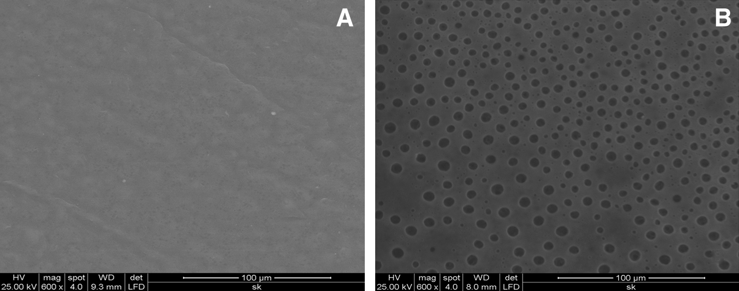
Environmental scanning electron microscopy images of scaffolds.
Release profile of the SNPs from the scaffold is shown in Figure 4. When known quantity of PEG-protected SNP was added into DI/W and kept for 24 h, the PEG dissolved and the SNP reacted with added HCl and developed turbidity. Measured turbidity was proportional to the quantity of SNP in the reaction mixture as seen from the calibration curve (Fig. 4A). Detection method showed linearity of measurement only from 2 to 12 μg/mL of SNP; so, the released SNPs were diluted to make it detectable and total release was calculated. To analyze release profile of the scaffold DI/W collected after different intervals of incubation with PCL scaffold was allowed to react with HCl and confirmed that SNPs got released continuously from the scaffold and the quantity was detectable using the analysis method developed (Fig. 4B). The starting total SNP concentration on PCL scaffold (4 mm diameter × 6 cm length) was 54 ± 5.43 μg per scaffold. It was seen that during the initial period of 24 h a major portion of the incorporated SNPs were released and by 72 h about 90% got released (Fig. 4C).

Quantification of SNP release.
Antimicrobial activity of SNPs migrated from PCL scaffold
Significant antibacterial activity was found in ZIA when the releasate from 0.1 Ag-PCL was loaded on the filter disc (Fig. 5) and gentamicin placed in the middle showed a large zone of inhibition, but the zone is diffused as compared to the clear zones around the SNP-loaded discs. While quantity of gentamicin on the disc was 10 μg, estimated quantity of SNP on 0.6 Ag disc was 25 ng, on 0.2 Ag disc it was 8 ng and on 0.1 Ag disc it was 4 ng. Quantity of SNP loaded on the disc was calculated assuming that 100% of SNP from the scaffolds were leached into the medium in 7 days period. Out of 3 mL medium in which scaffolds were immersed for mobilizing SNPs, only 50 μL was used for loading on the discs used for ZIA. Response pattern of E. coli (Fig. 5A) and S. aureus (Fig. 5B) are shown. Zone of inhibition was found to be dose dependent in Gram-positive and Gram-negative bacterial inhibitions, with released SNPs from 0.6 Ag PCL showing the widest inhibition zone. Up to 7 days, SNPs were detected in the medium, indicating that there was continuous and slow migration, and later it was undetectable.

Representative patterns of zone of inhibition assay.
Cell growth and survival
By actin staining of cells, their attachment and spreading in various Ag-containing scaffolds could be distinguished. Cells were well attached and spread on 0.1 Ag-PCL and the morphology of EC was comparable with that on control (0 Ag-PCL). On the other hand, cell attachment and spreading were found to be less at 0.2 Ag-PCL scaffolds compared to control samples (Fig. 6A–C). Cell survival, necrosis, and apoptosis on all scaffolds were quantified using flow cytometric analysis of cells harvested from the culture after 72 h of seeding. All cells including the floating cells in the medium were used for staining. Dead cells are propidium iodide-positive cells in quadrant1 (Q1), apoptotic cells positive for Annexin V-Alexa flour 488 are in Q4, whereas dual-positive apoptotic and dead cells are in Q2. In Figure 7, representative dot plots of each scaffold type is shown. Four such experiments were done in each case and mean ± S.D. is shown in Figure 7F. Consistent results with low SD was found on each type of scaffold. When the live/dead/apoptotic cell percentage on 0 Ag-PCL was compared against the other SNP-containing scaffolds, significant difference (p ≤ 0.05) was seen in the case of 0.2 and 0.6 Ag-PCL, whereas EC on 0.1 Ag-PCL was comparable with control.

Actin-stained human umbilical vein endothelial cells (ECs) on scaffolds. Texas Red Phalloidin staining for actin was done after 48 h cell culture on scaffolds.

Apoptosis assay of EC on PCL scaffolds. Viability of ECs on SNP-incorporated PCL scaffolds by Annexin V-fluorescein isothiocyanate and propidium iodide staining after 72 h.
Regulation of mRNA
If cells on the scaffold have changed their phenotype and become more prothrombotic, it can lead to graft thrombosis when tissue-engineered grafts are implanted. Therefore, analysis of selected molecules was carried out to ensure phenotype stability of EC when cultured on Ag-PCL scaffold. Even though 0.6 Ag-PCL seemed to have high antimicrobial effect, because number of live cells on this scaffold was relatively very low, these were not included for phenotype evaluation. Quiescent expression of prothrombotic vWF was comparable on 0.1 and 0 Ag-PCL (Fig. 8); however, statistically significant upregulation on 0.2 Ag-PCL samples was observed at 24 h as well as 72 h. Fold change was calculated with respect to expression of vWF in cells grown on tissue culture polystyrene harvested for seeding on scaffolds. Expression of tPA was found to be downregulated on 0.2 Ag-PCL at both periods of study (Fig. 9) as compared to the EC obtained before seeding on scaffolds. Expression of eNOS was significantly downregulated in 0.2 PCL-Ag scaffolds (Fig. 10) after 24 h, but by 72 h the difference between scaffolds was insignificant. Both the antithrombotic molecules were quiescent in EC grown on 0 and 0.1 Ag-PCL. Thus, there is an optimum concentration of SNPs at which maintenance of normal EC phenotype was established, whereas excess concentration of SNPs is shown to affect the cells adversely at molecular level.

Quantification of mRNA expression for von Willebrand factor. mRNA was isolated from EC grown for 24 and 72 h on each type of scaffold. mRNA from cells grown on tissue culture polystyrene was used as control and used for fold increase of expression. *Significant difference when compared with control (p < 0.05).

Quantification of mRNA expression for tissue plasminogen activator. mRNA was isolated from EC grown for 24 and 72 h on each type of scaffold. mRNA from cells grown on tissue culture polystyrene was used as control and used for fold increase of expression. *Significant difference when compared with control (p < 0.05).

Quantification of mRNA expression for endothelial nitric oxide synthase. mRNA was isolated from EC grown for 24 and 72 h on each type of scaffold. mRNA from cells grown on tissue culture polystyrene was used as control and used for fold increase of expression. *Significant difference when compared with control (p < 0.05).
Discussion
Objective of this study was to validate incorporation of SNPs with biodegradable scaffold to impart antimicrobial properties for tissue engineering applications. Before the scaffold is used for in vitro tissue engineering, it is essential to identify an optimum concentration of SNPs that would show antimicrobial activity at the same time not having any cytotoxic effect on the cells of the targeted tissue. The porous PCL scaffold was earlier investigated by our group and found to have adequate physicochemical properties for vascular tissue construction. Therefore, the modified scaffold with impregnated SNPs was investigated using ECs specifically to identify noncytotoxic dose range. Chemical reduction with PEG was used as a convenient method to protect nanoparticle from aggregation. Since PEG is miscible with polymer mixture and being water soluble, it migrates into tissue culture medium and exerts its antimicrobial effect during cell culture. In addition, as the PEG-protected SNPs migrate into medium with time, long-term detrimental effect on cells is unlikely to occur.
It is known that PEG can protect SNPs through the reduction of the hydroxyl group to aldehyde group. It is reported that the monomer ethylene glycol is not showing any reduction of silver as it requires an additional polymer such as PVP for the nanoparticle formation.24,25 The reducing activity of PEG is very sensitive to its M.W., the longer polymer chain of PEG exhibiting higher activity. 26 Recently, it was reported that PEG was able to act both as reducing agent and stabilizer. 27 In one study PEG was used as a stabilizing and reducing agent to form SNPs in the presence of UV light. 28 It has been reported that PEG form the most stabilized SNPs at 80°C. 29 In another report 30°C was found to be the correct temperature needed for the synthesis of SNPs. 30 PEG not only protects the nanoparticles from precipitation but also plays a critical role in the size, size distribution, morphology, and the biocompatibility of the resulting SNPs. In certain cases the stabilizing agent itself acts as reducing agent resulting in a one pot synthesis of SNPs. In this report we have used both low M.W. as well as long chain PEG for stabilizing the SNPs formed. The result is promising with uniform-sized (~20 nm) stable particles, which were easily incorporated with PCL.
In recent years interest grew toward the utilization of SNPs rather than other antibiotics due to its broad antimicrobial activities against Gram-negative and Gram-positive bacteria, ability to inhibit polymicrobial colonization, and the minimal possibility of developing bacterial resistance. It is demonstrated that SNPs inhibit the growth and multiplication of bacterial strains, including multidrug-resistant strains. 16 This study also investigated the antimicrobial property of prepared SNPs against both Gram-positive and Gram-negative bacteria and found to be effective against both types. Investigation of the effect of size of SNPs on antibacterial property using four types of Gram-negative bacteria—E. coli, Vibrio cholerae, Pseudomonas aerugenosa and Salmonella typhi—revealed that the bactericidal activity of the particles increases with decrease in size of the particles. 30 The larger the surface area (smaller the particle size), the greater will be the binding ability and hence exhibit greater bactericidal effect. The mechanism of action of SNPs is still not well understood. According to previous reports, interaction between SNPs and constituents of bacterial membrane causes structural changes and damage to membranes, finally leading to cell death. 31 It is speculated that sulfur-containing proteins in the membrane or inside the cell and phosphorous-containing elements like DNA/RNA are likely to be the preferential site for SNP binding. 14 In this study we demonstrate changes in the membrane structure as observed by actin staining experiments and at molecular level by mRNA studies when higher concentration of SNPs are exposed to EC.
SNPs can be used as effective growth inhibitors against various microorganisms, making them applicable to diverse medical devices and antimicrobial control systems. 18 Many attempts have been made to incorporate SNPs into the biomaterial for continuous release to impart antibacterial activity. Nanoparticulate bone cement shows high effectiveness even against multidrug-resistant bacteria without in vitro cytotoxicity. 32 Significant reduction in bacterial adhesion was observed on titanium implants coated with silver-hydroxyapatite layer. 33 In vitro antibacterial and biological activity of silver-incorporated bioglass system was evaluated and appears to be a promising material for dental applications. 34 Attempts were made to investigate the feasibility of SNPs impregnation into polymeric scaffolds for various antimicrobial applications.35,36 SNP-coated polyurethane foams were evaluated as an antimicrobial water filter and was found to be promising. 37
The present study investigated incorporation of SNPs into PCL scaffolds to impart antimicrobial property through release into the culture medium. The clear zone of inhibition indicates the strong antimicrobial property of the SNPs released from the scaffolds. As per the release study results up to 40 μg/mL was released during the first 24 h and at later period about 10 or 5 μg was released into 1 mL water or culture medium. Concentration as low as 5 μg/mL in water was found sufficient for imparting antimicrobial effect against both Gram-positive and Gram-negative microbes. Along with PEG, SNPs are expected to migrate because of the solubility of the former in aqueous medium, and the SEM images further confirm that PEG has migrated out, thus making the scaffold more porous. Using TEM analysis, small aggregates (two to three particles) were detected in the medium, but there were many particles that remained as single entity also. Because antimicrobial effect was studied using the SNPs that migrated into the culture medium, it is confirmed that these particles that are seen in TEM have sufficient activity. We have successfully grown EC on 0.1 Ag-PCL for a period of 12 days without any microbial contamination even though no antibiotic was added into the culture medium (data not shown). It is promising to observe that in spite of ~90% release of SNPs in 72 h of incubation in DI/W contamination control of the cultures were possible up to 12 days when EC was grown on these newly developed scaffolds.
High concentrations of silver have been found to be toxic to human cells—10 mg/l has been found to be the maximum toxic dosage. 38 It was found that higher concentrations of SNPs reduce the number of fibroblast and alter their fission under in vitro culture conditions. 39 So, while modifying biomaterial surfaces, the concentration of silver needs to be selected so that it can reduce bacterial adhesion and proliferation with minimal tissue toxicity. 10 The maximum concentration of SNPs that was present in our cultures during initial 24 h amounted to 40–50 μg/mL (0.1 Ag-PCL), which is a safe range for EC, but when 80–90 μg SNP (0.2 Ag-PCL) was present in the medium, toxicity was clearly evidenced at cellular and molecular level.
Maintenance of normal phenotype of endothelium determines the ultimate success of tissue-engineered vascular graft and it is modulated by expression of prothrombotic and antithrombotic molecules. We investigated expression of these molecules in the presence of SNPs. From qPCR data, it is evident that on 0.1 Ag-PCL, expression of prothrombotic factor (vWF) and antithrombotic factors (tPA and eNOS) is comparable to that of controls, whereas on 0.2 Ag-PCL the regulation has been shifted to a prothrombotic state.
The current study focused on in vitro toxicity of SNPs, but as scaffold is proposed for implant application, a question may be posed about the systemic toxicity of SNPs. Roe et al. investigated subcutaneous implantation of SNP-coated plastic catheters in mice to evaluate systemic toxicity by measuring silver biodistribution. 40 It was found that ~50% of released silver was excreted in the feces and ~20% remains with tissues in direct contact with the catheters with no significant accumulation in other major organs. There was a 4000-fold silver gradient over a distance of ~3–10 mm, suggesting a very limited diffusion of released silver through tissues. In another study, silver nanocrystals capped with bovine serum albumin were intraperitonially injected in rats for in vivo pharmacokinetic studies in tissue from all major organs. 41 Significant accumulation of Ag in liver, spleen, and kidney was observed proposing possible urinary and biliary excretion routes for nanostructured silver. Sadauskas et al. used gold nanoparticles as a model system when administered intravenously and intraperitoneally. 42 Rapid accumulation of gold nanoparticles was found in liver macrophages, whereas uptake in spleen macrophages was moderate. None of these studies indicated the carcinogenic or immunopathological phenomenon associated with nanoparticle accumulation in organ system. The biodistribution of SNPs in in vivo conditions is not well studied and is a concern for potential therapeutic applications. However, in our system we aim to utilize the antimicrobial property of SNPs during in vitro VTE. As major portion of the impregnated SNPs leach out to the medium during in vitro culture period, possibility of in vivo toxicity after implantation is minimal or absent.
Conclusions
This study demonstrates that use of PEG, as a reducer and stabilizer for the preparation of SNPs, is a convenient method for PCL scaffold preparation. Advantages of PEG are the biocompatible nature and miscibility with aqueous and nonaqueous solvent, which enables migration of PEG into medium. By leaching of PEG into medium it serves as a porogen to impart scaffold porosity and simultaneously removes SNPs from the scaffold to eliminate long-term toxicity to cells. The efficacy of SNP-incorporated PCL scaffold to support the EC growth, survival, and normal phenotype maintenance was proved in addition to the antimicrobial property. It was also verified that higher concentrations of SNPs adversely affect EC growth and survival. This study demonstrates that a controlled concentration of SNP-PCL hybrid scaffold has the potential to act as VTE scaffold with antimicrobial property. Since major portion of the SNP is released out during the initial phase of the cell culture for the in vitro tissue engineering, postimplantation toxicity to the recipient is unlikely. For in vitro tissue-engineered vascular graft, this scaffold is found suitable because in 12 days of dynamic culture, construct becomes ready for implantation and during this period contamination control is found to be effective even though the standard method of antibiotic supplementation was avoided.
Footnotes
Acknowledgments
The authors acknowledge the Director, SCTIMST, and the Head, BMT Wing, SCTIMST, for the facilities provided. We would like to acknowledge funding support from the Department of Biotechnology, Government of India, and the individual fellowship support from the CSIR, Government of India (Ragaseema). Technical support obtained from Mr. Unnikrishnan S. (JRF), Mr. Pradeep (Technical Assistant) for ZIA, Mr. Rejin (Technical Assistant) for scaffold preparation, and Dr. Annie John (Scientist) and Ms. Susan Mani (JRF) for TEM analysis is appreciated.
Disclosure Statement
No competing financial interests exist.