




Abstract
In this study, novel fibrous matrices were developed as a depot to store and liberate growth factors in a controlled manner. Specifically, heparin was covalently conjugated onto the surface of fibrous matrices (composites of poly[caprolactone] and gelatin crosslinked with genipin), and basic fibroblast growth factor (bFGF) was then reversibly immobilized. The immobilization of bFGF was controlled as a function of the amount of conjugated heparin. The sustained release of bFGF from the fibrous matrices was successfully achieved over 4 weeks whereas physical adsorption of bFGF released quickly. The bFGF released from the fibrous matrices significantly enhanced in vitro proliferation of human umbilical vein endothelial cells. From the in vivo study, the group implanted with a higher amount of immobilized bFGF significantly facilitated neo-blood vessel formation as compared with other implantation groups. These results indicate that the sustained release of bFGF is important for the formation of blood vessels and that our fibrous matrices could be useful for regulation of tissue damage requiring angiogenesis. Further, our system can be combined with other growth factors with heparin binding domains, representing a facile depot for spatiotemporal control over the delivery of bioactive molecules in regenerative medicine.
Introduction
Blood vessel formation is required for regeneration of ischemic myocardium or hind limb as well as for survival of transplanted tissues of large size.4–7 In particular, the interactions of ECM molecules with growth factors such as vascular endothelial growth factor (VEGF) and basic fibroblast growth factor (bFGF) are important in the formation of blood vessels. 8 Formation of stable blood vessels takes several weeks to months, and administering high levels of VEGF via bolus injection was shown to result in a hemangioma-like assembly, indicating that these bioactive factors should be available in the local area at an appropriate concentration during the entire angiogenic process. 9 Although ischemic conditions act to moderate in vivo over-expression of VEGF, the controlled liberation of these growth factors is primarily dependent on their binding to ECM components.10,11 Both VEGF and bFGF have high affinity to heparin through their heparin binding domains, which are rich with positively charged amino acids. 12 Presumably, the binding of growth factors to heparin controls their release in response to cellular demands and, further, protects them from degradation by proteases. 13 It was reported that use of a heparin-modified hydrogel with human mesenchymal stem cells increased local availability and sustained autocrine signaling of a model heparin-binding growth factor that was secreted by the encapsulated cells without any exogenous treatment. 14
During the past decade, many efforts to induce therapeutic angiogenesis have included attempts to deliver VEGF and bFGF by mimicking the biological processes of the ECM.1,15 Heparin has been conjugated to synthetic and natural polymers to immobilize bFGF or VEGF via affinity binding, and these conjugates have been further formulated as nanoparticles, porous scaffolds, fibers, and hydrogels.16–21 In addition, both the partial sulfation of a natural polymer and the employment of the perlecan domain I as an engineered growth factor binding domain have been investigated as alternatives to heparin.22,23 Although the duration of continuous release and activity of released growth factors could vary depending on specific experimental conditions, these strategies have been effective in minimizing the initial burst of growth factor release and maximizing the local availability of growth factors needed to improve cell proliferation and blood vessel formation.24–26
Electrospinning is an attractive method for the fabrication of fibrous matrices from many biodegradable polymers, because it provides control over the fiber diameters within a nanometer range. 27 This method has the ability to provide large surface areas, increased porosity, and a highly interconnective pore network of electrospun fibers. For these reasons, electrospinning has been utilized to prepare cell culture substrates and tissue engineering scaffolds that have shown to be effective for cell adhesion and growth as well as for local and sustained delivery of bioactive signals.28–31 Although currently developed electrospun fibers showed a moderate ability to mimic the three-dimensional fibrous structure of ECM, recapitulation of certain biophysical events of natural ECM including intercellular signaling by cell-specific binding and modulation of growth factor liberation has not yet been fully achieved.
Our ultimate goal is to develop an artificial ECM that provides a biomimetic environment to promote or suppress specific biological signals as natural ECM does. Given that, we developed novel fibrous matrices that are able to control the release of an angiogenic growth factor, bFGF, by exploiting the reversible binding of bFGF and heparin. The objectives of our study were as follows: (1) to investigate the effect of heparin concentration on the amount of affinity-bound bFGF, (2) to monitor the in vitro release kinetics and activity of the bound bFGF, and (3) to assess the effect of the amount of released bFGF on the formation of new capillary network using athymic mice. The bFGF released from the fibrous matrices significantly enhanced in vitro proliferation of human umbilical vein endothelial cells (HUVECs) and in vivo neo-blood vessel formation, suggesting that our system could be useful for regeneration of tissue loss requiring angiogenesis.
Materials and Methods
Materials
Poly(caprolactone) (PCL) (MW = 80,000) and gelatin type B (from bovine skin) were purchased from Sigma-Aldrich. Genipin (MW = 226.23) and 1,1,1,3,3,3-hexafluoro-2-propanol (HPLC grade) were purchased from Wako Pure Chemical Industries. Polyoxyethylene bis(amine) (PEG-diamine, MW = 3350), 2-(N-Morpholino) ethane sulfonic acid (MES) sodium salt (MW = 217.22), 1-ethyl-3-(3-dimethylaminopropyl)-1-carbodiimide hydrochloride (EDC), N-hydroxysuccinimide (NHS), toluidine blue O (TB, MW = 305.83), and ethylenediaminetetraacetic acid (MW = 372.24) were purchased from Sigma-Aldrich. Heparin sodium salt was purchased from Acros Organics. bFGF (17.3 kDa) was purchased from Peprotech. Fluorescein isothiocyanate (FITC)-labeled heparin was purchased from Invitrogen. Tris-base (MW = 121.14), 2-mercaptoethanol, glycerol, sodium deoxycholate, triton X-100, dimethyl sulfoxide, 2-bromophenol blue sodium salt, and sodium dodecyl sulfate (SDS) were purchased from Sigma-Aldrich. Coomassie brilliant blue was purchased from Bio-Rad. Dulbecco's phosphate-buffered saline, trypsin, and penicillin-streptomysin were purchased from Gibco BRL. Endothelial cell growth medium-2 (EGM®-2) was purchased from Lonza group Ltd. Sucrose (MW = 342.24), bovine serum albumin (MW = 66 kDa), 3-(4,5-dimethylthiazol-2-yl)-2,5-diphenyl tetrazolium bromide (MTT), glycine (MW = 75.07), and SDS (MW = 288.38) were purchased from Amresco. Transwells (pore size: 8 μm) and culture plates were purchased from Costar® (Corning Inc.). Antibodies against smooth muscle (SM) α-actin and von Willebrand Factor (vWF) and proliferating cell nucleus antibody (PCNA) were purchased from Abcam. A secondary antibody conjugated with FITC was purchased from Jackson ImmunoResearch Laboratories.
Preparation of bFGF-immobilized PCL-gelatin fibrous matrices
Figure 1A presents the schematic illustration of the preparation of bFGF-immobilized PCL-gelatin fibrous matrices. PCL and gelatin were separately dissolved in 1,1,1,3,3,3-hexafluoro-2-propanol (6% w/v), and the PCL-gelatin blend with a weight ratio of 50:50 (PCL:gelatin) was prepared by thorough stirring. PCL-gelatin blend solution was then injected from a stainless steel needle connected to a syringe using syringe pumps (KD Scientific) at a rate of 1 mL/h. The applied voltage between the syringe needle and the grounded collector was 15 kV, and the collector was placed 15 cm from the tip of the needle to collect the fibrous matrices produced. The prepared fibrous matrices were dried overnight at room temperature and then crosslinked for 24 h by immersion in 2.0% (w/v) genipin as previously described. 32 The PCL-gelatin fibrous matrices crosslinked with genipin (PG) were then rinsed with ethanol thrice and dried overnight at 50°C. For immobilization of bFGF, the carboxylic groups on PG were first activated by a reaction with EDC or NHS solution (0.1 M, pH 5.0 MES buffer), which was subsequently reacted with PEG-diamine for 6 h (PG-D), and then rinsed with MES buffer thrice for 12 h. Then, various concentrations of heparin were activated with the EDC or NHS solution, which had been reacted with the PG-D. After conjugation of heparin on PG-D (PG-D-H) for 3 h, all unreacted heparin was eliminated by rinsing the matrices with MES buffer thrice. For immobilization of bFGF, the PG-D-H fibrous matrices were reacted with 100 ng/mL bFGF, 0.1% bovine serum albumin, 5% sucrose, and 0.01% ethylenediaminetetraacetic acid for 12 h at room temperature. After the whole process, the morphology of fibrous matrices was investigated using scanning electron microscopy (JEOL, JSM-6300).

(
Quantification of heparin
To quantify the amount of negatively charged ionic groups on the fibrous matrices, TB assays were conducted as previously described. 33 Briefly, the fibrous matrices were immersed in TB solution (0.1 mM TB, NaCl 1.25 wt% dissolved in pH 2.0 HCl) for 3 h at room temperature, and the ionic complexes of TB on the fibrous matrices were dissolved by a reaction with a mixture of 0.1 M NaOH and ethanol (1:4 v/v). The optical density (OD) of each sample was then measured at 640 nm using a plate reader (Spectra Max M2e; Molecular Devices). The standard calibration curve was obtained using known concentrations of TB solution.
Analysis of the immobilized bFGF on the fibrous matrices
SDS-polyacrylamide gel electrophoresis was conducted to detect the presence of bFGF on the fibrous matrices. bFGF from the substrate was eluted using 2% SDS solution, and the protein-eluted solution was mixed with sample buffer (50% glycol, 10% SDS, 1 M Tris-base [pH 6.8], 2-mercaptoethanol, 1% bromophenol blue sodium salt), boiled at 98°C for 10 min, and separated by 12% SDS-polyacrylamide gel electrophoresis. The SDS gels were stained using coomassie brilliant blue to visualize the separated proteins. The amount of bFGF on the fibrous matrix was indirectly analyzed using an enzyme-linked immunosorbent assay (ELISA; Invitrogen) kit while following the manufacturer's guide. The bFGF was immobilized on the matrices, and the remaining bFGF in the reaction solution was quantified.
Heparin and bFGF release experiment
To investigate the release of heparin from the fibrous matrices, 1.0 mM FITC-labeled heparin was conjugated to the matrices. The release kinetics of FITC-labeled heparin from fibrous matrices were determined by incubating the sample in 5 mL of phosphate-buffered saline at 37°C under continuous agitation. At 1, 3, and 5 days of incubation, 100 μL of sample was collected; and OD was measured at 490 nm using a plate reader. The release of bFGF from the fibrous matrices was examined under the same condition. During 4 weeks of release study, the sample was collected at 1, 2, 3, 5, 7, 10, 13, 18, 23, and 28 days after incubation. The cumulative amount of released bFGF in the collected solution was also determined using the same ELISA kit as described earlier; and the relative release was calculated by normalization with the amount of bFGF before the release study.
Evaluation of in vitro bioactivity of the released bFGF
The biological activity of the bFGF released from the fibrous matrices was measured using HUVECs (American Type Culture Collection). Briefly, HUVECs were maintained in EGM®-2 medium under standard culture conditions (37°C, 5% CO2). The cells that reached ∼70% confluency were enzymatically lifted and seeded onto the culture plates at a density of 2 × 10³ cells/cm2. After 1 day of cell culture, the media were changed to basal medium without cytokines; and the transwells containing circular fibrous matrices (1 × 1 cm) sterilized with 70% ethanol and UV irradiation were then placed on the top of culture plates. The MTT assay was conducted at designated time points (1 and 5 days) to investigate the relative metabolic activity of HUVECs. The relative metabolic activity of HUVECs was normalized by OD value of PG groups at 1 day.
In vivo angiogenesis assay
The angiogenic activity of bFGF released from the fibrous matrices was further investigated using a previously described animal model. 21 The fibrous matrices were implanted in subcutaneous tissues on the dorsal site of eight female athymic mice (4-week-old, 20 g body weight; Orient, Seoul, Korea) that were housed in a specific pathogen-free barrier facility and screened regularly for pathogens. Mice used in our experiment were anesthetized with xylazine (10 mg/kg) and ketamine (100 mg/kg) before any surgical procedures. Fibrous matrices of rectangular form (size: 5 × 5 mm) were prepared and piled together, with 16 samples being piled up for one site of implantation to adjust 100 ng of immobilized bFGF. The matrices were then implanted to four sites of dorsal subcutaneous tissues by minimal skin incision. 6 The skin incisions were sutured using 6-0 silk suture (Ethicon). All animals received humane care in compliance with “the Guide for the Care and Use of Laboratory Animals” published by the National Institutes of Health (NIH publication No. 85-23, revised 1996). Skin samples were harvested from mice at 28 days after implantation, embedded in optimal cutting temperature compound (TISSUE-TEK® 4583), frozen, and cut into 20-μm-thick sections at −22°C. For staining of arterioles and capillaries near the fibrous matrices, sections were immunofluorescently stained with anti-SM α-actin and anti-vWF, respectively, which were then reacted with FITC-conjugated secondary antibodies, counterstaining with 4′,6-diamidino-2-phenylindole (DAPI). For quantification of arterioles and capillaries near fibrous matrices, 30 randomly selected fluorescence images of sections per skin sample were counted using a fluorescence microscope (TE-2000; Nikon Corp.). To detect newly formed arterioles and capillaries near the fibrous matrices, sections were immunofluorescently double stained with PCNA combined with anti-vWF or anti-SM α-actin. Due to strong red fluorescence (an effect of genipin) raised from the fibrous matrices and FITC signals from capillaries and arterioles, PCNA positive (+) cells were detected using blue fluorescence conjugated secondary antibody. To avoid signal interference, the DAPI counterstaining step for preexisting cell nucleus staining was blocked. The amount of PCNA (+) arterioles and capillaries was calculated as percentages of the total number of SM α-actin and vWF-positive arterioles and capillaries, respectively, which were obtained from the 30 randomly selected fluorescence images.
Statistical analysis
Quantitative data were obtained in triplicate and are reported as means ± standard deviation, where indicated. Statistical analysis was performed using a one-way and two-way analysis of variation, followed by Tukey honestly significant difference (HSD) for multiple comparisons, and a p-value of <0.05 was considered statistically significant.
Results
Morphology of fibrous matrices
During the overall process, PG appeared as a continuous and nanofibrous structure with three-dimensional open pores by fiber-to-fiber bonding. These pores were homogeneously distributed throughout samples and were maintained in PG-D-H as shown in Figure 1B–D. Additionally, the average fiber diameter (400–500 nm) of PG was maintained in PG-D-H (data not shown).
Quantification of heparin
As shown in Figure 2, the amount of TB taken up by PG was 1.07 ± 0.14 mM, and this amount decreased to 0.38 ± 0.04 mM after the reaction with PEG-diamine. The amount of TB taken up increased again after the conjugation of heparin to PG-D fibrous matrices. For example, when the fibrous matrices were reacted with 0.1 mM heparin, the TB uptake by PG increased from 0.38 ± 0.04 to 1.09 ± 0.23 mM. The reaction of PG with 1.0 mM heparin resulted in a further increase in TB uptake to 1.74 ± 0.01 mM, which was the saturation point. Higher concentration of heparin above 1.0 mM showed similar amounts of TB uptake by PG.

Quantification of carboxylic acid groups present on the PG, PG-D, and PG-D-H fibrous matrices, analyzed by toluidine blue assay.
Analysis of the immobilized bFGF on the fibrous matrices
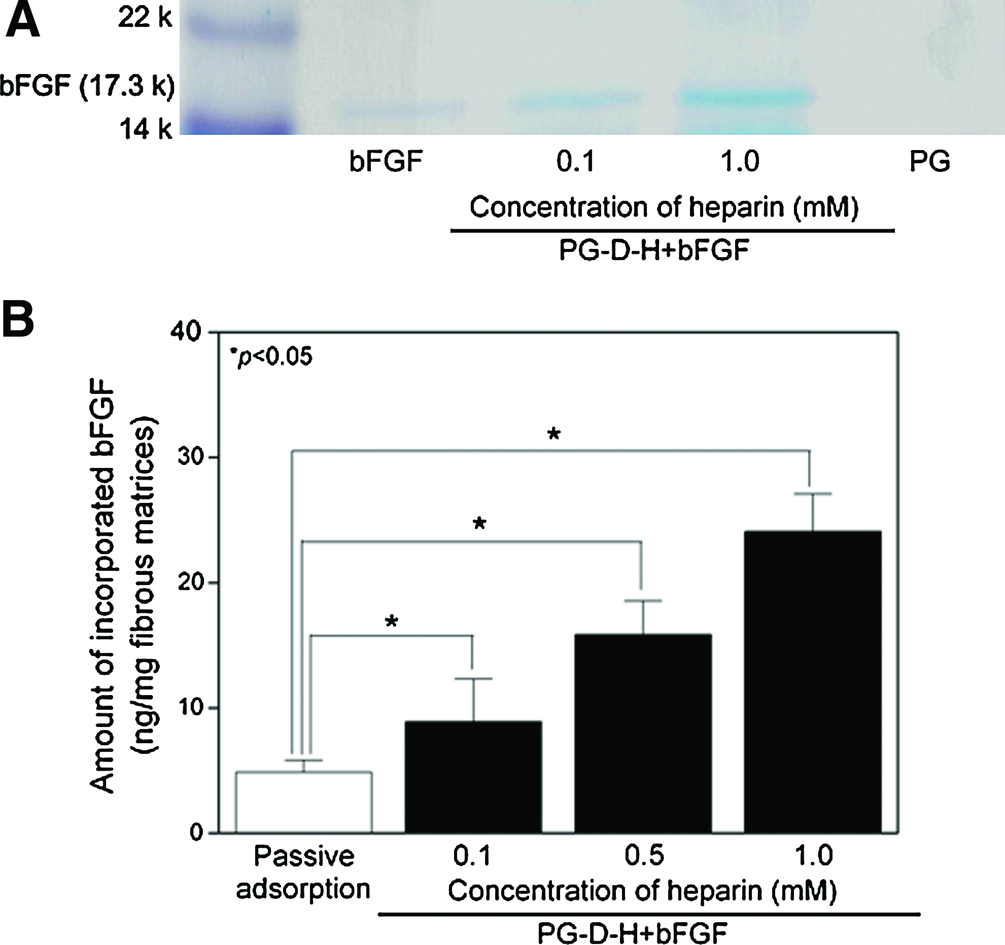
As shown in Figure 3A, bFGF (17.3 kDa) bands were detected from PG-D-H+bFGF samples, and band intensity increased with increasing concentrations of heparin from 0.1 to 1.0 mM whereas no band was observed from passive PG samples. The positions of bands from the fibrous matrices were consistent with that for native bFGF.
(
Quantification of bFGF on the fibrous matrices using ELISA analysis showed a similar trend. As shown in Figure 3B, the amount of immobilized bFGF on PG-D-H was 4.86 ± 0.94 ng when bFGF was passively absorbed. bFGF immobilization on the heparin-conjugated fibrous matrices was greatly affected by the concentration of the heparin on the surface. For example, when the concentration of heparin in the stock solution for modification was increased from 0.1 to 0.5 and 1.0 mM, the amount of bFGF on the fibers that reacted with the corresponding heparin solution increased from 8.83 ± 3.41 to 15.85 ± 2.62 and 24.06 ± 3.01 ng, respectively.
Release of conjugated FITC-labeled heparin and bFGF
Figure 4A shows the release of conjugated FITC-labeled heparin from the fibrous matrices. The cumulative release of heparin was 0.22 ± 0.03 and 0.24 ± 0.02 ng at 1 and 5 days, respectively. The release profiles of bFGF from the fibrous matrices are shown in Figure 4B and C. In particular, the passive adsorption samples showed an initial burst release in which 45.61% ± 8.77% and 75.43% ± 14.21% of bFGF was released on days 1 and 5, respectively, during the release study. We then observed no further releases of bFGF from this group. However, PG-D-H+bFGF samples showed sustained release of bFGF over the course of 28 days. For example, immobilized bFGF on PG-D-H was released at rates of 3.04% ± 1.12% and 2.15% ± 0.85% at day 1 and 17.70% ± 5.45% and 26.64% ± 3.05% at day 5 from 0.1 and 1.0 mM heparin groups, respectively. The release of bFGF from PG-D-H+bFGF appeared to be linear with minimal burst release, regardless of the amount of bFGF immobilized on the fibers. During the 28 days of the in vitro release study, 82.71% ± 5.10% and 71.92% ± 2.85% of immobilized bFGF was released in each sample. Figure 4D shows a scanning electron microscopy image of fibrous matrices after the 28 days of testing. The average diameter of fibrous matrices appeared to be similar to the original shape, although the relative amount of three-dimensional pores was decreased due to partial dissolution compared with those from Figure 1D.
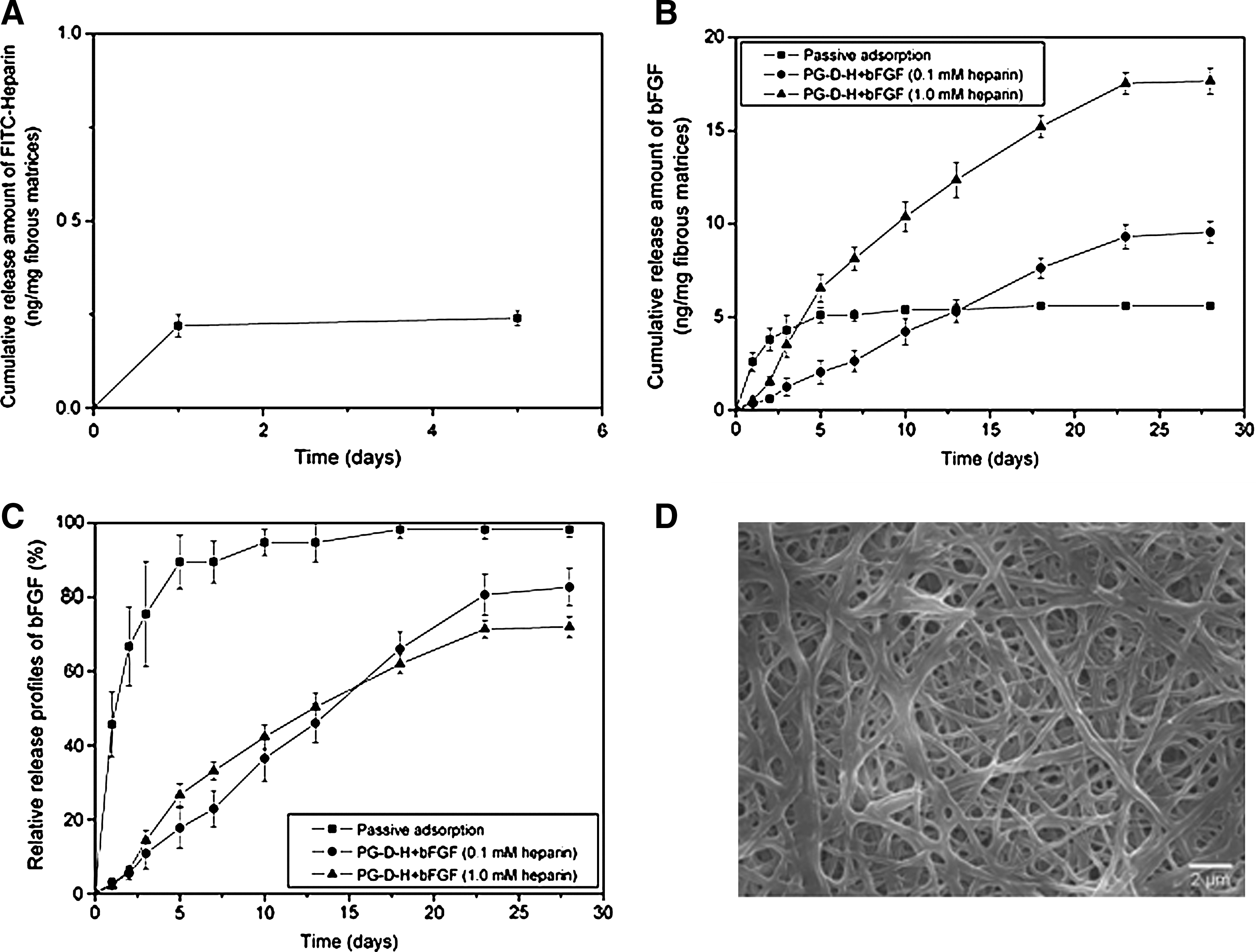
(
Evaluation of in vitro bioactivity of the released bFGF
After 1 day, the relative metabolic activity of HUVECs treated with conditioned media incubated with passive adsorption fibrous matrices was 1.43 ± 0.15, which was significantly higher than other samples (0.97 ± 0.17, 1.07 ± 0.16, and 0.99 ± 0.19 for PG-D-H, PG-D-H+bFGF reacted with 0.1 and 1.0 mM heparin, respectively) in Figure 5. However, the long-term proliferation of HUVECs was influenced by the prolonged release of bFGF. On day 5, the relative metabolic activities of HUVECs from PG-D-H+bFGF reacted with 0.1, and 1.0 mM heparin samples were increased to 2.97 ± 0.72 and 3.31 ± 0.91 from 1.07 ± 0.16 and 0.99 ± 0.19, respectively. These values were significantly greater than those achieved from the conditioned media incubated with PG (0.65 ± 0.10), passive adsorption (1.81 ± 0.27), and PG-D-H 1.0 mM heparin (0.72 ± 0.14).
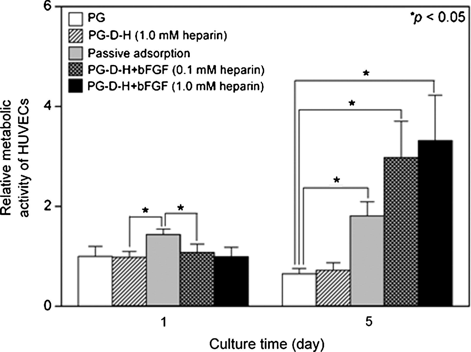
Relative metabolic activity of HUVECs during 5 days of culture supplemented with conditioned media. The conditioned media presumably contained bFGF released from different samples of passive adsorption and PG-D-H + bFGF (0.1 and 1.0 mM heparin). HUVECs, human umbilical vein endothelial cells.
In vivo angiogenesis assay of the bFGF released from fibrous matrices
At 28 days postimplantation of fibrous matrices, tissue samples (containing fibrous matrices) were obtained from mice as shown in Figure 6. From photomicrographic images, we observed that transparent, thin layers of tissue surrounded the surface of all samples and that tissue layers on PG-D-H+bFGF samples (Fig. 6C, D) were richer in blood vessels as compared with other groups. Additionally, more dense blood vessels had formed in the group implanted with PG immobilized with a larger amount of bFGF.
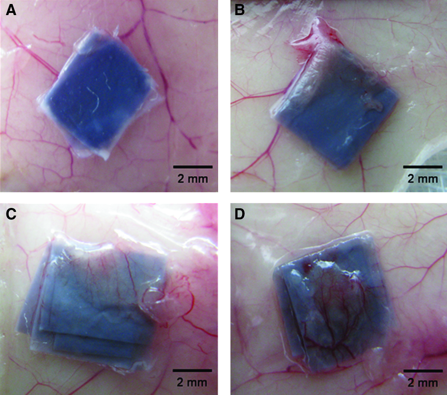
Photomicrographic images of tissues at 28 days postimplantation with (
Immunohistological analysis was conducted to quantify the number of arterioles around the implanted fibrous matrices as shown in Figure 7. Figure 7A–D shows representative fluorescent images, as red, green, and blue colors represent fibrous matrices, SM α-actin, and nucleus, respectively. Approximately 3–4 arterioles were found in the PG-D-H+bFGF (1.0 mM heparin) group whereas no arterioles were observed in other samples. Also, PCNA-positive arterioles were only found in samples from the group implanted with PG-D-H+bFGF (inset fluorescent images) fibers. The quantification of newly formed arterioles supported a role for bFGF from the fibrous matrices in the generation of new arterioles. For example, the number of arterioles were 0.90 ± 0.28 and 3.04 ± 0.35 for PG-D-H with low and high amounts of bFGF, respectively; and both these values were significantly greater than those found in other groups. Further, the percentage of PCNA (+) arterioles was also higher for the group implanted with the higher quantity of heparin PG-D-H+bFGF (62.30 ± 21.01 and 81.42 ± 2.75).
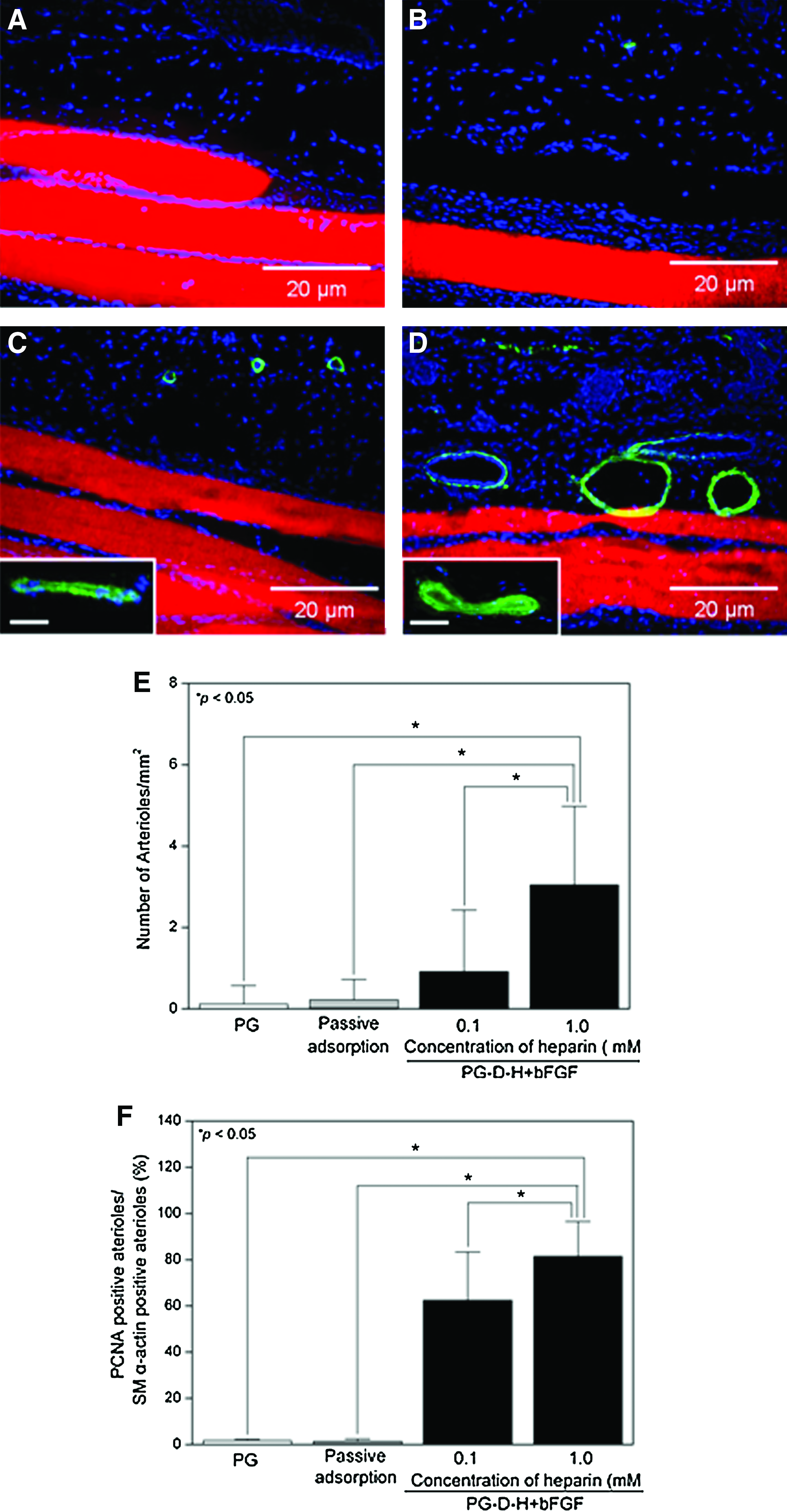
Fluorescent cross-sectional images of tissues implanted for 28 days with (
Figure 8A–D shows representative fluorescence images where the green fluorescence represents vWF of capillaries, the red shows fibrous matrices, and blue indicates nucleus. Similarly to the results found with SM α-actin staining, four to six capillaries were found only in the PG-D-H+bFGF implantation group as compared with other samples, as shown in Figure 8A–D. The inset fluorescent images show that these capillaries were also PCNA (+). For further investigation of the capillaries, the number of capillaries and the percentages of PCNA (+) capillaries were quantified from 30 randomly selected fluorescence images. As shown in Figure 8E and F, larger numbers of capillaries and higher percentages of PNCA (+) capillaries were found in PG-D-H + bFGF samples as compared with the other samples. For example, the number of capillaries counted was 13.65 ± 1.32 and 16.57 ± 2.05 for PG-D-H with low and high amounts of bFGF, respectively. The percentages of PCNA (+) capillaries were 18.35% ± 11.82% and 43.33% ± 12.51% for PG-D-H+bFGF created with 0.1 and 1.0 mM heparin, respectively. Similar to results seen with arterioles, the number of capillaries and higher percentages of PNCA (+) capillaries were directly related to the amount of immobilized bFGF.

Analysis of capillary formation from the implantation with (
Discussion
The ultimate goal of our research is to develop an artificial ECM, mimicking the fibrous structure, as well as the chemical and physiochemical functions of native ECM for applications in regenerative medicine. As an initial step toward our goal, we prepared electrospun fibrous matrices using biocompatible polymers, and the function of ECM as a depot to store or liberate soluble growth factors was then engineered by the immobilization of heparin to control interactions with growth factors. Subsequently, bFGF, a model angiogenic factor, was incorporated into the artificial ECM and its biological activity was evaluated with in vitro as well as in vivo conditions.
To quantify the amount of the conjugated heparin on the fibrous matrices, we used TB, which can form ionic complexes through reactions with negatively charged ionic groups (COO−, SO32−) of polysaccharides. 34 As shown in Figure 2, the amount of TB complexes decreased after the conjugation of PCL or gelatin nanofibers with PEG-diamine and increased after the immobilization of heparin. These results suggest that the conjugation of PEG-diamine was undergone by the consumption of carboxylic groups in the gelatin; and the subsequent increase in ionic groups from the next step is attributed to the successful immobilization of heparin. In addition, it should be noted that a controllable amount of heparin can be immobilized on the fibrous matrices, which can allow us to control the amount of growth factors on the matrices. The amount of TB complexes appeared to reach saturation on reaction with solutions with over 1 mM heparin. This limit may be due to the repulsion of highly negatively charged heparin molecules when used at higher concentrations in aqueous conditions. 35
Many growth factors are subject to quick degradation on intravenous injection and, therefore, the localized retention of growth factors at therapeutic concentrations is clinically crucial. 36 In the native environment, many components of ECM are involved in the storage and liberation of growth factors. In particular, heparin and heparin sulfate are known to bind to several growth factors with high affinity and specificity. 37 In the case of bFGF, these interactions are primarily due to electrostatic binding between positively charged amino groups of bFGF rich with arginine and lysine and negatively charged sulfonyl and carboxyl groups. 38 These specific interactions can also prevent bFGF from rapid degradation. In a previous study exploiting the interactions between bFGF and heparin, passively adsorbed bFGF in polyamide fibrous matrices was more easily degraded as compared with bFGF in heparin-conjugated fibrous matrices at room temperature. 39 In addition, heparin-conjugated microcapsules, nanoparticles, porous scaffold, and fibrin gel have been fabricated to deliver bFGF.18,19,23,26,40–43 As shown in Figure 4A, a tiny amount of conjugated FITC-labeled heparin was released from the fibrous matrices, indicating that covalent binding between heparin and the matrices is very strong. The burst release of bFGF was observed in the group treated with passively adsorbed bFGF, whereas the release of bFGF in the group treated with PG-D-H was sustained over 28 days in a linear pattern with minimal burst release. Our results indicate that heparin immobilized on the fibrous matrices controlled the release of bFGF and also strongly supports a role for heparin in preventing the degradation of bFGF. Moreover, the release profiles of bFGF from the fibrous matrices were similar, regardless of the concentration of bFGF used to form the matrices, suggesting that the reversible interaction between heparin and bFGF was the limiting factor and was not affected by the concentration of bFGF tested in our study.
bFGF has been generally implicated in the enhancement of in vitro formation of focal adhesion and proliferation of various types of cells in a concentration-dependent manner. 9 We confirmed that the bFGF concentrations greater than 5 ng/mL were effective for stimulating HUVECs proliferation consistent with previous reports. 40 The results in Figure 5 suggest that the released bFGF maintains its biological activity for over 5 days. On day 1, it appeared that the passively adsorbed group showed higher proliferation of HUVECs than the other groups, which may be due to the burst release of passively adsorbed bFGF on the first day. These results are in good agreement with the release profiles of bFGF shown in Figure 4. However, the proliferation stimulated in the group treated with passively adsorbed bFGF was significantly lower than that from the PG-D-H + bFGF group after 5 days of culture, suggesting that sustained release of bFGF is required for long-term in vitro activation of HUVECs. The heparin released from the fibrous matrices seemed to be too small to affect the proliferation of HUVECs for 5 days. It was reported that even a larger amount of soluble heparin was not effective for proliferation of HUVECs. 44
The effect of bFGF released from fibrous matrices was further confirmed using an animal model, which has been well-established by other reports.19,41,42 As shown in Figure 6, it was apparent from the images of implantation areas that blood vessels were formed at a higher density in PG-D-H + bFGF groups. We found that the number of arterioles and capillaries was significantly enhanced in PG-D-H+bFGF groups, suggesting that the released bFGF affected formation of blood vessels near implanted samples for 28 days. PCNA (+) cells were found to be localized within newly formed blood vessels, indicating that blood vessel formation was mediated by proliferation of endothelial cells. The formation of blood vessels is controlled by active bFGF, which stimulates the over-expression of matrix metalloproteinases and, subsequently, induces the proliferation and migration of endothelial cells. 41 Our results also support that the appropriate amount of bFGF and its retention at local area are necessary for stimulation of blood vessel formation. The amount of bFGF used in our experiment is likely lower than that used from previous studies; and, therefore, optimization of required bFGF concentration with our fibrous matrices should be further pursued in future. 43
Overall, our results suggest that bFGF-immobilized fibrous matrices can be used to treat tissue damage requiring blood vessel formation such as skin wounds and ischemic myocardial diseases. Further, heparin-conjugated fibrous substrates may be useful to store and release other types of growth factors that contain heparin binding domains.
Conclusion
In this study, heparin-immobilized fibrous matrices based on PCL and gelatin crosslinked with genipin were fabricated to utilize the specific interactions between heparin and growth factors that have heparin binding domains. As a model system, we immobilized bFGF and demonstrated that this growth factor maintained its bioactivity when stored and released from the fibrous matrices over 28 days. The released bFGF significantly enhanced in vitro proliferation of HUVECs. An in vivo study in mice revealed that the group implanted with matrices holding a higher concentration of bFGF showed significantly increased formation of arterioles and capillaries. These results suggest that bFGF-immobilized fibrous matrices may hold promise for use as a substrate to stimulate blood vessel formation. Further, our heparin-immobilized matrix system could be combined with other types of growth factors that contain heparin binding domains to actively control their bioactivity.
Footnotes
Acknowledgments
This work was partially supported by a grant of the Korea Healthcare Technology R&D Project, Ministry of Health & Welfare, Republic of Korea (A080189), and by a pioneer research program for converging technology through the Korea Science and Engineering Foundation, Ministry of Education, Science, and Technology (M10711060001-08M1106-00110) (to H. Shin).
Disclosure Statement
No competing financial interests exist.