




Abstract
Introduction:
In this study, thick polyethersulfone (PES) nanofibrous scaffolds were prepared by fine tuning of electrospinning parameters and was evaluated for wound dressing applications.
Materials and Methods:
Scanning electron microscopy and Brunauer-Emmett-Teller methods were used for PES nanofibers characterization. The interaction between fibroblasts and nanofibers was studied in vitro. Further, a mouse model was used to evaluate the effectiveness of the PES scaffold in wound healing. Vaseline gauze dressing and a conventional gas permeable bandage were used as a control. The wound repair process was evaluated by histological examination and immunohistochemistry staining using antibodies to cytokeratin 10 (CK10), proliferating cell nuclear antigen (PCNA), and alpha-smooth muscle actin (alpha-SMA).
Results and conclusion:
The characterization of nanofibers showed that the PES membrane has nanoscle, porous, high surface area structure. These properties conferred higher exudate absorption capacity for the PES scaffold which is essential for effective wound healing. In vitro results indicated that the PES scaffold can support fibroblast proliferation similar to that with tissue culture polystyrene. Epithelial regeneration was expeditiously accelerated under PES as compared with Vaseline gauze. Greater fibroblast maturation, improved collagen deposition and faster edema resolution were the superior properties of PES over the commercial dressing. Based on these results we conclude that the biocompatible PES nanofibers can effectively be used as a dressing to accelerate wound healing.
Introduction
Electrospinning has been known for about eight decades. 4 It is one of the methods used for fabrication of synthetic and natural materials into fibrous nanostructures.5–8 Electrospun nanofibrous scaffolds have potential characteristics of an ideal wound dressing including high gas permeability and wound protection from contamination and dehydration. 9 Nanofibrous matrices have been shown to increase the rate of epithelialization and improve dermal reconstruction. 10 They can also support the attachment and proliferation of dermal fibroblasts by providing a high level of surface area. 9 Nanofibrous matrices from different materials such as chitosan, polyurethane, gelatin collagen, and PLGA have been constructed and shown to accelerate wound healing effectively.10–13 Although many in vitro studies have evaluated the potential use of electrospun nanofibers for wound dressing,1,14–18 only a few in vivo studies have investigated their utility in wound healing.13,19–21
Polyethersulfone (PES) is a biocompatible, nondegradable, synthetic polymer (Fig. 1) which has been widely used in biomedical applications like hemodialysis, filtration, ultrafiltration, and bioreactors.22–26 Recently, PES has attracted increasing attention for utilization as a scaffold in tissue engineering.27–31 It has been also shown that an electrospun PES scaffold has a biocompatible porous structure5,32 which makes it a good candidate to be used as a wound dressing.
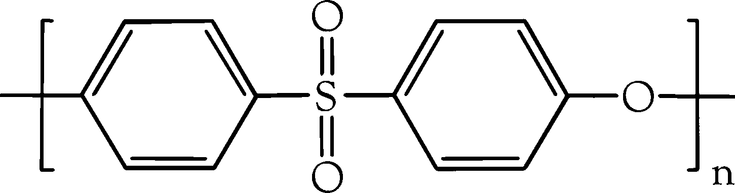
Chemical structure of PES. PES, polyethersulfone.
In this study, a nanofibrous PES scaffold was fabricated by the electrospinning technique. Unlike usual thin scaffolds obtained from electrospinning by other researchers,33,34 thick scaffolds were fabricated by the fine tuning of process parameters. For the first time, the biocompatibility and wound healing effect of PES nanofibers were evaluated both in vitro and in vivo systems.
Materials and Methods
Nanofiber fabrication
Electrospinning was performed to construct the PES (Ultrason E6020P; BASF) nanofibrous scaffold. Briefly, 4 mL of PES solution (26% wt/wt) in dimethylformamide (DMF) (Sigma), was placed in a 5-mL syringe. A syringe pump was used to feed the solution through an extension tube ending in a blunted 21-gauge needle. To collect the PES nanofibers, a cylindrical collector was located at a distance of 20 cm from the needle. High voltage potential (20 kV) was applied between the needle and the collector. PES solution was forced to leave the needle and collected as nanofibers on the steel cylinder. The nanofibrous mat was separated from the collector surface and used for further analysis and application.
Nanofiber characterization
Morphology
For evaluation of nanofiber morphology, the specimens were gold coated using a sputter coater and then scanned by a scanning electron microscope (SEM; Philips XL30). The diameter of fibers was determined from scanning electron microscopy (SEM) micrographs using image analysis software.
Porosity
For porosity determination, four randomized circular samples with the diameter of 20 mm were used and the estimated porosity of each sample was calculated by the following equation:
Specific surface area
The specific surface area of PES nanofibers were measured through the Brunauer-Emmett-Teller (BET) method 35 by BELSORP-mini apparatus (BEL Japan, Inc.). Nitrogen adsorption-desorption isotherms of nanofibers were obtained and the surface area was calculated from the BET plot of isotherms by BELSORP-mini software.
Isolation of mouse skin-derived fibroblasts
Fibroblasts were isolated from the dorsal skin of new born Balb/c mice. Briefly, dorsal skin was cut and the epidermis was separated from the dermis after an overnight incubation with 0.1% dispase in Dulbecco's modified Eagle's medium (DMEM) at 4°C. The remaining dermis was minced into small parts, trypsinized and cultured in DMEM (supplemented with fetal bovine serum [FBS] 10%, 100 U/mL penicillin, 100 μg/mL streptomycin, at 37°C with 5% humidified CO2). After cell adherence, fibroblasts were trypsinized and cultured in DMEM containing 10% FBS. Passage 3 cells were used for MTT assay (Sigma).
Biocompatibility of nanofibers
Cell viability
The standard MTT assay was used to evaluate cell viability on PES nanofibers. 36 PES nanofibrous scaffolds were sterilized using a 70% ethanol solution for 12 h which was followed by immersion in a solution containing penicillin, streptomycin, and amphotericin B for 24 h. Scaffolds were placed in a 12-well culture plate and seeded with 120 μL of the fibroblast cell suspension with a cell density of 12.5 × 10⁴ cells/mL. The cell-seeded scaffolds were incubated at 37°C, 5% CO2 for 2 h followed by the addition of 500 μL of DMEM 10% FBS. After 6, 48, 72, and 96 h of cell seeding, 50 μL of MTT solution (5 mg/mL in DMEM) was added to each well (n = 4). For conversion of MTT to formazan crystals by mitochondrial dehydrogenases of living cells, the plate was incubated at 37°C for 3 h. For dissolution of the dark-blue intracellular formazan, the supernatant was removed and a constant amount of an appropriate solvent was added. The optical density was read spectrophotometrically at a wavelength of 570 nm. The same procedures were performed for cultured cells in tissue culture polystyrene (TCPS) as control.
Cell morphology
The morphology of cells seeded on PES nanofibrous scaffolds was investigated using SEM images. Sterilized scaffolds were placed in 24-well plate followed by the addition 75 μL of fibroblast suspension with a cell density of 2 × 10⁵ cells/mL. After 2 and 6 days of incubation the upper medium was removed and the scaffolds rinsed carefully with phosphate-buffered saline. Finally, the cells on the scaffolds were fixed with 2.5% glutaraldehyde for 1 h, and then subjected to graded alcohol dehydrations, air-dried overnight, sputter-coated with gold, and examined with SEM.
Excisional wound model
All animal experiments were carried out in accordance with the Stem Cell Technology Research Center (Tehran, Iran) guidelines. Male Balb/c mice (Razi Institute, Karaj, Iran) weighing 25–30 g were housed under standard condition in a controlled temperature (20°C) and a light/dark cycle (12/12 h). Mice were individually anesthetized via intraperitoneal injection of ketamine (20 mg/kg) and xylazine (2 mg/kg) and inhaled mixture of 20%v/v isoflurane and propylene glycol. The dorsal surface was shaved with an electric hair clipper and sterilized by 10% povidone-iodine. A round full thickness wound with a diameter of 15 mm was excised from the back using curved blade surgical scissors. The wound was dressed with sterile PES mat. The wounds covered with Vaseline gauze or TIELLE™ Xtra (Johnson & Johnson) were used as a control. The dressed wound was then covered with an elastic cotton bandage and treated mice were placed in individual cages.
Wound closure
On day 7, 10, 12, and 15 postsurgery, the mice were anesthetized with intraperitoneal injection of ketamine and sacrificed by cervical dislocation. The dressing was removed and several pictures were taken with a digital camera. The rate of wound closure was determined by the reduction in the wound size on digital photography using image analysis software (n = 3, for each group, on each day). Finally, the wound site was excised, and the tissue was processed for histological evaluation.
Histopathology
The excised tissues were fixed in 10% buffered formaldehyde solution, processed, and embedded in paraffin. Thick sections (3–5 μm) were stained with hematoxylin and eosin (HE) and Masson's trichrome stain. For quantification of pathologic findings of HE stained samples, a modified scoring system was used based on a scoring system introduced by Abramov et al. 37 Briefly, three and five criteria were scored for epidermis and dermis, respectively (Table 1). For each criterion the scoring system was based in order of improving signs from zero to three, that is, the higher score indicates more desirable finding for wound repair. Two pathologists separately scored each sample. The sum of these eight scores (epidermal and dermal) was reported for each group. In addition, the number of high-power fields in which the new epithelium has formed, were considered to evaluate the length of the epithelial tongue.
Unordinary proliferation of cells in granular layer of epidermis.
Masson's trichrome staining was used for a better assessment of collagen formation. The extension of collagen deposition and the morphology of collagen bundles were considered in the evaluation. Each sample was scored for these two different criteria (Table 1) and the sum of scores was reported. Results of Masson's trichrome staining were further evaluated by fluorescence microscopy for detection of autofluorescence of collagen fibers in HE stained sections.
Immunohistochemistry
Proliferating cell nuclear antigen (PCNA) (Dako), cytokeratin 10 (CK10) (Dako), and alpha-smooth muscle actin (alpha-SMA) (Abcam) antibodies were used for identification of dermal proliferating cells, terminal differentiated keratinocytes and myofibroblasts, respectively. Briefly, paraffin embedded tissues were sectioned at 4 μm and placed on poly L-lysin coated glass slides and heated to 60°C in an oven for 1 h. Sections were deparaffinized using xylene and rehydrated in ethanol with decreasing concentrations. To inactivate the endogenous peroxidase, the sections were incubated in a 0.5% hydrogen peroxide solution in methanol. After washing, antigen retrieval was performed in citrate buffer (pH = 6) for 30 min in boiling water followed by blocking of nonspecific binding with goat serum for 20 min. Sections were then incubated for 1 h with primary antibodies as mentioned previously. Staining was visualized using the Envision kit (Dako) followed by immersion with liquid 3,3′-diaminobenzidine (DAB) for 5 min and counter stained with hematoxyline for 1 min. To quantify cell proliferation in dermis, the number of PCNA positive cells in four different high power fields was counted for each sample by using image-Pro Plus 6.0 software (Media Cybernetics).
Statistical analysis
All data is reported as the mean ± standard deviation. Statistical significance was determined by Mann–Whitney U test as a nonparametric equivalent of student's independent sample t-test for analysis of MTT assay findings. Simple one-way analysis of the variance and its nonparametric equivalent (Kruskal–Wallis test) were used for analysis of PCNA staining and wound closure results, respectively. All analyses were performed using SPSS 17.0 software. p-Values <0.05 were considered significant. Nonparametric tests were chosen because of the small sample sizes and distributions that may not be normal.
Results
Nanofiber characterization
As shown in Figure 2, the electrospun PES scaffold has a nanofibrous and highly porous structure. Image analysis of 200 randomly selected nanofibers demonstrated that they had an average diameter of 492 ± 106 nm (Fig. 3). The porosity of mats was calculated as 76.27% ± 1.24%. The measured BET specific surface area of nanofibers was found to be 39 m2/g. Moreover, the scaffold thickness was measured to be about 3 mm.
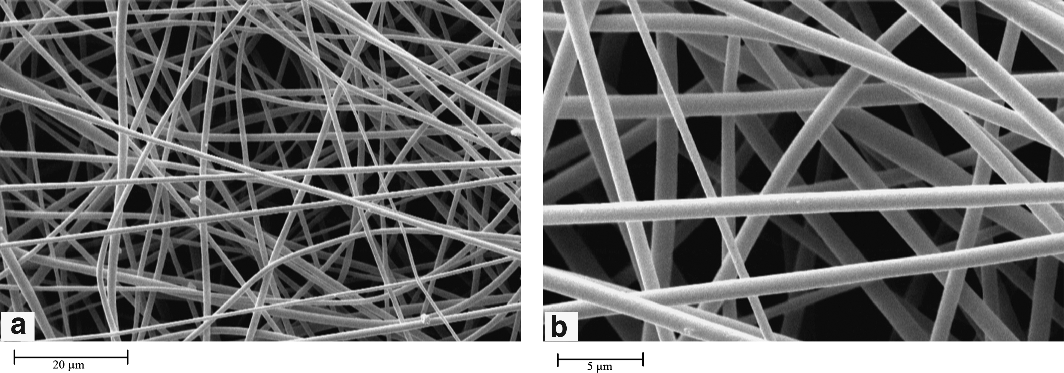
Scanning electron micrographs of the PES nanofibers at different magnifications

Frequency distribution of PES nanofiber diameters.
Biocompatibility of nanofibers
As shown in Figure 4, 6 h after cell seeding, there was no significant difference of cell viability on PES compared with TCPS. After 48, 72, and 96 h of cell seeding, cell viability on PES and TCPS significantly increased (p-value <0.05) and no meaningful difference was found between two groups at each time point. This finding implied that PES nanofibers as well as TCPS, support fibroblast proliferation.

Fibroblast viability on PES and TCPS during a 6-day culture period. Data represent mean ± SD (n = 4). TCPS, tissue culture polystyrene; SD, standard deviation. Asterisk shows significant difference with p < 0.05.
As shown in Figure 5a, fibroblasts easily attached to the PES nanofibers and showed flattened and polygonal morphology as reported previously.16,38–40 After a 4-day culture period, proliferated cells had clearly spread over the surface of the nanofibers (Fig. 5b).
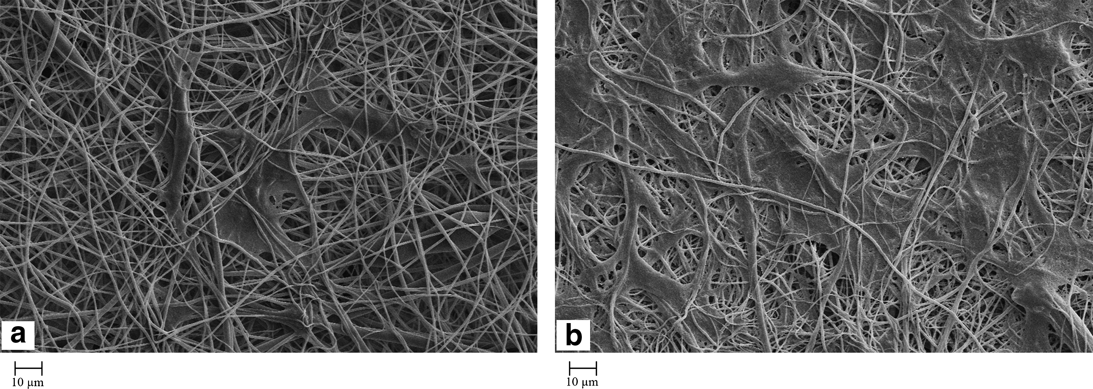
Scanning electron micrographs of cell seeded PES membrane after
Wound healing
As demonstrated in Figure 6, PES nanofibers could effectively accelerate wound closure. About 90% wound closure was achieved by PES after 10 days and it was nearly completed by day 12.
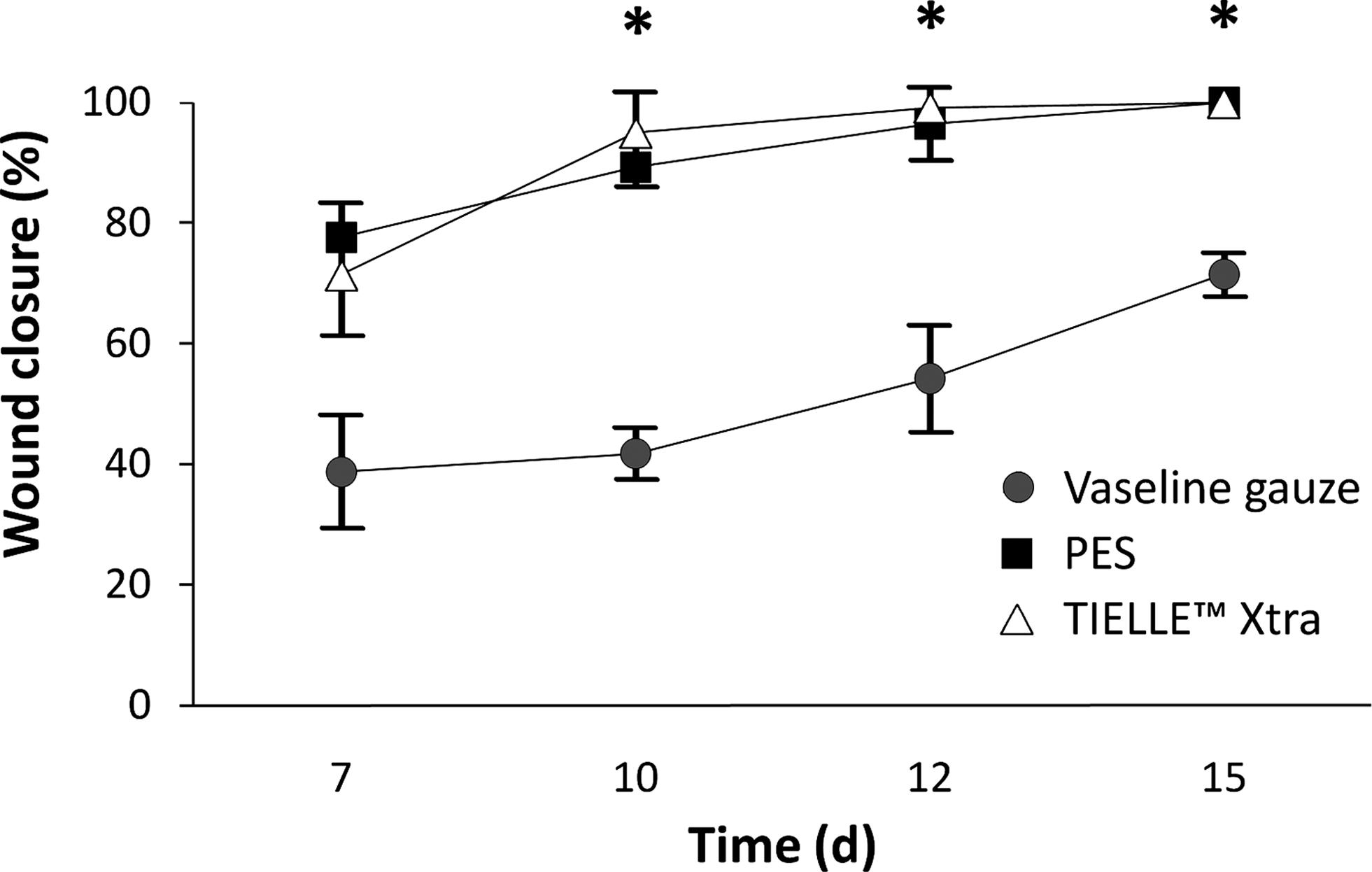
Percent wound closure for the in vivo wound healing experiments. Results are shown for the three experimental groups: Vaseline gauze, PES and TIELLE™ Xtra groups (mean ± SD, n = 3). Asterisk shows significant difference with p < 0.05.
On day 7, the number of high power fields in which the wound was epithelialized was significantly higher in PES and TIELLE Xtra groups compared with the Vaseline gauze (Fig. 7).

Length of the epithelial tongue on day 7(mean ± SD, n = 3). Asterisk shows significant difference with p < 0.05.
Using the scoring system for histological examination of wounds on postoperative days 7, 12, and 15, more collagen deposition and fibroblast maturation were observed at each time point in the PES group in comparison with the other groups (Fig. 8a–c). Edema and depth of inflammation were the least in the PES group at all time points (Fig. 8b). Crust formation and the PMN amount were more prominent in the Vaseline gauze group. Spongiosis (intercellular edema of the epidermis) which was not observed in PES group, was the other remarkable pathologic finding in the Vaseline gauze group. Hypergranulosis was seen prominently in PES and TIELLE Xtra groups (Fig. 8b, c). In summary, PES group had the best pathologic score at all three time points (Fig. 9).

Histological findings of wound healing:
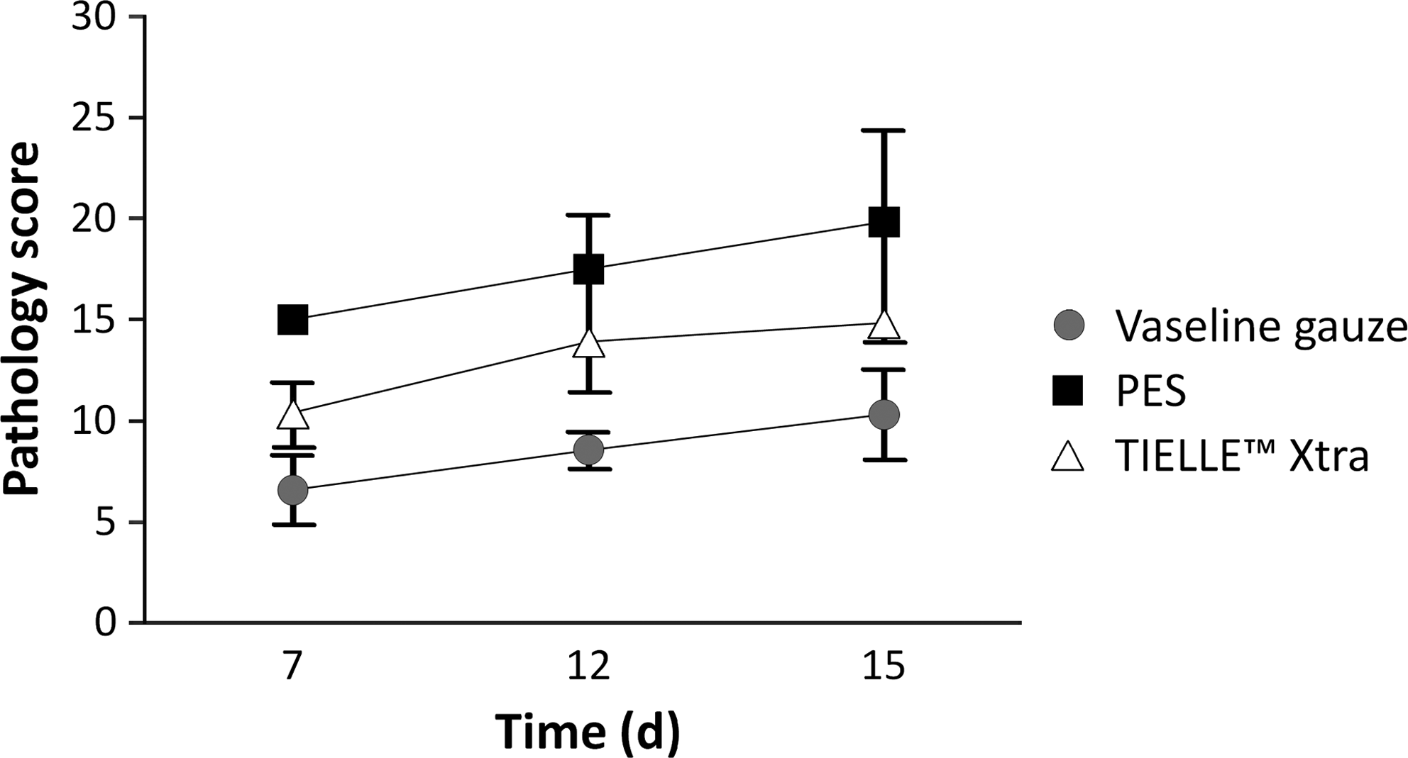
Pathology score (mean ± SD, n = 3).
Mason's Trichrome staining on postoperative days 10 and 15 demonstrated that collagen deposition was more extensive in the PES group and was mostly composed of thick wavy collagen fibers on day 15 (Figs. 8d–f and 10). These findings were in concordance with the results of the fluorescence microscopy of collagen fibers (Fig. 8g–i).

Massons' trichrome staining score (mean ± SD, n = 3).
As demonstrated in Figure 11, the number of PCNA positive cells was significantly higher in PES and TIELLE Xtra groups on day 7 but was considerably decreased on day 15 in both of these groups.

PCNA staining (mean ± SD, n = 12). Asterisk shows significant difference with p < 0.05.
Alpha-SMA staining on postoperative showed that myofibroblasts were present on day 10 in all groups (Fig. 12a–c) but dramatically disappeared only in the PES group on day 15 (Fig. 12e).
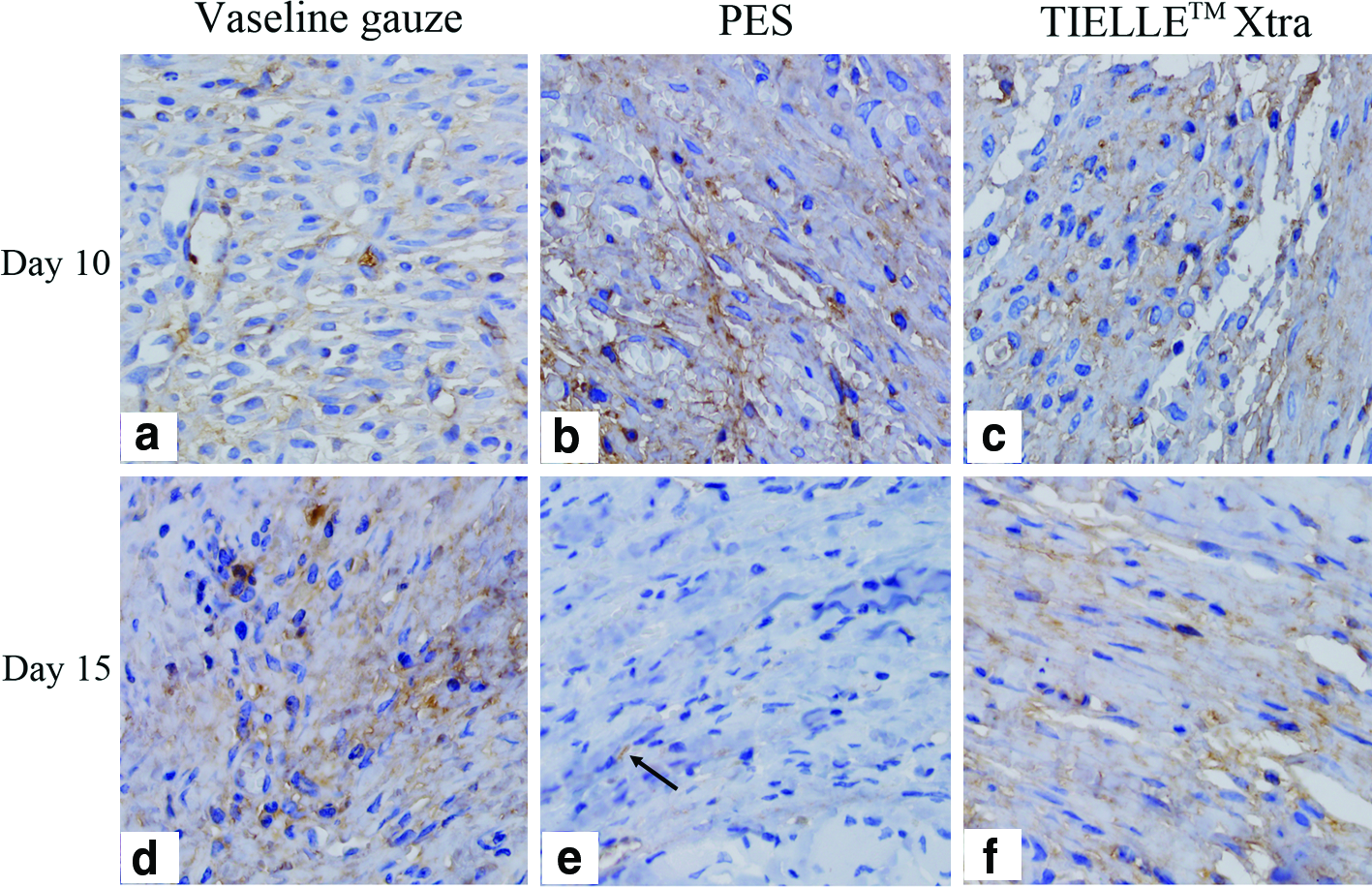
Alpha-SMA staining on day 10
CK10 staining showed suprabasal expression in all healed tissues without significant difference between the groups (Fig. 13).
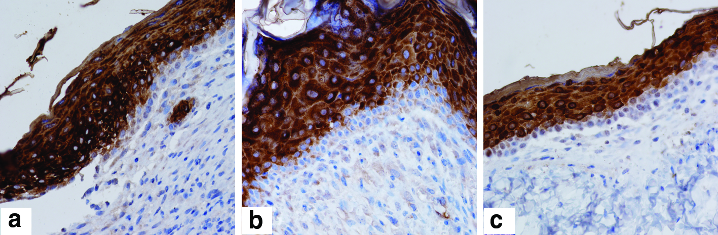
CK10 staining on day 12 in vaseline gauze
Discussion
Recently, several studies have focused on designing wound dressings which can create and maintain an optimal wound healing environment. Nanofibrous dressings have been shown to accelerate wound healing.13,19 Electrospinning, as a method of producing nanofibers, has become the focus of attention because of its simplicity and ability in mass production of one-by-one continuous nanofibers. 41 Electrospun nanofibers have distinct advantages including their high surface area and controlled porosity over the ones generated by most other techniques. 42
The results from this study showed that the electrospun PES scaffold has a three-dimensional and highly porous structure which allows efficient gaseous exchange. Gaseous exchange promotes wound healing by supplying oxygen 43 which is essential for energy provision, fibroblast proliferation, collagen synthesis, and polymorphonuclear cells function.44–46
Electrospinning of scaffolds thicker than 300 μm is difficult due to technical limitations but in current research, thick scaffolds were fabricated by the fine tuning of process parameters. The data illustrated that prepared nanofibrous scaffolds had large specific surface area and high thickness which led to a greater total surface area and consequently higher exudate absorption capacity. Absorption of extra exudate is essential because exudate accumulation under the dressing may lead to infection.
The interaction between electrospun PES scaffolds and stem cells (neural, hematopoietic, and unrestricted somatic stem cells) have been studied before. These in vitro studies have shown that electrospun PES scaffolds support stem cell proliferation and differentiation.5,30,32 In this study the results of the MTT assay demonstrated that the viability trend of the mice skin-derived fibroblasts on PES was similar to that on TCPS. This finding together with well adherence and typical normal morphology for fibroblasts on PES nanofibers is in agreement with the suggestion that the electrospun PES nanofibrous scaffold is biocompatible and supports cell proliferation.
In vivo study of electrospun PES nanofibers as a wound dressing has not been reported before. Our in vivo results in the current study demonstrated that accelerated wound healing could be achieved by application of electrospun PES as compared with Vaseline gauze which is still among the most widely used dressings for the nonsurgical treatment of burns worldwide. 47 About 75% wound closure was achieved on postoperative day 7 by PES application comparing to about 39% for Vaseline gauze. According to the microscopic finding, the epithelialization process was significantly improved in PES than the Vaseline gauze group, implying that accelerated epidermal regeneration has an important role in the acceleration of wound closure under PES nanofibers. Hypergranulosis and suprabasal expression of CK10, both are representative of a well-organized mature structure of newly formed epidermis under PES dressing. Spongiosis as a remarkable pathologic finding in the majority of Vaseline gauze treated cases could be a sign of contact dermatitis addressing an adverse effect for this material.
Our comparison of the PES with the TIELLE Xtra dressing (as a conventional gas permeable dressing) revealed that PES can effectively accelerate wound closure similar to the TIELLE Xtra dressing in gross observation. The better pathologic score of wound healing resulted from greater collagen deposition, more fibroblastic maturation, and improved edema resolution in the PES treated group, indicate the superiority of PES nanofibers over the TIELLE Xtra dressing.
During normal wound healing, fine collagen fibers of early granulation tissue gradually are replaced by thick mature collagen fibers. 48 Extensive deposition of thick mature collagen fibers on day 15 under the PES dressing favor that PES nanofibers can effectively accelerate improve of dermal reconstruction. A remarkable reduction of PCNA expression on day 15, after showing high expression on day 7, is in favor of near termination of dermal repair process on this day.
It is well established that myofibroblasts play an important role in wound contraction and matrix formation during normal wound healing. 49 Remarkable decrease of myofibroblasts by apoptosis from neo-tissue characterizes the terminal phase of wound healing. 50 Failure of myofibroblast elimination would lead to greater scar tissue formation. 49 Lower SMA staining in the PES group on day 15 may suggest the greater ability of this material for expeditious elimination of myofibroblasts and exclusion of scar formation.
Until now, a few polymeric biomaterials such as chitosan, collagen, gelatin, silk, alginate and polyurethane, cellulose and its derivates have been used in wound dressing applications. Herein, a new wound dressing biomaterial based on PES nanofibers was introduced and shown to hold superior characteristics for the acceleration of wound healing and greater potential for clinical application. Although PES nanofibers were shown to have a noticeable superiority in the improvement of wound healing, there remain several issues to be addressed including ease of use, patient acceptance and pain on removal which cannot be assessed in animal models.
Conclusion
Based on the fact that gas exchange and excess exudate absorption is essential for optimal wound healing, a nanofibrous PES scaffold with a porous structure was prepared using electrospinning. A highly porous structure of the PES scaffold together with a high surface area of nanofibers allows appropriate gas exchange and exudate absorption. In vitro and in vivo results of this study suggest that the PES nanofibrous scaffold is nontoxic and nonallergenic and its application can accelerate wound healing by better supplying oxygen, absorbing excess exudate and protecting the wound from further contamination.
Footnotes
Acknowledgments
This work was financially supported by Stem Cell Technology Research Center (Tehran, Iran). We are grateful to Professor Alireza R. Rezaie from St. Louis University School of Medicine, for valuable comment. We also thank Dr. Issa Jahanzad, Seyed mahmoud Hashemi, Sara Soudi, and Naser Ahmadbeigi for helpful advice.
Disclosure Statement
No competing financial interests exist.