




Abstract
Over the last several years, our in vitro and in vivo studies have focused on optimizing the use of fibrin to deliver cells. We have shown that some three-dimensional (3D) fibrin constructs with specific fibrinogen and thrombin concentration support robust proliferation of normal human dermal fibroblasts, whereas different fibrinogen and thrombin concentrations support high mesenchymal stem cell proliferation in 3D fibrin constructs. In this article, we found that normal human epithelial keratinocytes proliferate well in 3D fibrin constructs consist of fibrinogen concentration ranging from 17 to 33 mg/mL and thrombin concentration of 1 U/mL. Further, using a new proliferation assay, we studied the proliferation of fibroblasts and keratinocytes cocultured in various 3D fibrin constructs of different fibrinogen and thrombin concentrations. We found that 3D fibrin constructs with a range of fibrinogen concentration (5–34 mg/mL) and a thrombin concentration of 1 U/mL produce an optimal cell proliferation for both cell types when cocultured. This profile of proliferation is different from that seen when keratinocytes or fibroblasts are incorporated separately in 3D fibrin constructs. In conclusion, we found that one needs to choose the fibrinogen and thrombin concentration carefully depending on the cell type to deliver; that is, different fibrin constructs with different fibrinogen and thrombin concentration are required to deliver fibroblasts or keratinocytes alone or to codeliver both cell types. Moreover, there seems to be a cross-talk between keratinocytes and fibroblasts when they are cointroduced in 3D fibrin constructs. This feedback could be due to the effects of growth factors produced by the two cell types in the 3D fibrin constructs.
Introduction
Materials and Methods
Harvesting cells
T-75 flask (Sarstedt, Newton, NC) containing normal human dermal fibroblasts (NHDF) or normal human epithelial keratinocytes (NHEK) (Clonetics–BioWhittaker, Walkersville, MD) was rinsed with 10 mL prewarmed Hank's balanced salt solution (Clonetics–BioWhittaker), and incubated with 4 mL of prewarmed Trypsin (Clonetics–BioWhittaker) for up to 2 min. Trypsin was discarded and the flask was examined under the microscope to make sure the cells were detached. After the cells were detached, 10 mL of warm serum-containing medium (Clonetics–BioWhittaker) was added to the flask and the cell suspension was transferred form the flask to a 50 mL centrifuge tube. The T-75 flask was rinsed with 5 mL of the same medium and the rinse was added to the centrifuge tube. The sample was centrifuged at about 2000 rpm for 6 min and the supernatant was removed. The cell pellet was washed with the 5 mL warm serum-free medium and centrifuged at about 2000 rpm for 6 min, and the supernatant was removed. The cell pellet was resuspended in 1 mL of the prewarmed serum-free medium and the cells were counted using a hemacytometer.
Labeling cells with antifibroblast microbeads
Seventy microliters of the serum-free medium was added to centrifuge tubes and followed by 30 μL of antifibroblast microbeads. To another set of tubes, 100 μL of the serum-free medium was added. This tube was used as a control. The above tubes were incubated at room temperature for 30 min. The correct concentration of cells in 0.5 mL was added to tubes with or without microbeads. For blank microtubes, 0.50 mL of the serum-free medium was added. The microtubes were rotated for 30 min at room temperature. Positive selection column (Magnetic Activated Cell Separation) was placed in the magnetic field of MACS separator and washed with 0.50 mL of the serum-free medium. The cell suspensions from the various tubes were poured onto the column. The negative fraction (cells not bound to the beads) was allowed to pass through, and then the column was rinsed three times with 0.50 mL of the serum-free medium. The collected negative fraction was transferred to a 5 mL centrifuge tube and centrifuged at about 1600 rpm for 5 min. The supernatant was removed and the cell-bead pellet was resuspended in 300 μL of the serum-free medium. The column was removed from the separator and placed on a collection tube, and 0.50 mL of the serum-free medium was added onto the column. Using the plunger supplied with the column, the positive cells (cells bound to the beads) were forced out of the column into a 5 mL centrifuge tube. The procedure was repeated twice more. The column was washed three times with 1 mL of the serum-free medium and flushed out using the plunger. The wash was collected in a 15 mL centrifuge tube. The washes were placed on a culture dish and checked under a microscope for any visible cells. The collected cells were centrifuged at 1600 rpm for 5 min. The supernatant was removed and the cell-bead pellet was resuspended in 300 μL of the serum-free medium. For the microtubes containing cells without microbeads, the microtubes were centrifuged at 6500 rpm for 5 min. The supernatant was removed and the cells were resuspended in 300 μL of the serum-free medium. Fibroblasts bind to the antifibroblast beads, such that the positive fraction contains the fibroblasts.
Labeling cells with Live/Dead stain
The fractions collected above were transferred to 24-well plates. Live/Dead dye solution (Molecular Probes, Eugene, Oregon) was prepared as follows: 8 mL fibroblast basal medium (FBM) and 40 μL EthD-1 were spun down on minicentrifuge and then vortexed for 30 s, and spinning repeated; 10 mL Calcein.AM was spun down on minicentrifuge and then vortexed for 30 s, and spinning repeated; the contents were mixed very well and vortexed for 1–2 min; 300 mL of the Live/Dead stain was added to all wells; the plates were kept in dark at room temperature for 50 min; the plates were then read using a Fluorescence plate reader (Precision Scientific, Winchester, VA).
Formation of cell—3D fibrin constructs
Seven flasks containing NHEK or NHDF were rinsed twice with 10 mL of the prewarmed serum-free medium, once with 6 mL prewarmed Trypsin and then incubated with 6 mL prewarmed Trypsin in a 5% CO2 incubator at 37°C until the cells started to detach. After the cells were detached, 12 mL of Trypsin Neutralizing Solution was added to the flasks and the cell suspension was transferred to a 50 mL centrifuge tube. The flasks were rinsed with 10 mL of the serum-free medium and the rinse was added to the centrifuge tube. The tube was, then, centrifuged at about 2000 rpm for 6 min and the supernatant discarded. The cell pellet was washed with 5 mL of the serum-free medium, centrifuged at about 2000 rpm for 6 min, and resuspended in 1 mL of prewarmed tris-buffered saline (TBS). The cells were counted using a hemacytometer. Fibrinogen and thrombin solutions were reconstituted as described in the Tisseel® product insert. Briefly, fibrinogen component was reconstituted in Aprotinin solution, and thrombin component was reconstituted in 30 mM CaCl2. Both solutions were diluted to the appropriate concentrations by diluting fibrinogen in TBS and thrombin in 30 mM CaCl2 in TBS. Fifty thousand of NHEK and of NHDF cells were mixed with the fibrinogen and diluted using TBS. The final number of combined NHDF and NHEK cells was 100,000 cells per 3D fibrin construct. One hundred fifty microliters of the fibrinogen solution with cells was added to each well of the 24-well plates. The contents in the wells were mixed well by tapping and tilting the plates. Four plates were prepared for days 0, 1, 3, and 5 with three wells per day. One hundred fifty microliters thrombin solution was added and the wells were mixed well by tapping and tilting the plates. The plates were incubated at room temperature for 2 h. Three-dimensional fibrin constructs were washed twice with 1 mL of the warm serum-free medium. One milliliter of the serum-containing medium was added to each well and plates were transferred to a 5% CO2 incubator (Nuaire, Plymouth, MN) at 37°C. The medium was replaced on days 1 and 3 with 2 mL of the serum-containing medium.
Cell proliferation within the 3D fibrin constructs
Three-dimensional fibrin constructs containing cells were washed twice with 1 mL of the serum-free medium. Three-dimensional fibrin constructs from four-wells of one culture plate were dislodged from the walls of the wells for cell quantification. Two 3D fibrin constructs of each run remained in the plates for recording digital micrographs. About 0.5 mL of Trypsin solution was added to each 3D fibrin construct in the four wells. Then, the plates were placed in a 5% CO2 incubator at 37°C until the 3D fibrin constructs were dissolved. The dissolved 3D fibrin constructs were transferred to 2 mL minicentrifuge tubes. The dissolved 3D fibrin construct-cell solutions were transferred to 1.5 mL microcentrifuge tubes. Wells were washed with 0.5 mL of the serum-free medium and the washes also transferred to the microcentrifuge tubes. The tubes were spun down for 5 min at 6500 rpm, and supernatant was discarded. The cell pellet was washed again with 0.5 mL of the serum-free medium and then resuspended in the serum-free medium as follows: for formulations containing NHDF or NHEK alone, cells were resuspended in 150 μL of the serum-free medium and transferred to a new 24-well plate. The tubes were again rinsed with 150 μL of the serum-free medium and the washes were transferred to the same wells. Cells were labeled with Live/Dead stain as indicated above. For formulations containing both NHDF and NHEK, cells were resuspended in 0.50 mL of the serum-free medium and separated as described above.
Results
Human keratinocyte proliferation in 3D fibrin constructs
We have shown previously 10 that human fibroblasts proliferate better in some 3D fibrin constructs than others. The difference among the various constructs was the final fibrinogen and thrombin concentration in the 3D fibrin constructs. In this study, we have examined human keratinocytes proliferation in various formulations (Table 1) of 3D fibrin constructs. Figure 1 summarizes the data for keratinocyte proliferation in various formulations of 3D fibrin constructs. A robust keratinocyte proliferation occurs in a fibrinogen concentration of 17–33 mg/mL and a low thrombin concentration of about 1 U/mL. On the other hand, human fibroblasts 10 proliferate robustly in 3D fibrin constructs that consist of fibrinogen concentration of 5–17 mg/mL and thrombin concentration of 1–167 U/mL. In conclusion, human fibroblasts and keratinocytes prefer different fibrin formulations for their proliferation.
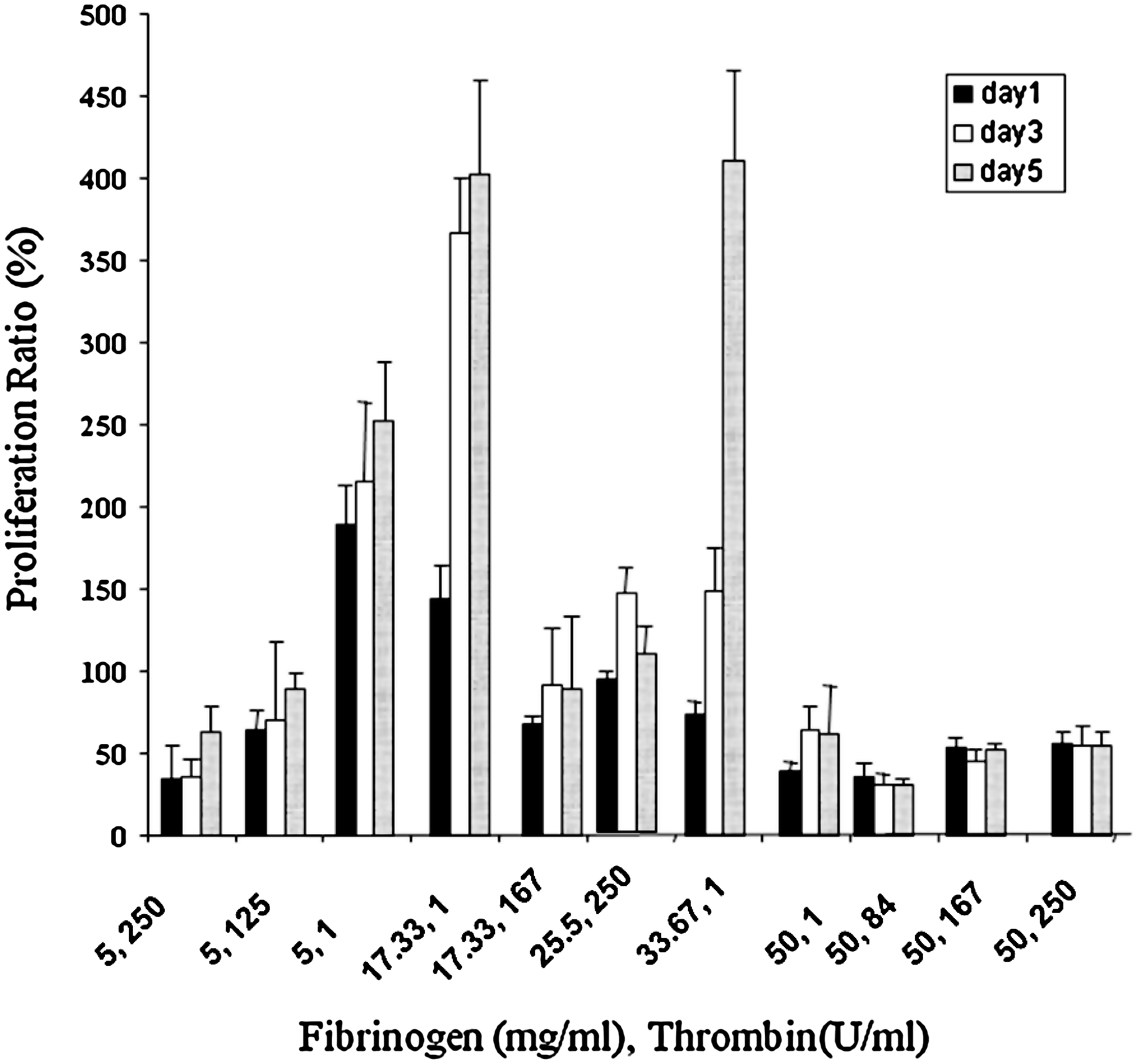
Proliferation of keratinocytes in various three-dimensional (3D) fibrin constructs with different fibrinogen and thrombin concentrations.
Fibrinogen or thrombin solutions were diluted as described in the Materials and Methods section to obtain the final concentrations in the final clots below, that is, after mixing the two solutions. In subsequent experiments, cells were mixed with the fibrinogen solution before mixing with the thrombin solution.
Optimizing growth medium for both NHDF and NHEK
We are interested in studying the cell proliferation of human fibroblasts and keratinocytes when they are cocultured in 3D fibrin constructs. To study the cell proliferation for both NHDF and NHEK in the same fibrin constructs, we needed first to determine the growth medium in which both cells proliferate well in a 24-well culture dish. For that purpose, new growth media were generated by mixing the NHDF and NHEK media at different ratios. Then, the cell proliferation (Fig. 2A) and cell morphology (Fig. 2B) of NHDF and NHEK were examined.

Human keratinocytes grew well in all growth media concentrations except fibroblast growth medium (FGM)-2 (100%). The overall trend for keratinocytes proliferation from highest to lowest were keratinocyte growth medium (KGM) and FGM as follows: KGM (100%) > KGM (75%) FGM-2 (25%) > KGM (50%) FGM-2 (50%) > KGM (25%) FGM-2 (75%) > FGM-2 (100%). This indicates that all growth medium concentrations, except FGM-2 (100%), were favorable for NHEK proliferation, with KGM (100%) being the most favorable.
Human fibroblasts grew well in all growth media. This indicates that the five growth medium concentrations were favorable for NHDF proliferation, with FGM-2 (100%) being the most favorable. According to the above data, the growth media that would be most favorable for both NHEK and NHDF proliferation were KGM (75%) FGM-2 (25%), and KGM (100%). These two growth media were used for future experiments measuring proliferation of NHEK and NHDF coincorporated in 3D fibrin constructs.
Optimization of NHDF and NHEK separation with antifibroblasts microbeads
To separate the fibroblasts and keratinocytes after they are comixed in the 3D fibrin constructs and allowed to grow for a few days, we have established a new assay using antifibroblast magnetic microbeads to separate the fibroblasts from keratinocytes. In Figure 3A, we incubated fibroblasts with antifibroblast microbeads, and then using the magnetic plate as described in Materials and Methods, we determined the specificity of the beads. We found that almost all fibroblasts were pulled down with the microbeads (positive fraction) and very few cells remained in the negative fraction or the wash medium.
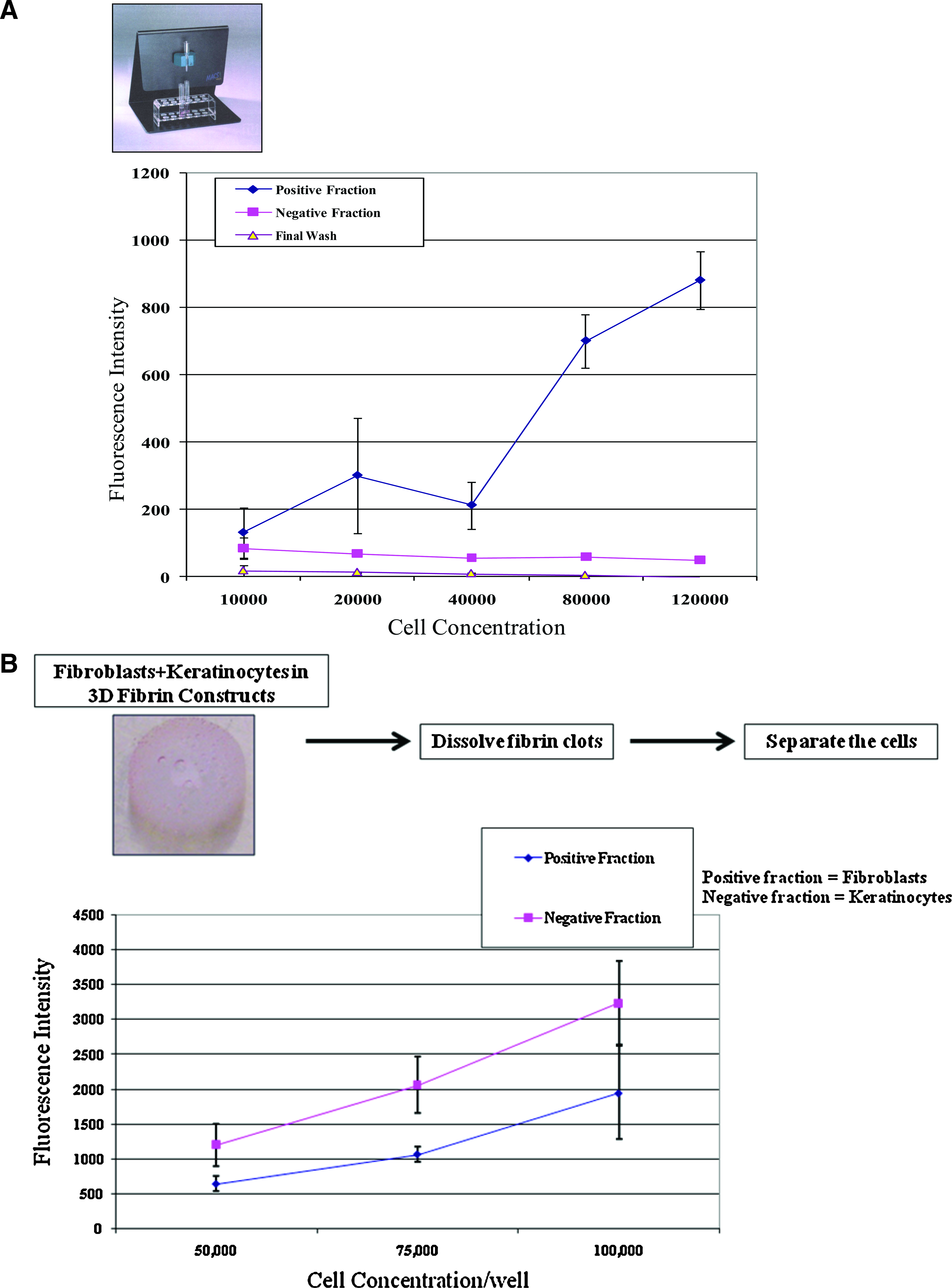
To further test this approach, the antifibroblast microbeads were used to successfully separate cocultured fibroblasts and keratinocytes incorporated inside fibrin constructs as shown in Figure 3B. The separated cells were then allowed to adhere and proliferate on a separate culture plates to examine their morphology to further support that the separation was complete and that there was no cell contamination in the two fractions, that is, no keratinocytes in the positive fraction and no fibroblasts in the negative fraction (Fig. 3C).
In conclusion, we have established a new approach to separate fibroblasts from keratinocytes that were initially incorporated in 3D fibrin constructs.
Keratinocyte and fibroblast proliferation in same 3D fibrin constructs
We used the established assay described above to measure the proliferation of keratinocytes and fibroblasts incorporated inside the 3D fibrin constructs over a 5-day period. We used formulations that were found to provide the optimal proliferation for either keratinocytes alone (Fig. 1) or fibroblasts alone. 10 These formulations were 3, 4, 6, 7, 8, and 11 from Table 1. Here we detail how both cell types proliferated in these formulations.
Formulation 3 (Fib = 25 mg/mL; Thr = 250 U/mL)
Our previous studies have shown that in this formulation fibroblasts proliferated well 10 and keratinocytes proliferated poorly (Fig. 1). In the coculture experiment (Fig. 4A) fibroblasts proliferated much better in the 3D fibrin constructs, almost threefolds more than when they were incorporated alone. On the other hand, there was no change in keratinocytes proliferation from when they were incorporated alone in 3D fibrin constructs.
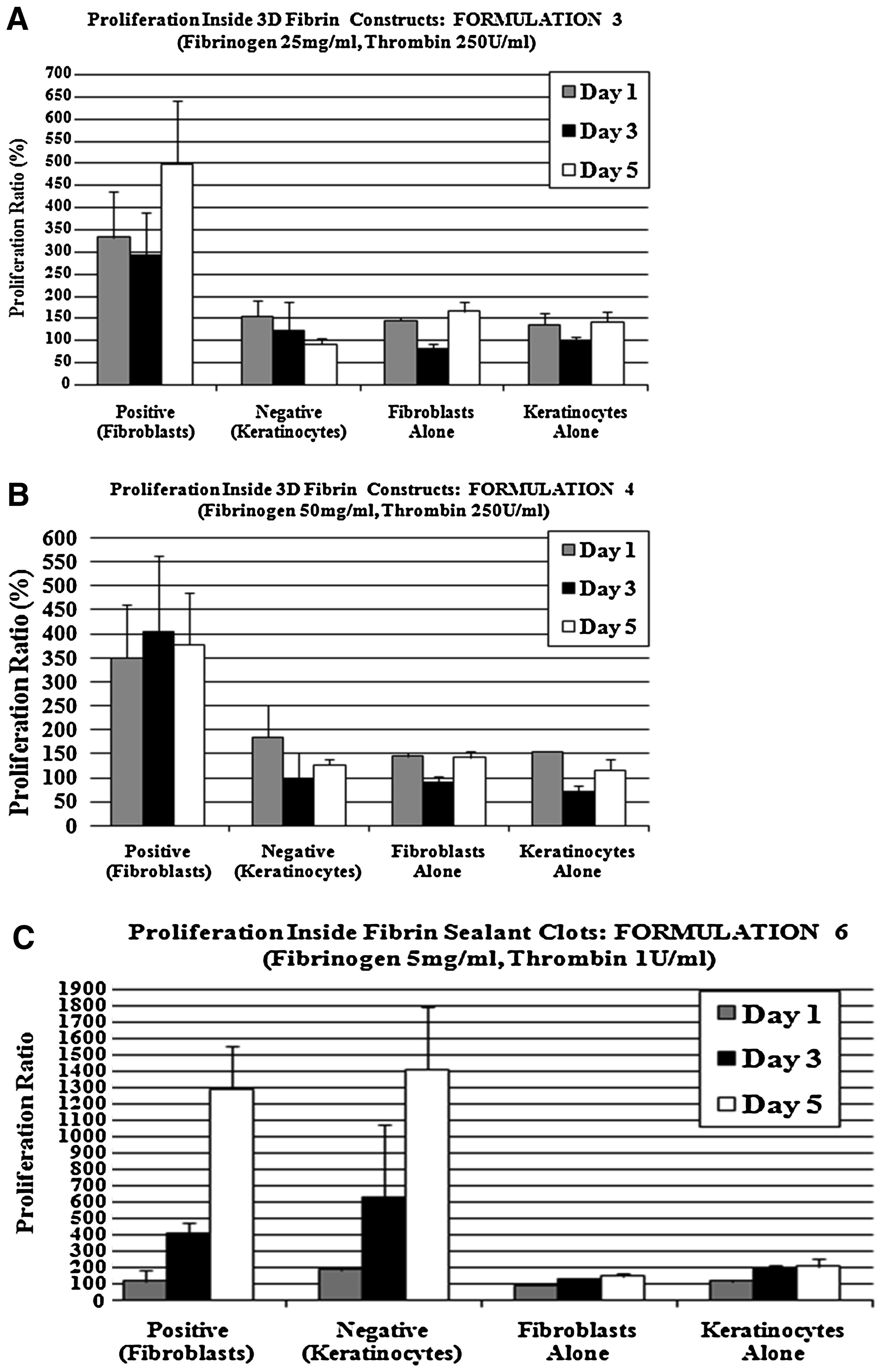
Cell proliferation of cocultured fibroblasts/keratinocytes versus fibroblasts alone or keratinocytes alone in 3D fibrin constructs. Positive (fibroblasts) and negative (keratinocytes) fractions from 3D fibrin constructs where both cell types were comixed. Fibroblasts alone or keratinocytes alone is when each cell type added alone to 3D fibrin constructs.
Formulation 4 (Fib = 50 mg/mL; Thr = 250 U/mL)
Our previous studies have shown that in this formulation fibroblasts proliferated well 10 and keratinocytes proliferated poorly (Fig. 1). In the coculture experiment (Fig. 4B), fibroblasts proliferated much better in the 3D fibrin constructs about 2.5-folds more than when they were incorporated alone in the fibrin constructs. On the other hand, keratinocytes proliferation was not different from when they were incorporated alone in 3D fibrin constructs.
Formulation 6 (Fib = 5 mg/mL; Thr = 1 U/mL)
Keratinocytes proliferated well in this formulation (Fig. 1) and fibroblasts proliferated poorly. 10 In the coculture experiment (Fig. 4C), both fibroblasts and keratinocytes proliferated much better in 3D fibrin constructs than when they were incorporated alone in the 3D fibrin constructs. Fibroblasts proliferated almost 13-folds better and keratinocytes almost 7-folds better in the cocultured experiment than when they were cultured alone in the 3D fibrin constructs.
Formulation 7 (Fib = 34 mg/mL; Thr = 1 U/mL)
Keratinocytes proliferated well in this formulation (Fig. 1) and fibroblasts proliferated to some extent. 10 In the coculture experiment (Fig. 4D), both fibroblasts and keratinocytes proliferated much better in 3D fibrin constructs than when they were incorporated alone in the 3D fibrin constructs. Fibroblasts proliferated almost 3-folds better and keratinocytes almost 2.5-folds better in the cocultured experiment than when they were cultured alone in the 3D fibrin constructs.
Formulation 8 (Fib = 17 mg/mL; Thr = 1 U/mL)
Our previous study had shown that in this formulation both keratinocytes (Fig. 1) and fibroblasts 10 proliferated well. In the coculture experiment (Fig. 4E), both fibroblasts and keratinocytes proliferated much better in the 3D fibrin clots when they were mixed than when they were incorporated alone in the 3D fibrin clots. Fibroblasts proliferated almost threefolds better and keratinocytes almost fourfolds better in the cocultured experiment than when they were cultured alone in the 3D fibrin constructs.
Formulation 11 (Fib = 17 mg/mL; Thr IIa = 167 U/mL)
Our previous studies have shown that in this formulation, fibroblasts proliferated well 10 and keratinocytes proliferated poorly (Fig. 1). In the coculture experiment (Fig. 4F), both fibroblasts and keratinocytes proliferated slightly better in 3D fibrin constructs when they were mixed than when they were incorporated alone in the 3D fibrin clots. Both fibroblasts and keratinocytes proliferated twofolds better in the cocultured experiment than when they were cultured alone in the 3D fibrin constructs.
Discussion
Many attempts were made over the last two decades to produce skin-substitute products to treat chronic wounds. These products consist of bioactive therapeutics or cell-based technologies. None of these products have proven to be efficient in treating the different types of chronic wounds. Thus, there are unmet needs for different and novel products. Many studies have shown fibrin to be a good delivery vehicle for bioactive substances or cells.1–6 We have shown that fibrin is an excellent delivery vehicle for human dermal fibroblasts10,17 as well as for human mesenchymal stem cells.11,12 In this article, we examined the cell behavior of human fibroblasts and keratinocytes when cocultured in 3D fibrin constructs.
In this article, we clearly show that human keratinocytes proliferate differentially depending on the final fibrinogen and thrombin concentration in 3D fibrin constructs. The optimal fibrinogen and thrombin concentration for keratinocytes proliferation in 3D fibrin constructs ranges for fibrinogen concentration between 5 and 17 mg/mL and for thrombin concentration of 1 U/mL. This range for keratinocytes proliferation is very different from the optimal fibrinogen and thrombin concentration for fibroblast proliferation as shown in our previous work. 17
We were, then, interested in introducing both fibroblasts and keratinocytes into 3D fibrin constructs, the reason for that is to determine the optimal formulation for fibroblast and keratinocyte proliferation in 3D constructs—an optimal formulation that allows us to codeliver both cell types into a wound defect. Before we could perform such an experiment, we needed to optimize two assays. First is to dissolve 3D fibrin constructs containing both cell types and be able to separate the two cell types to determine cell number (proliferation). Second is to determine a new growth medium where both fibroblasts and keratinocytes proliferate well. Fibroblast proliferation, in our lab, require the addition of fetal bovine serum, but keratinocytes do not need fetal bovine serum. As shown in Figure 2, we have addressed the later assay by serial dilution of fibroblast medium with keratinocyte medium. We determined that KGM (75%) FGM-2 (25%) is the optimal growth medium to grow both cell types. We have also optimized separation of fibroblasts from keratinocytes after they were allowed to grow in 3D fibrin constructs (Fig. 3).
The optimization of the above two assays allowed us to perform the main experiment of this article, that is, determine the proliferation of the two cell types cocultured in 3D fibrin constructs for various periods of time. Interestingly, we found that both cell types seem to proliferate well in 3D fibrin formulations in the presence of each other versus when they are incorporated alone. In general, we determined that 3D fibrin constructs with fibrinogen concentration of 5–34 mg/mL and very low thrombin concentration of 1 U/mL presented the best environment for cell proliferation for both cell types.
Other similar work showed that skin could be reconstructed from cultured human keratinocytes and fibroblasts on a collagen-glycosaminoglycan biopolymer substrate. 18 Work by Dai et al. 19 shows the development of composite skin substitute based on collagen and poly(ɛ-caprolactone). We have also shown in our previous work that there is a correlation between initial cell seeding, cell proliferation, composite (clot) degradation in relation to fibrinogen, and thrombin final concentration in the 3D fibrin constructs. 9 We have also examined these characteristics in animal studies in work we published recently. 15
In conclusion, we found that the cell proliferation profile of fibroblasts and keratinocytes, when they are codelivered in the 3D fibrin constructs, is very different from the cell proliferation profile (more cell proliferation) of these cells when they are introduced separately in the 3D fibrin constructs, suggesting a cross-talk of cell-specific growth factors between the two cell types. This type of cross-talk was also observed by Sun et al., 20 where keratinocytes were found to retard fibroblasts migration into 3D fibrin constructs compared to fibroblasts cultured alone.
Footnotes
Disclosure Statement
N.S., M.C., and B.T. are employed by Baxter Healthcare Corporation.