




Abstract
Biocements are clinically applied materials for bone replacement in non-load-bearing defects. Depending on their final composition, cements can be either resorbed or remain stable at the implantation site. Degradation can occur by two different mechanisms, by simple dissolution (passive) or after osteoclastic bone remodeling (active). This study investigated both the passive and active in vitro resorption behavior of brushite (CaHPO4 · 2H2O), monetite (CaHPO4), calcium-deficient hydroxyapatite (CDHA; Ca9(PO4)5HPO4OH), and struvite (MgNH4PO4 · 6H2O) cements. Passive resorption was measured by incubating the cement samples in a cell culture medium, whereas active resorption was determined during the surface culture of multinuclear osteoclastic cells derived from RAW 264.7 macrophages. Osteoclast formation was confirmed by showing tartrate resistant acid phosphatase (TRAP) activity on CDHA, brushite, and monetite surfaces, as well as by measuring calcitonin receptor (CT-R) expression as an osteoclast-specific protein by Western blot analysis for struvite ceramics. An absence of passive degradation and only marginally active degradation of <0.01% were found for CDHA matrices. For the secondary calcium phosphates brushite and monetite, active degradation was predominant with a cumulative Ca2+ release of 2.02 (1.20) μmol during 13 days, whereas passive degradation released only 0.788 (0.04) μmol calcium ions into the medium. The struvite cement was the most degradable with a passive (active) release of 9.26 (2.92) Mg2+ ions and a total weight loss of 4.7% over 13 days of the study.
Introduction
Degradation of ceramic biomaterials is based on two different mechanisms. Passive resorption by simple chemical dissolution occurs if the solubility product of the ceramic is several times higher than the corresponding ion concentrations in the surrounding body fluid, which is the case, for example, for β-TCP or secondary, protonated calcium phosphates like monetite (CaHPO4) and brushite (CaHPO4
This study investigated both the passive and active in vitro resorption behavior of various cement matrices. Four materials were tested: brushite (CaHPO4
Methods and Materials
Scaffold fabrication
TCP (Ca3(PO4)2) were prepared by sintering CaHPO4 (Mallinckrodt-Baker) and CaCO3 (Merck) in a molar ratio of 2: 1 for 5 h at 1100°C to obtain β-TCP or 1400°C for α-TCP. Farringtonite (Mg3(PO4)2) was synthesized by using a 2:1 molar ratio powder mixture of MgHPO4 · 3H2O and Mg(OH)2 (Fluka-Sigma-Aldrich), which was sintered at 1100°C for 6 h. The sintered cakes were crushed and were passed through a 125-μm sieve followed by ball milling (PM400; Retsch) for 10 min at 200 rpm (β-TCP), 2 h (α-TCP), and 1 h (Mg3(PO4)2).
Samples for testing in cell culture were produced using silicone rubber molds with a diameter of 6 mm and a height of 2 mm (cytotoxicity testing and staining experiments) or a diameter of 35 mm and a height of 2 mm (Western blots). Brushite cements were produced by mixing the β-TCP powder in an equimolar ratio with monocalcium phosphate monohydrate (Ca(H2PO4)2·H2O, Sigma-Aldrich) in a coffee grinder followed by mixing these powders with 0.05 M citric acid at a constant powder-to-liquid ratio (PLR) of 3.0 g/mL on a glass slab for 20 s. The cement paste was then transferred into the mold and allowed to set for 24 h at room temperature. Monetite scaffolds were produced by hydrothermal treatment by autoclaving the brushite samples for 20 min at 121°C. CDHA samples were obtained by mixing the α-TCP powder with a 2.5 wt% Na2HPO4 (Merck) solution at a PLR of 3.0 g/mL. Struvite samples were produced by mixing 1.1 g Mg3(PO4)2 and 2.2 g struvite powder (obtained by the reaction of Mg3(PO4)2 in ammonium phosphate solution (Merck) which was then reacted with 1.6 mL of a 3.5 M (NH4)2HPO4 (Merck). All samples were stored in phosphate-buffered saline (137 mM NaCl, 2.7 mM KCl, 7.0 mM Na2HPO4
Cell culture
RAW 264.7 (American Type Culture Collection [ATCC] No. CRL-2278) were cultured in Dulbecco's modified Eagle's medium (ATCC Order-No. 30-2002) supplemented with 10% fetal bovine serum, 100 U/mL penicillin, and 100 μg/mL streptomycin (Invitrogen). Cells were used up to passage 18. Confluent cell layers were scraped in 10 mL culture medium and resuspended. For osteoclast formation RAW 264.7 cells were seeded onto glass cover slips placed into the wells of a 24-well plate and, as control, in the wells directly at a density of 104cells/cm2. Calcium and magnesium phosphate cements (CDHA, brushite, monetite, and struvite) were placed into the wells of a 96-well plate and incubated with cells at a density of 2 × 10⁴ cells/cm2. Cell number was examined by a CASY cell counter (Schärfe Systems). Twenty-four hours after seeding the medium was supplemented with 50 ng/mL RANKL (Peprotech), then changed every 48 h, replaced by medium containing 50 ng/mL RANKL, and collected for inductively coupled plasma–mass spectroscopy (ICP-MS; Varian) to examine the biodegradation of different calcium phosphate cements. Cultures were maintained up to 16 days.
The biological response of the cell cultures to the calcium and magnesium phosphate cements was determined by cell counting and a cell activity test after 3, 6, 10, and 13 days of culture on the surfaces as described earlier. 14 Briefly, cell proliferation was analyzed by electronic cell counting using a CASY 1 TTC cell analyzer (Schärfe System). Cell activity was analyzed by using cell proliferation reagent WST 1 (Roche Diagnostics). This reagent is metabolized in mitochondria to a yellowish substance giving an indication for the amount of adenosine triphosphate production in the cell. After incubating the cells for 30 min with the WST reagent 1:10 in supplemented Dulbecco's modified Eagle's medium at 37°C, the adsorption of the supernatant was quantified in a Tecan spectra fluor plus photometer (Tecan). For each analysis the samples were examined in triplicate; the average and standard deviation were calculated.
TRAP staining was used to identify RAW 264.7 cells differentiated into osteoclasts by using a commercial TRAP-staining kit (Sigma; Cat No. 387).
Besides a TRAP-positive staining, osteoclasts had to contain multiple nuclei (at least three or more) and form an actin sealing ring. These features were examined by fluorescence microscopy. As the calcium and magnesium phosphate cements are self-fluorescing, this analysis was performed on glass to see whether osteoclasts formed in these conditions. The samples were fixed for 5 min in 2% paraformaldehyde (PFA) at 4°C and permeabilized for 5 min in 0.2% Triton-X-100 at different time points. The cells were incubated with fluorescein isothiocyanate-conjugated Phalloidin for 30 min to detect actin. Nuclei were stained with 4′,6-diamidino-2-phenylindole for 7 min. Images of labeled structures were recorded with a Zeiss Axiophot (Carl Zeiss) equipped with epifluorescence optics and a charge-coupled device (CCD) camera.
Western blot
Cells were scraped into 1 mL Laemmli sample buffer, 15 and the proteins were precipitated in six times the volume of acetone (Sigma-Aldrich) at −20°C over night and resuspended in Laemmli sample buffer again. The proteins were electrophoretically separated 16 and transferred to nitrocellulose as previously described. 17 After blocking in 5% nonfat dry milk in TBST (140 mM NaCl, 10 mM Tris-HCl, and 20% Tween-20, pH 7.4) over night at 4°C, the membrane was incubated with anti-CT-R (1:200 in blocking solution, H-80; Santa Cruz Biotechnology) for 2 h. After washing in TBST, incubation with the secondary antibody followed (anti-rabbit peroxidase 1:5000 in blocking solution, Dianova; 1 h at room temperature). Bound antibodies were observed after wash steps in TBST using the ECL™ system (Amersham) according to the manufacturers instructions. The membrane was stripped of the bound antibodies and relabeled with anti-tubulin (1:200 in blocking solution; Sigma-Aldrich) and the corresponding secondary antibody (anti-mouse peroxidase, 1:5000 in blocking solution; Dianova). The optical density of the signals was calculated using ImageJ 1.4G (NIH) software.
ICP-MS analysis
Ion concentrations (Mg2+, Ca2+, and HPO42−) in the cell culture medium were determined using ICP-MS (Varian) after 1:10 dilution against standard solutions of 5 and 10 mg/L (Merck). Since these values represent the sum of both the passive degradation of the matrices in serum by chemical dissolution and the active resorption by the osteoclastic cells, reference experiments were performed by incubating the ceramic samples in corresponding volumes of the cell culture medium without cells to determine only the chemical dissolution of the materials. The eluates were collected at the days of medium change in cytotoxicity experiments.
Statistical analysis
Three samples of each calcium phosphate modification were analyzed and the results used for calculating the mean and standard deviation. Statistical variance was analyzed using a two-tailed t-test using Excel (Microsoft). Differences were judged significant when p < 0.05.
Results
X-ray diffraction revealed that the ceramic matrices consisted of >95% of CDHA, brushite, monetite, or struvite with only small amounts of unreacted cement raw materials (Fig. 1). The surface morphology of HA scaffolds (Fig. 2A) showed the appearance of plate-like crystals in the micrometer range, which are known to be clusters of monometer-scale plate-like structures formed by the hydrolysis of α-TCP. 18 Brushite and monetite surfaces (Fig. 2B, C) had a more irregular structure and were formed by monolithic crystals in the range ∼1–5 μm. In contrast, struvite surfaces (Fig. 2D) seemed to be quite different with large crystals of >20 μm size. The total porosity of the samples was calculated to be 19% (struvite), 34% (brushite), and 49% (monetite and CDHA) (Table 1).

X-ray diffraction patterns of ceramic samples; the patterns correspond to the PDF reference patterns for brushite (PDF 09-0077), monetite (PDF 09-0080), hydroxyapatite (PDF 09-0432), and struvite (PDF 15-0762) with only small peaks of unreacted cement raw material [a, α-TCP; b, β-TCP; c, Mg3(PO4)2].
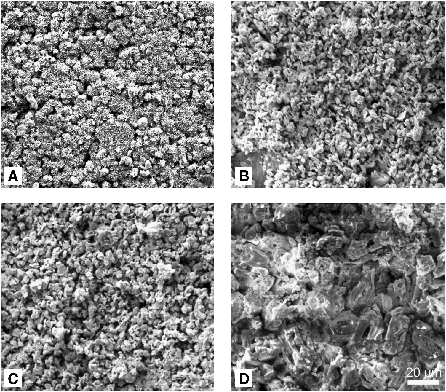
Surface morphology of low temperature ceramic surfaces determined by scanning electron microscopy: (
Values are calculated based on the measured ion concentrations in the culture medium (200 μL) and the average weight of the samples.
CDHA, calcium deficient hydroxyapatite.
Cytotoxicity testing
After 3 days of culture, cell activity and cell number reached the maximum and dropped at day 6 to the minimum on the polystyrene (PS) control (Fig. 3A, B). On these control surfaces cell activity and cell number increased again, but cell activity reaches only the value of day 3, indicating that fewer cells show higher metabolic activity. This was confirmed by calculating the activity per single cell for PS. Cell activity per cell reaches its maximum on day 10 on PS. Cells on CDHA show increasing activity over the whole period. The cell number on these surfaces has its maximum on day 10. On brushite surfaces a similar pattern was observed where cell activity increased over the culture time, but cell number has its maximum on day 6. The cell activity of cells grown on the monetite samples showed less increase, and the cell number rose until day 10. For struvite, both parameters, cell activity and cell number, increased over the whole period with maxima on day 13. Compared to the already used bone replacement material brushite, cell activity (p < 0.05) as well as cell number (p < 0.01) on struvite was significantly increased on day 13. Also, on monetite samples the cell number reached its maximum on day 10 and was significantly higher (p < 0.05) than that of brushite at the same time point. Comparing the activity per cell at day 13, similar results (p > 0.05) were observed on CDHA and brushite. Cell activity on monetite and struvite was significantly (p < 0.05) less when cultured on CDHA or monetite.
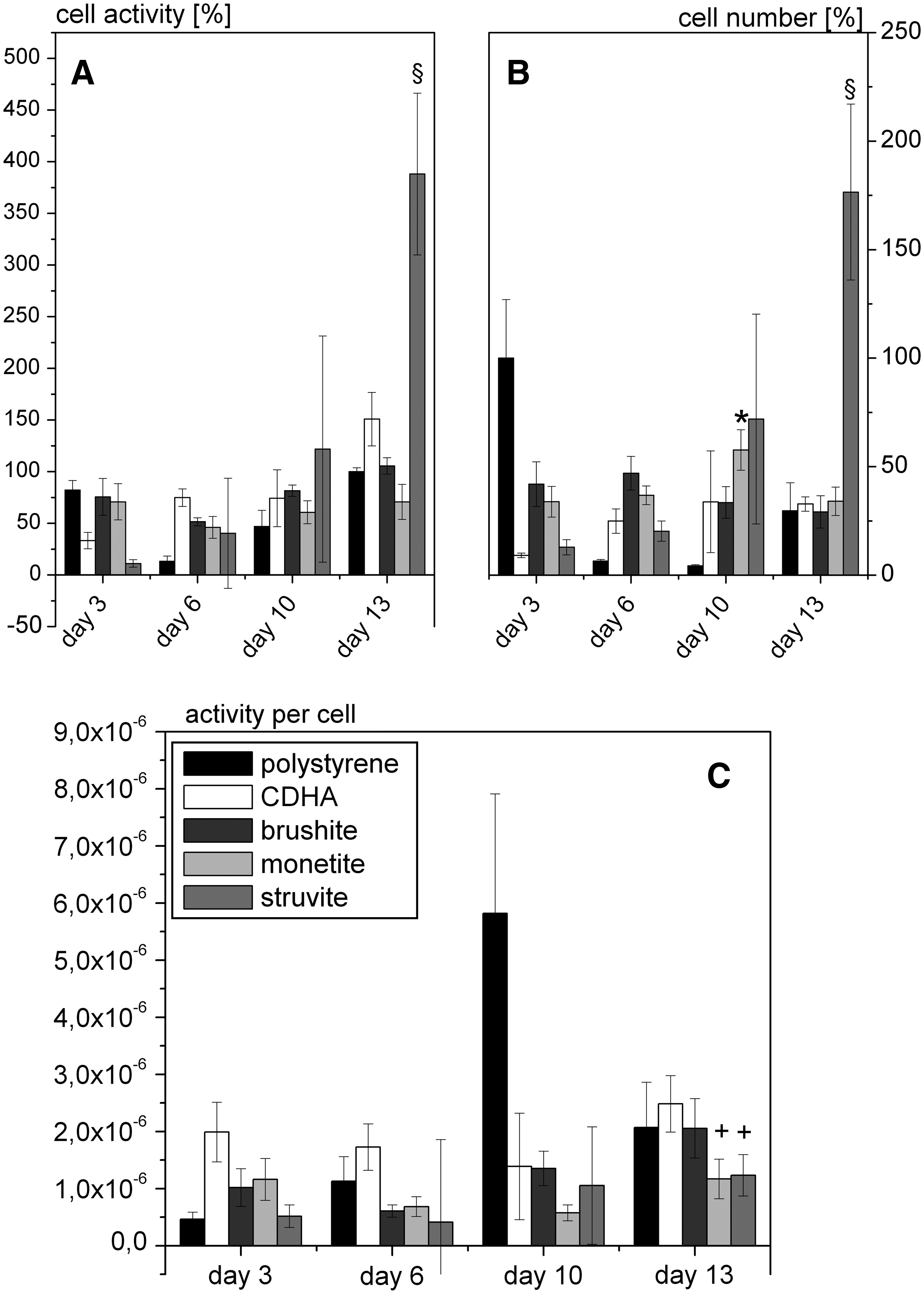
Cell activity (
Fluorescence microscopy
To verify osteoclast formation under the experimental conditions used here, the generation of an actin sealing ring and the number of nuclei per cell were determined using staining with fluorescein isothiocyanate-labeled phalloidin as actin marker and 4′,6-diamidino-2-phenylindole for DNA staining and evaluation of the samples using fluorescence microscopy. As the calcium phosphate samples showed very high background fluorescence, these experiments were performed on glass surfaces. Figure 4 showed the formation of large multi-nucleated cells expressing an actin sealing ring after 3 days of culture in medium containing 50 ng/mL RANKL (Fig. 4, B–B″), indicating the differentiation into osteoclast like cells under the used culture conditions.

Fluorescence microscopy of Phalloidin-stained RAW 264.7 cells. After 3 days of culture the control cells (
TRAP-staining
A purple/red staining of the samples indicates TRAP-positive cells. After 3 days a staining could be observed on glass, CDHA, brushite, and monetite, which becomes more intense over the time (Fig. 5), whereas on struvite surfaces, no TRAP staining could be detected during the whole culturing period. On day 16 the staining on brushite and monetite decreased. The TRAP activity is a characteristic for osteoclast cells; together with the formation of the actin sealing ring, this feature strongly indicates the existence of osteoclast-like cells on the different surfaces with the used differentiation medium. Additionally, Western blot analysis of the osteoclast-specific protein calcitonin receptor (CT-R) was performed and this protein could be detected in all of the osteoclast cells cultivated on the cement surfaces as well as on PS (Fig. 6). In Figure 6A the normalization of the CT-R signals to tubulin β is shown and in Figure 6B the corresponding CT-R signals are given. CT-R expression is highest on the brushite, monetite, and also on the TRAP-negative struvite cements on day 13.
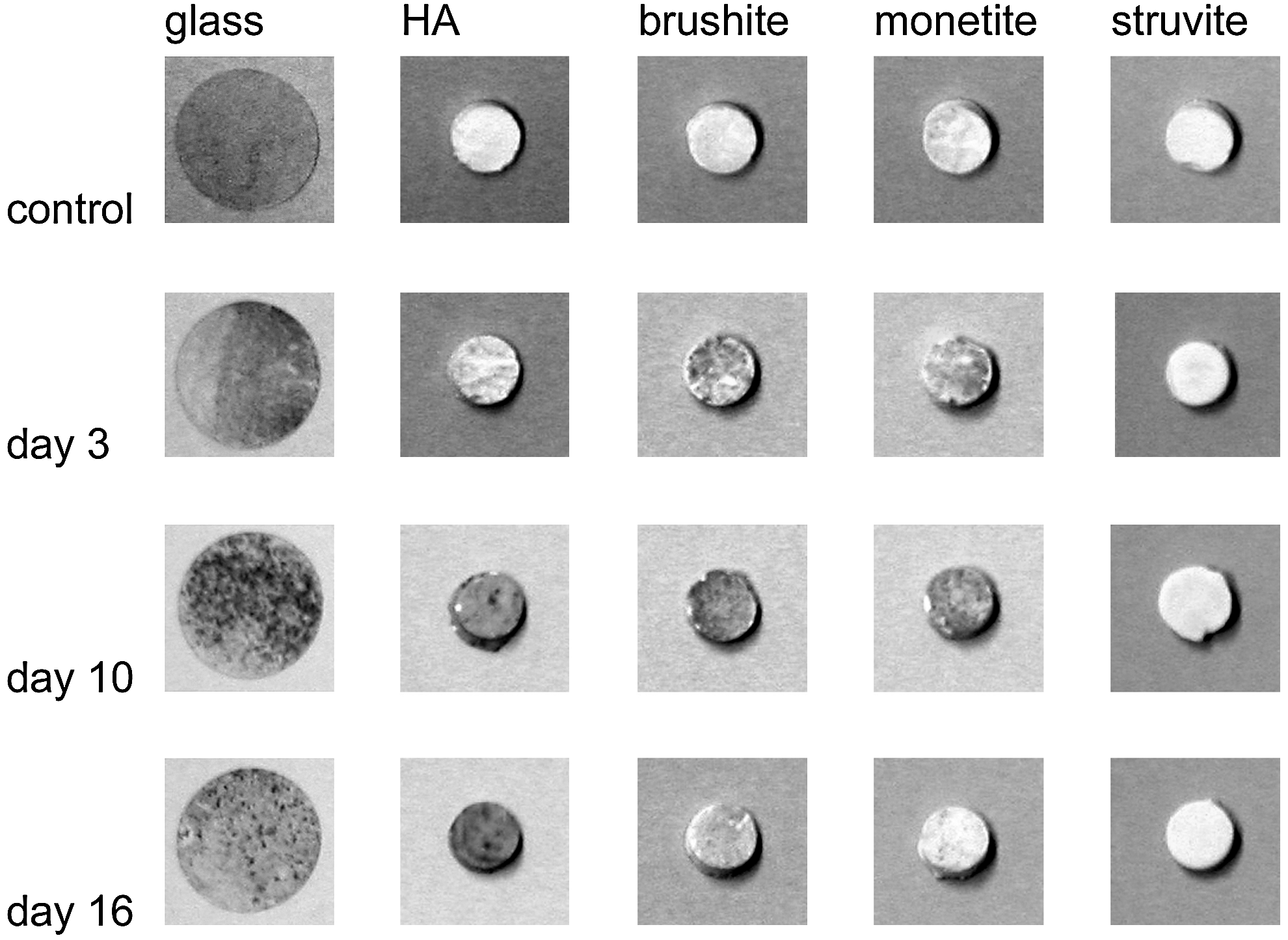
Cytochemical detection of TRAP activity on glass, HA, brushite, monetite, and struvite samples. From day 3 on a red/brown (appears gray in figure) staining can be observed on the glass, HA, brushite, and monetite samples, indicating the formation of proper osteoclasts. TRAP, tartrate resistant acid phosphatase.

Analysis of osteoclast-specific CT-R expression on different cement surfaces by Western blot. (
Ion release from cements during degradation
The concentration of ions released from the samples was determined by means of ICP-MS. Relevant for analysis of the resorption process was the release of Ca2+-ions for CDHA, brushite, and monetite; for struvite, the release of Mg2+-ions was followed. Figure 7 shows the results for resorption of the CaP scaffolds and the MgP cement. It is noticeable that Ca2+ is depleted from the medium by the HA surface (Fig. 7A). The best resorbed CaP surface is brushite, whereas the struvite cement shows nearly three times the resorption of brushite. After calculating the active osteoclastic resorption (Fig. 7C) by subtracting the passive resorption from total resorption again struvite shows the highest values.

Total (
Discussion
In vivo degradation of ceramic biocements occurs by both passive dissolution (brushite and struvite) and osteoclast activity (CDHA). Although various studies have been published concerning the degradation of biocements in vivo19–22 and in vitro,23,24 none of the works clearly distinguished and quantified the active and passive degradation rate of the cements. The current study combined the measurement of the simple dissolution of the cements with the cellular mediated degradation by osteoclasts derived from RAW264.7 cells. Differentiation into osteoclasts was induced by adding 50 ng/mL RANKL to the cell culture medium after 24 h of culture. 10 Osteoclast formation was confirmed by showing TRAP activity on CDHA, brushite, and monetite surfaces; however, there was no TRAP staining obtained on the struvite surface (Fig. 5). This was surprising since the detection of CT-R expression as an osteoclast-specific protein by Western blot analysis also indicates osteoclast formation in this case (Fig. 6A, B). A possible explanation for these results is the multiple washing steps during TRAP staining, which likely caused disintegration and removal of the struvite surface including the attached osteoclastic cells.
The measurement of ion concentrations (Ca2+ and Mg2+) in the cell culture medium with and without osteoclastic cells clearly showed strong differences among the various cement matrices and the degradation regime. Simple immersion of the cement samples in the culture medium resulted in no changes for monetite, while brushite and struvite increased the Ca2+ (Mg2+) by the factor of 1.4 (8) compared with the medium reference level. Interestingly, the immersion of CDHA samples decreased the overall calcium concentration in the medium, likely due to a recrystallization of the calcium-deficient apatite into more stoichiometric HA consuming dissolved calcium ions from the medium. For all materials, passive degradation was constant over time. Osteoclastic activity increased dissolution of the calcium phosphate ceramics; in comparison, more degradation of the struvite samples occurred passively (Table 1). Calcium release into the medium was highest on day 10 of cell culture for CDHA, brushite, and monetite surfaces, whereas biologically mediated magnesium release from the struvite samples continuously increased until the end of the experiment. These dissolution results correspond well with the cell number and activity of cells where a maximum was found on day 10 for the calcium phosphates and a strong increase on day 13 for struvite (Fig. 3). This increase on the magnesium phosphate cements may be due to an adaptation process of the cells to this material after which a boost in cell growth takes place. Eventually, there is an initial retarding effect of the released Mg2+ and NH4+ ions on cell growth and proliferation.
The overall degradation of the matrices by both passive and active degradation was estimated on the basis of the cumulative ion release during the experiment and found to be in the range CDHA < monetite < brushite < struvite. The absence of passive degradation and an only marginal active degradation of <0.01% for CDHA matrices is as expected. However, the values for brushite and monetite (0.23%–0.61%) are quite low compared with other in vitro dissolution studies. Grover et al. 24 have shown a dissolution of ∼20% of a brushite cement in a serum-containing electrolyte after 13 days of immersion at 37°C; however, these results were obtained at a liquid-to-cement volume ratio (LCVR) of 60. The study did also show a strong decrease of the brushite degradation when a lower LCVR of 6 mL/mL and serum-free phosphate-buffered saline was used. At this condition, only 4% of the matrix was dissolved within the same time period. In contrast, our study used an LCVR of only 3.5 mL/mL for immersion to compare the results with the biological testing regime. Other reasons are likely differences in the composition and the physical properties of the tested cements. Whereas we used phase-pure brushite cement mixed at a high powder-to-liquid ratio of 3.0 g/mL resulting in a porosity of only 34%, cements in the above-mentioned study had porosities of 42%–65% due to a PLR of 0.75–1.75 g/mL and they contained only 34% brushite due to the preparation conditions from β-TCP and 2 M phosphoric acid. All these parameters clearly enhanced cement degradation due to a better fluid exchange within the microporous cement and the higher ion dissolution capacity of the larger amount of immersion liquid.
A higher passive and active degradation behavior was found in our study for struvite forming cement leading to a total cement degradation of ∼4.7% within 13 days. Struvite is a pathological formed biomineral and the main component in kidney stones formed after a bacterial infection. 25 Compared with calcium phosphate cements, only few struvite-forming matrices are described in literature.26–30 A major advantage of these degradable materials is the setting reaction at a physiological pH value similar to (slowly degrading) HA cements. This would be beneficial for a further cement modification with pH-sensitive drugs, for example, BMPs, which are noncompatible with the strong acidic setting environment of brushite cements. Indeed, all studies concerning drug modification of brushite cement either used low-pH-sensitive drugs, for example, vancomycin, doxycyclin, or chlorhexidine,31–33 or the drug loading was performed after setting and repeated rinsing with a buffer solution.34,35
The results from this study may aid in the clinical translation of the materials by enabling a more accurate prediction of degradation rate in vivo. Brushite-based materials have been shown to degrade about 52%–98% in orthotopic implantation models within 4–12 months depending on the site of implantation, the size of the implant and the animal model.19,22,36–38 According to these results we would expect a similar degradation rate in vivo for monetite and an even faster degradation for struvite ceramics. However, long-term in vivo behavior may include changes of the phase composition of bioceramics; for example, it is known that brushite can transform in part into low soluble HA, 39 whereas monetite ceramics degraded more consistently without any phase transformation after intramuscular implantation into rats. 40 Struvite cement was more soluble than brushite or monetite in our study, and degradation releases a large amount of magnesium ions into the surrounding of an implant. Since the latter are a strong inhibitor of HA crystal growth, 41 we would not expect phase transformation into a lower solubility phase and would therefore estimate a total (in vivo) degradation period of <12 months for this material.
Conclusions
RAW 264.7 cells were cultivated for up 16 days on different low temperature cement matrices like HA, brushite, monetite, and struvite. Cells differentiated into osteoclasts after addition of 50 ng/mL RANKL and showed an active resorption of the materials. Mechanistically, osteoclasts acidify the resorption lacuna to a pH of about 4.5 at which brushite and struvite resorption was found to be the highest under these conditions. Since HA is the most stable calcium phosphate phase at a pH >4.2, the lowest resorption activity was observed on CDHA surfaces. The magnesium phosphate cement is promising for future applications as it resorbed the most rapidly and set under physiological pH conditions.
Footnotes
Acknowledgments
This work was supported in part by a Quebec-Bavarian support grant from the “Developpement economique, innovation et exportation” of Quebec, Canada, by the Canadian Institute of Health Research and a Canada Research Chair (J.B.) and by the Deutsche Forschungsgemeinschaft (DFG GB1/7-2).
Disclosure Statement
No competing financial interests exist.