




Abstract
Heart failure remains the leading cause of death in many industrialized nations owing to the inability of the myocardial tissue to regenerate. The main objective of this work was to develop a cardiac patch that is biocompatible and matches the mechanical properties of the heart muscle for myocardial infarction. The present study was to fabricate poly (glycerol sebacate)/gelatin (PGS/gelatin) core/shell fibers and gelatin fibers alone by electrospinning for cardiac tissue engineering. PGS/gelatin core/shell fibers, PGS used as a core polymer to impart the mechanical properties and gelatin as a shell material to achieve favorable cell adhesion and proliferation. These core/shell fibers were characterized by scanning electron microscopy, contact angle, Fourier transform infrared spectroscopy, and tensile testing. The cell–scaffold interactions were analyzed by cell proliferation, confocal analysis for the expression of marker proteins like actinin, troponin-T, and platelet endothelial cell adhesion molecule, and scanning electron microscopy to analyze cell morphology. Dual immunofluorescent staining was performed to further confirm the cardiogenic differentiation of mesenchymal stem cells by employing mesenchymal stem cell-specific marker protein CD 105 and cardiac-specific marker protein actinin. The results observed that PGS/gelatin core/shell fibers have good potential biocompatibility and mechanical properties for fabricating nanofibrous cardiac patch and would be a prognosticating device for the restoration of myocardium.
Introduction
The choice of biomaterials is a vital step in the development of a heart patch for MI. Among the natural polymers, collagen, alginate, and gelatin have been under intensive investigation for myocardial tissue engineering.6–9 The elastomer poly(glycerol sebacate) (PGS) was recently developed for soft tissue engineering.10,11 Elastomer-based grafts may facilitate compliance of matching, thereby ameliorating the lifespan of the patients. 12 Scaffolds composed of PGS are elastic and reversibly deformable and are thereby conducive to contracting cardiomyocytes and engineered myocardium. 13 Other desirable properties of PGS include control of its mechanical properties, 14 the capacity to form a variety of geometries on the macro- 15 and microscales, 16 and low inflammatory response and fibrotic encapsulation, coupled with retention of mechanical strength during degradation in vivo.17,18 PGS represents a feasible biomaterial from the mechanical perspective; gelatin favors enhanced cell adhesion for tissue engineering. Fabricated PGS/gelatin fibrous scaffold is a unique extracellular matrix-like topography and has been suggested to be a potential biomaterial for MI.
The optimal cell source to create an engineered myocardial patch should be easy to harvest, proliferative, and nonimmunogenic, and has the ability to differentiate into mature, functional cardiomyocytes. Cardiomyocytes have the native contractile and electrophysiological properties of the heart muscle, they are difficult to obtain and expand, and are allogenic cells. Disadvantages of embryonic stem cells include their potential for transformation into teratocarcinoma and other malignancies. 19 In contrast, mesenchymal stem cell (MSCs) can be easily isolated, cultured, and nonimmunogenic, and can be readily expanded in the laboratory setting, 20 making them an attractive candidate for cardiac tissue engineering (CTE). After expansion, stem cells can be directed to differentiate into cardiomyogenic lineage for CTE.21–23
The primary objective of this work was to develop a biocompatible elastomeric cardiac patch from PGS and gelatin for CTE. In this study, we described a hybrid cardiac patch engineered from a synthetic elastomer, PGS, and a natural polymer, gelatin, supplemented with coculture of cardiomyocytes and MSCs, prioritizing on the pertinent considerations—biocompatibility and cell delivery. A suitable approach to deliver cells into the heart to promote its regeneration is to use tissue engineering concept, in which a biodegradable patch material is populated with cells in vitro and implanted into the myocardial infarct region. Our approach takes advantage of the ability of an elastomeric biomaterial sheet comprising of PGS/gelatin core/shell fibers to act as a flexible patch; with this approach, cells would remain adhered to the nanofibrous patch, preventing cell loss and providing a more site-directed repair mechanism for MI.
Materials and Methods
Fabrication of core/shell fibers
The materials used in this study were gelatin, 2,2,2-trifluoroethanol (TFE), and 1,1,1,3,3,3-hexafluoro-2-propanol (purchased from Sigma Aldrich Chemical Company, Inc.). PGS was synthesized by Wang et al.'s established procedure. 13 A mixture of glycerol and sebacic acid in the ratio of 1:1 was reacted at 120°C under nitrogen for 24 h. The pressure was then reduced to 40 m torr and the reaction held at 120°C for 48 h to synthesis PGS. Gelatin 10% (w/v) was dissolved in TFE solvent to form the shell solution. Similarly, PGS 15% was dissolved in 1,1,1,3,3,3-hexafluoro-2-propanol solvent to form the core solution. A two-fluid coaxial spinneret was set up for electrospinning. The inner tube had a diameter of 1 mm, whereas the outer tube had a diameter of 2 mm. The spinneret was designed such that the fluids are immiscible before exiting the nozzle. Fluid was provided to the nozzle by two syringe pumps (KD Scientific, Inc.) that provide a constant-volume flow rate of 0.3 mL/h for the core solution and 1.0 mL/h for the shell solution. A high-voltage electric field (DC high-voltage power supply from Gamma High Voltage Research) was applied at the tip of the spinneret at 12 kV. A Taylor cone was formed at the coaxial tip with an outer droplet surrounding the inner one. The outer droplet was then transformed into a jet, because of the surface charges creation, whereas there was no creation of surface charges on the inner droplet owing to the rapid evaporation of the solvent which reduces the solvents mixing during this process, thereby producing core/shell fibers. A collector plate was placed at a distance of 15 cm from the tip of the spinneret to collect the core/shell fibers. Besides, gelatin nanofibers were also fabricated using 12% w/v solution in TFE. The electrospinning conditions used were 1.0 mL/h flow rate, 12 cm distance between the needle tip and collector plate, and 15 kV voltage supply. The fibers produced were subsequently vacuum dried so as to remove any residual solvents. The fibers were then cross-linked using 50% glutaraldehyde (Sigma) vapor for 24 h to improve its mechanical stability suitable for CTE.
Material characterization
The surface morphology of electrospun nanofibrous scaffolds was studied under a scanning electron microscope (JEOL JSM–5600LV) at an accelerating voltage of 15 kV, after gold coating (JEOL JFC-1200 fine coater). For measuring the fiber diameter of electrospun fibers from the scanning electron microscopy (SEM) images, n=10 fibers were chosen at random on each of the scaffolds. For each scaffold material n=5 samples were chosen for analysis of fiber diameter. The average fiber diameter was then calculated along with SD using image analysis software (Image J; National Institutes of Health). Functional groups present in the scaffolds were analyzed using Fourier transform infrared spectroscopic analysis on Avatar 380 (Thermo Nicolet), over a range of 400–4000 cm−1 at a resolution of 4 cm−1. The hydrophobic or the hydrophilic nature of the electrospun fibers was measured by sessile drop water contact angle measurement using VCA Optima Surface Analysis system (AST Products). Tensile properties of electrospun nanofibrous scaffolds were determined using a tabletop tensile tester (Instron 3345) at 10 N load capacities. Rectangular specimens of dimensions 10 mm×20 mm were used for testing, at a rate of 5 mm/min. The data were recorded at room conditions 25°C and 34% humidity. The data of only those samples that have failed in the center, giving a dog bone-like appearance and not those that have failed at the grips where the load was being applied, were employed for calibrating the stress–strain curves. Thereby, uncertain data were avoided. The tensile stress strain values obtained from the instrument were plotted using an Excel sheet.
Cell culture on the electrospun scaffolds
The electrospun fibers collected on round glass cover slips of 15 mm in diameter were placed in a 24-well plate with a stainless steel ring to prevent swelling. The fibers were sterilized under ultraviolet light for 2 h, washed thrice with phosphate-buffered saline (PBS) for 15 min each to remove any residual solvent, and subsequently immersed in Dulbecco's Modified Eagle's Medium (DMEM) overnight before cell seeding. The rabbit cardiomyocytes were isolated using collagenase treatment. The aorta of the animal was cannulated and perfused using a bicarbonate perfusion fluid composed of NaCl 118.5, NaHCO3 25.0, KCl 4.7, MgSO4 1.2, KH2PO4 1.2, glucose 11.0, and CaCl2 2.5 (mM). After perfusion, the rabbit heart was washed thrice thoroughly with antibiotic solutions made in PBS at a concentration of 2× and 3×, for 30 min each, and fragmented into fine pieces. It was subsequently followed by treatment of 1% collagenase in PBS for 30 min at 37°C. The dispersed cells were harvested after each 5 min of incubation by decantation. The cells harvested from first incubation usually contain a high percentage of damaged cells and hence were not employed for the present study. The isolated heart cells from second and third incubation were centrifuged at 1000 rpm for 5 min and cultured in DMEM supplemented with 10% fetal bovine serum (Gibco Invitrogen) and 1% antibiotic and antimycotic solutions (Invitrogen Corporation) in a 75 cm2 cell culture flask, to isolate the cardiomyocytes. A high yield of isolated heart cells was obtained by this technique. The yield of myocytes was determined by actinin expression from flow cytometry procedure. We obtained positive actinin expression corresponding to myocytes yield of 82%±12% (data not shown). The MSCs (PT-2501; Lonza) were cultured in low-glucose DMEM supplemented with 10% fetal bovine serum (Gibco Invitrogen) and 1% antibiotic and antimycotic solutions (Invitrogen Corporation) in a 75 cm2 cell culture flask. Cells were incubated in CO2 incubator at 37°C at 5% CO2. The culture medium was changed every alternate day. Before seeding, the cells were detached by adding 1 mL of 0.25% trypsin containing 0.1% EDTA. Detached cells were centrifuged, counted by Trypan blue assay using a hemocytometer, and seeded on the scaffolds. MSCs and adult cardiomyocytes were used as negative and positive controls, respectively.
The scaffolds were separated into three groups: the control tissue culture plate (TCP), gelatin nanofibrous scaffolds, and the PGS/gelatin core/shell fibers. These were further segregated into coculture scaffolds, onto which both MSCs and cardiomyocyte cells were seeded in the ratio of 1:1 at a seeding density of 10,000 cells per well (5000 MSCs:5000 cardiomyocytes); positive control scaffolds, onto which cardiomyocytes were seeded at the same seeding density of 10,000 cells per well; and the final batch comprised of the negative control scaffolds, onto which MSCs were seeded at the same seeding density of 10,000 cells per well.
Cell proliferation
Cell proliferation on the nanofibrous substrates was determined using the colorimetric 3-(4, 5-dimethylthiazol-2-yl)-5-(3-carboxymethoxyphenyl)-2(4-sulfophenyl)-2H tetrazolium (MTS) assay (CellTiter 96 AQueous One solution; Promega). The reduction of yellow tetrazolium salt [3-(4, 5-dimethylthiazol-2-yl)-5-(3-carboxymethoxyphenyl)-2(4sulfophenyl)-2H tetrazolium] in MTS to form purple formazan crystals by the dehydrogenase enzymes secreted by mitochondria of metabolically active cells formed the basis of this assay. The formazan dye shows the absorbance at 492 nm and the amount of formazan crystals formed is directly proportional to the number of cells. After 5 days of cell seeding, the medium was removed from the 24-well plates and the scaffolds were washed in PBS. The scaffolds were then incubated in a 1:5 ratio mixture of MTS assay and serum-free DMEM medium for 3 h at 37°C in a 5% CO2 incubator. After the incubation period, the samples were pipette out into 96-well plates. The absorbance was then calibrated at 490 nm using a microplate reader (Fluostar Optima; BMG Lab Technologies). The same procedure was repeated for days 10 and 15 of the cell-cultured samples.
Cell morphology
The cell morphology was analyzed using SEM. After 10 days of seeding the scaffolds with cells, the medium was removed from culture wells and the samples were fixed with 3% glutaraldehyde in PBS for 3 h. The scaffolds were then rinsed with distilled water for 15 min and then dehydrated with a series of ethanol gradients starting from 30% to 50%, 75%, 90%, and 100% (v/v). Subsequently, the samples were treated with hexamethyldisilazane (Sigma) solution and allowed to air-dry at room temperature in the fume hood. The samples were then gold coated and the cells morphology was analyzed using SEM.
Immunofluorescent staining
To observe whether the MSCs cocultured with cardiomyocytes have undergone cardiogenic differentiation, immunofluorescent staining of the selected proteins of cardiomyocytes–actinin and troponin and endothelial cell marker protein platelet endothelial cell adhesion molecule (PECAM) were performed. For cardiac protein analysis, cells cultured for 10 days on the scaffolds were fixed with 100% ice-cold methanol for 15 min. The samples were then washed with PBS once for 15 min and incubated in 0.5% Triton-X solution for 5 min to permeabilize the cell membrane. Nonspecific sites were blocked by incubating the cells in 3% bovine serum albumin (Sigma) for 1 h. The primary antibodies α-actinin, troponin-T, and PECAM conjugated with FITC (Sigma) were added separately into each well, in the dilution 1:100, and incubated for 90 min at room temperature. The samples were then washed thrice with PBS for 15 min each to remove the excess unbound primary antibodies. This was followed by incubation for 60 min with Alexa Fluor 488 secondary antibodies (Invitrogen) in the dilution of 1:250 at room temperature. The samples were again washed thrice with PBS for 15 min. Negative controls were also employed in each analysis to delete the primary antibody. The cell nuclei were stained using 1:5000 dilution of 4,6-diamidino-2-phenylindole hydrochloride (DAPI; Invitrogen) for 30 min at room temperature. The cells were again washed with PBS thrice to remove any excess staining. The samples were then removed and mounted over glass slides using H-1000 Vectashield mounting medium (Vector Laboratories). The edges of the samples were sealed using Fluoromount. The glass slides were then viewed using fluorescence microscopy (Olympus FV 1000).
Double immunofluorescent staining was further performed on the coculture scaffolds to confirm the differentiation of MSCs into cardiomyocytes. The MSC-cardiomyocyte coculture cells plated on TCP, gelatin nanofibers, and PGS/gelatin core/shell fibers for 10 days were stained with MSC-specific marker CD 105 (Abcam) in the dilution 1:100 for 90 min at room temperature, before which the nonspecific sites were blocked with 3% bovine serum albumin. This was followed by the addition of secondary antibody Alexa Fluor 488 (Green) in the dilution 1:250 for 60 min at room temperature. The samples were washed with PBS thrice to remove the excess staining. The samples were then treated with cardiac-specific marker protein actinin in the dilution 1:100 for 90 min at room temperature. This was followed by the addition of the secondary antibody Alexa Fluor 594 (Red) (Invitrogen) in the dilution 1:250 for 60 min at room temperature. The samples were washed with PBS thrice to remove the excess staining and then incubated with DAPI in the dilution 1:5000 for 30 min at room temperature. The samples were then removed and mounted over a glass slide using Vectashield mounting agent and examined under the fluorescent microscope (Olympus FV 1000).
Statistical analysis
For each experiment, n=5 samples were tested to calculate the mean±SD; t-test and analysis of variance using SPSS12.0 software (SPSS) were employed to calculate statistical significance. Differences were considered statistically significant at p≤0.05 and p≤0.001.
Results and Discussion
Electrospinning is a versatile method for bio-mimicking nanofibers for the native environment for better cell adhesion and tissue growth. 24 It has been proved that electrospun fibrous scaffolds provide both flexibility and guidance for cardiomyocyte growth and used to obtain structurally and functionally competent constructs. 25 Moreover, it was known that the extracellular matrix provides both physical support and outside-in signals that regulate many cellular functions, such as adhesion, migration, proliferation, and differentiation, and shall be maintained for optimal cellular benefits. 26 Gelatin is a derivative of collagen, acquired by denaturing the triple-helix structure. 27 Gelatin exhibits excellent biodegradability, nonantigenicity, cost efficiency, cellular biocompatibility, and antithrombogenicity.28–33 The SEM images of electrospun fibers as shown in Figure 1 revealed uniform beadless gelatin fibers and PGS/gelatin core/shell fibers of diameter 405±47 nm and 1±0.125 μm, respectively. Gelatin nanofibers and PGS/gelatin core/shell fibers with shell material as gelatin were not stable in DMEM and PBS without cross-linking in glutaraldehyde. The scaffolds remained stable for 1 month in cell culture at the time of proliferation and differentiation. Moreover, for the infarcted myocardium to regenerate the patch material has to remain stable for a stipulated period in vivo to enable homing of MSCs and cardiomyocytes to regenerate the infarcted myocardium. The cross-linking with glutaraldehyde improves the mechanical stability of the nanofibers.

Scanning electron microscopy (SEM) images showing the fiber morphology of
The contact angle results of gelatin and PGS/gelatin are shown in Table 1. PGS, being a hydrophilic and low-molecular-weight polymer, could serve as a plasticizer, and was protected by a shell with the natural polymer-like gelatin to augment their hydrophilicity and softness, to make them more suitable for cell attachment and contraction, and hence the low contact angle of PGS/gelatin of 7±1.4, compared to gelatin fibers, which had a contact angle of 20.1±2.7. The hydroxyl groups present in gelatin form hydrogen bonds with water molecules, thus imparting the relevant hydrophilicity.
PGS, poly(glycerol sebacate).
Functional groups present in nanofibers were analyzed using Fourier transform infrared spectroscopy (Fig. 2) in which characteristic bands of gelatin were observed in gelatin and PGS/gelatin scaffolds. Amide bands of gelatin were obtained for these scaffolds, where amide I band for C=O stretch, amide II peak for N–H bend coupled with C–N stretch, and amide III peak for N–H bend pertaining to triple helical structure of gelatin were obtained at 1645.83, 1538.17, and 1233.16 cm−1, respectively. Similarly for PGS/gelatin amide I band for C=O stretch, amide II peak for N–H bend coupled with C–N stretch and amide III peak for N–H bend were noticed at 1642.24, 1541.76, and 1251.10 cm−1, respectively.
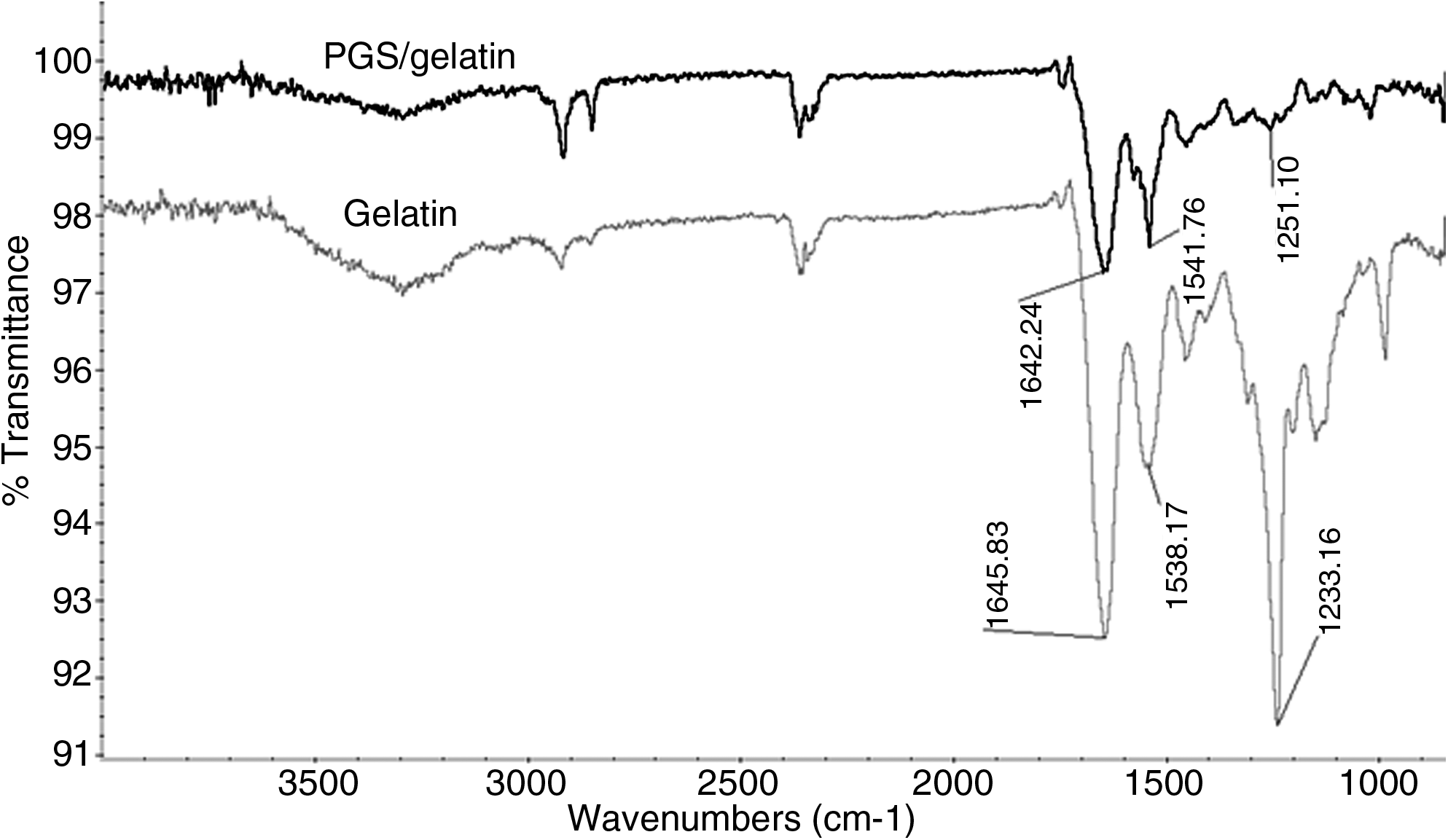
Fourier transform infrared images. Blue curve: gelatin fibers showing characteristic amide peaks at 1645.83, 1538.17, and 1233.16 cm−1. Red curve: PGS/gelatin core/shell fibers showing characteristic amide peaks at 1642.24, 1541.76, and 1251.10 cm−1.
The tensile properties of gelatin and PGS/gelatin are also tabulated in Table 1. The stress–strain curves of the fibers are shown in Figure 3. The Young's modulus of gelatin nanofibers was 72.15±3.08 MPa. The young's modulus was calculated using Secant modulus method as the curve is nonlinear; here a tangent was drawn to the curve from the origin. The slope of this tangent gives the Young's modulus. It can be noticed that upon the incorporation of PGS to gelatin, the Young's modulus has shown a tremendous decrease from 72.15±3.08 MPa to 6.08±0.49 MPa. The mechanical property of the core/shell fibers is chiefly dictated by the core-material PGS, whereas the shell polymer offers external functions such as adhesion and proliferation. Li et al. 34 observed the culture of fetal rat ventricular muscle in Gelfoam to form cardiac grafts. These grafts were cultured in vitro for 7 days, forming a beating cardiac tissue in vitro and were then implanted onto myocardial infarcted site in a cryoinjured rat heart. It was found that the cells within the grafts survived and formed junctions with the recipient heart cells. However, no improvement of ventricular function in hearts receiving either cell-seeded or unseeded gelatin grafts was noticed. To engineer the myocardial tissue, which beats synchronously and constantly throughout the life, the biomaterial should be as soft and elastic as heart muscle. An elastomer-like PGS with gelatin is a desirable biomaterial to reproduce the mechanical characteristics of the heart muscle, as evidenced by its mechanical properties. Li et al. 34 reported bioengineered cardiac grafts made of fetal cardiac cells and 3D gelatin mesh formed cardiac-like tissue and contracted spontaneously. However, this approach had major limitations: (1) limited ability of cardiomyocytes to proliferate, and (2) unlike MSCs, the fetal and neonatal cells are prone to immune rejection after transplantation, and are relatively sensitive to ischemic injury. 35 The use of stem cells derived from adult tissues overcomes these disadvantages. Figure 4 showed that the rate of cell proliferation was significantly (p≤0.05) higher on gelatin and PGS/gelatin scaffolds compared to TCP on day 15. Statistically significant difference (p≤0.05) was observed between the coculture groups of PGS/gelatin core/shell fibers and gelatin fibers on day 15. Additionally, a significant (p≤0.05) difference was also noticed between the cardiomyocytes group cultured on the core/shell fibers compared to gelatin nanofibers on days 10 and 15, indicating increased rate of cardiomyocyte proliferation on the core/shell fibers compared to gelatin nanofibers. It has been reported that core/shell fibers comprising of collagen as shell and polycaprolactone (PCL) as core promoted greater human dermal fibroblasts proliferation, compared to PCL nanofibers coated with collagen, confirming that core/shell fiber architecture is more favorable for cell proliferation compared to coated nanofibrous scaffold. 36 A significant level of difference (p≤0.001) was observed using SPSS12.0 software for calibrating the analysis of variance compared between gelatin and PGS/gelatin core/shell fibers for culturing cardiomyocytes and MSCs coculture on days 10 and 15 (Fig. 4). Additionally, the cell proliferation of cocultured cells was found to increase by 219.45% and 132.21% from day 5 to 15 on PGS/gelatin and gelatin scaffolds, respectively. The rate of proliferation of cardiomyocytes was also augmented by 171.34% and 101% from day 5 to 15 on PGS/gelatin and gelatin scaffolds, respectively. The rate of proliferation of cardiomyocytes was not limited on gelatin scaffolds as reported earlier by Li et al. 34 This may be because the presence of MSCs induces paracrine signaling molecules into the environment, thereby preventing apoptosis of cardiomyocytes. It has been reported that MSCs secrete insulin-like growth factor I and vascular endothelial growth factor, which have a cardioprotective effect. Insulin-like growth factor-I mediates an anti-apoptosis effect, whereas angiogenesis is mediated by vascular endothelial growth factor. 37 Exogenous expression of these factors at the transplantation site may elevate tissue integration and survival. MSC transplantation might act in a paracrine or cell–cell signaling fashion to affect cardiac repair via cytokine-induced enhancement of endogenous cardiac cell function. 38 Moreover, studies have reported that cardiomyocyte adhesion and organization into a contractile tissue have been far superior on natural scaffolds compared to synthetic scaffolds.7,39 Failure to produce new myocardial tissue in clinically significant numbers was ascribed to cell death occurring after engraftment and inability of engrafted myoblasts to differentiate and integrate within the host myocardium 40 ; Dai et al. proposed that the retention of MSCs in the infarcted region can be improved by implanting cells in a collagen matrix. The MSCs were labeled with isotopic colloidal nanoparticles and its distribution was analyzed by measuring the nanoparticle radioactivity in the lung, liver, spleen, and kidney. It was found that collagen matrix significantly reduced the relocation of transplanted MSCs to remote organs and noninfarcted myocardium. 41 A clinical study was evaluated to determine the potential of a 3D type I collagen construct seeded with bone marrow cells onto the infarcted region. It was determined that the scar area thickness progressed from 6±1.4 to 9±1.5 mm (p≤0.005) and ejection fraction improved from 25%±7% to 33%±5% (p≤0.04). 42 We have applied an alternative approach, by the encapsulation of MSCs and cardiomyocytes onto PGS/gelatin core/shell nanofibrous scaffold. This technique might prevent the loss of cells from the site of implantation and also induces the cardiogenic differentiation of MSCs.

Tensile stress–strain curves of gelatin fibers and PGS/gelatin core/shell fibers.

Cell proliferation study for days 5, 10, and 15 on tissue culture plate (TCP), PGS/gelatin core/shell fibers, and gelatin nanofibers using cardiomyocytes, mesenchymal stem cells (MSCs), and MSC-cardiomyocyte coculture. Bar represent means±SD. Asterisks (t-test and analysis of variance) indicate significant difference in the measurement when compared to gelatin and PGS/gelatin nanofibrous scaffold (*p≤0.05, **p≤0.001).
To observe the myocardiogenic differentiation of MSCs, the immunofluorescence stains of specific cardiomyocyte marker proteins like troponin-T and α-actinin43–45 were examined. Troponin-T is important for effective cardiomyocytes that contain contractile proteins, as it regulates the force and velocity of myocardial contraction, 43 and actinin is an important constituent of the contractile apparatus. Troponin-T is one of the essential proteins for contractile function and an indicator of differentiation in cardiomyocytes.46,47 The present study employed actinin and troponin-T to prove cardiogenic differentiation of MSC and PECAM to prove the MSC differentiation into endothelial lineage. Initially, at day 0, when the cells have not attached to the scaffold surface, striations were observed upon staining with actinin marker, as shown in Supplementary Fig. S1 (Supplementary Data are available online at www.liebertonline.com/tea). However, upon attachment to the scaffold surface the striations were not obvious as witnessed in Figure 5A. Figure 5C (a–f) shows that the expression of PECAM was preponderant on the PGS/gelatin scaffolds compared to the gelatin fibers, and also Figure 5A (a–f) and 5B (a–f) show that the expression of cardiomyocyte marker proteins was predominant in the coculture system [Fig. 5A (a–c) and 5B (a–c)] compared to the cardiomyocyte culture group [Fig. 5A (d–f) and 5B (d–f)]. This may be because MSCs that have undergone cardiogenic differentiation owing to the presence of cardiomyocyte in their close proximity also express the marker proteins. Moreover, it can also be observed that the expression of cardiac proteins was increased on PGS/gelatin substrates compared to gelatin scaffold because of the soft nature of the biomaterial than gelatin, due to the presence of elastomeric PGS. A softer substrate and the ability to tune the mechanical properties within a given range could be beneficial, as cell differentiation was shown to be affected by substrate stiffness. 48 As shown in Figure 5A (g–i), 5B (g–i), and 5C (g–i), the MSC did not express the marker proteins in their undifferentiated state, but they express DAPI alone, which stains the nucleus. Figure 6a, d, and g shows the expression of MSC-specific marker protein CD 105 by the MSCs cultured in the coculture environment on TCP, gelatin, and PGS/gelatin core/shell fibers. Figure 6b, e, and h shows the expression of cardiac marker protein actinin. The MSCs that have undergone cardiogenic differentiation express both CD 105 and cardiac-specific marker protein actinin. This results in dual expression of both CD 105 and actinin by the MSCs that have undergone cardiogenic differentiation, as shown in Figure 6c, f, and i. We observed that in PGS/gelatin core/shell fibers (Fig. 6i), more cells express both the MSC and the actinin marker, indicating that the differentiation is more in these scaffolds than in gelatin nanofibers (Fig. 6f). A number of studies suggested that bone marrow-derived MSCs could differentiate into cardiomyocytes both in vitro and in vivo.21,22,49–52 Short-term in vivo rat studies show that MSCs injected into damaged myocardium adopt characteristics of cardiomyocytes and occupy a major part of the damaged area. 53 Direct injections of isolated cells into myocardium have the advantage of avoiding an open-heart surgery; these have disadvantages attributed to massive cell loss from the site of injection. To address these problems, we proposed a suitable 2D cell delivery vehicle using a nanofibrous scaffold that has both biocompatibility and mechanical strength suitable for CTE.
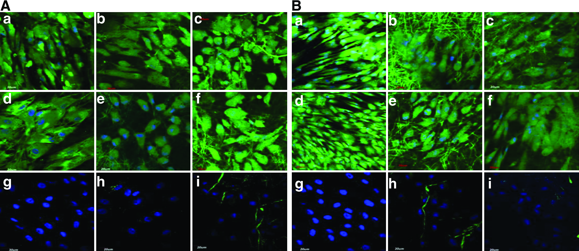

Dual immunocytochemical analysis for the expression of MSC marker protein CD 105
The ability of seeded cells to adhere, survive, and migrate within a scaffold is crucial when trying to regenerate a tissue in vivo. Previous reports of CTE using scaffolds noted that even if the surface layers of the construct were filled with cells, its interior was usually meagre in tissue regeneration. 54 We analyzed the migration of cells into the scaffold using Imaris software after staining cells of the coculture group with troponin-T. Figure 7 clearly observed that the cells migrated interior of the fibers. Moreover, it can be noticed that more cells have migrated within the core/shell fibers (upto 588 μm) compared to gelatin nanofibers (upto 505 μm). This may be because large fiber diameter of core/shell fibers ensures larger pore size, and more elastic nature of the scaffold favors interior migration of cells.

Three-dimensional image using Imaris software of MSC-cardiomyocyte coculture group stained with cardiac-specific marker protein troponin-T at 60× magnification on
The cell morphology was analyzed using SEM images as shown in Figure 8A and 8B. It was revealed that the nanofiber topography allowed the cardiomyocytes to make extensive use of provided cues for isotropic or anisotropic growth, and to some degree even to crawl inside the fibers, as evidenced in Figure 8A (a–c) and 8B (a–c). Day 10 observed cell-to-cell contact between the MSC and cardiomyocyte group, leading to cell fusion in the coculture group [Fig. 8A (d–f)]. This cell-to-cell interaction favored the cardiogenic differentiation of MSC. Terada et al. found that bone marrow stem cells can adopt the phenotype of other cells by spontaneous cell fusion. 55 MSCs have adopted the cardiomyocyte phenotype as revealed in Figure 8B (d–f) on day 15. The MSCs also showed favorable growth on the nanofibers with the extension of filopodia as shown in Figure 8A (g–i) and 8B (g–i).

Yang et al. observed that most of the implanted MSCs did not survive in the postinfarction myocardium, indicating that the local environment was not favorable for MSC engraftment after MI. 56 Their studies indicated that the local environment plays a critical role in cell engraftment. Therefore, we suggest that a suitable milieu provided by the PGS/gelatin nanofibrous patch material for the growth and differentiation of stem cells might overcome the above drawback. The intercellular interactions between the stem cells and cardiomyocytes might induce MSCs to acquire the phenotypical characteristics of cardiomyocytes and endothelial cells. Signals in local milieu are likely to be major factors determining their fates. 57 Our results strongly suggest that the close proximity between the cardiomyocytes and MSCs is a requisite to induce cardiogenic differentiation of MSCs. To guide the organization, growth, and differentiation of cells in tissue-engineered constructs, the biomaterial scaffold should be able to provide not only mechanical support for the cells but also the chemical and biological cues needed for stimulating the specific differentiation of cells in forming functional tissues. 58 Despite improvements in the delivery technologies, clinical success is baffled by maximum cell loss presumptively due to physical stress, myocardial inflammation, myocardial hypoxia, anoikis, absence of survival factors in the transplanted heart, disruption of cell-to-cell interaction, and insufficient vascular supply.59,60 The proposed core/shell nanofibrous patch material composed of PGS and gelatin is likely to overcome the above drawbacks associated with cell adhesion and cell delivery for the regeneration of infarcted myocardium.
Conclusion
The newly developed construct comprising of PGS and gelatin can provide the required mechanical strength to support seeded MSCs differentiation into cardiomyocytes for tissue regeneration to repair the infarcted myocardium and improve cardiac functions. An elastomeric material can resist the heart wall pressure while providing a structure that enhances the potential for cell proliferation and differentiation of MSCs into cardiomyocytes. In fact, many researchers agree that the implanted stem cells alone are not sufficient to directly affect myocardial regeneration and believe that the paracrine effects of the implanted cells are more likely to influence the growth of myocardium than any direct effect of the implanted stem cells. Our findings indicate that the coculture of MSCs and cardiomyocytes with a suitable elastomeric biomaterial combination has synergistic effects and are more effective than MSCs or cardiomyocytes alone. We have currently employed the use of coculture of stem cells and cardiomyocytes with scaffold microenvironments engineered to improve tissue survival and enhance differentiation. This combinatorial epitome might eventually bring CTE into the clinical forefront.
Footnotes
Acknowledgments
This study was supported by the NRF-Technion (R-398-001-065-592), Ministry of Education (R-265-000-318-112), and NUSNNI, National University of Singapore, Singapore.
Disclosure Statement
No competing financial interests exist.
References
Supplementary Material
Please find the following supplemental material available below.
For Open Access articles published under a Creative Commons License, all supplemental material carries the same license as the article it is associated with.
For non-Open Access articles published, all supplemental material carries a non-exclusive license, and permission requests for re-use of supplemental material or any part of supplemental material shall be sent directly to the copyright owner as specified in the copyright notice associated with the article.
