




Abstract
Tissue engineering is a promising approach for treatment of disc degeneration. Herein, we evaluated effects of rotating bioreactor culture on the extracellular matrix production and proliferation of human annulus fibrosus (AF) cells. AF cells were embedded into alginate beads, and then cultured up to 3 weeks in a rotating wall vessel bioreactor or a static vessel. By real-time reverse transcription–polymerase chain reaction, expression of aggrecan, collagen type I and type II, and collagen prolyl 4-hydroxylase II was remarkably elevated, whereas expression of matrix metalloproteinase 3 and a disintegrin and metalloproteinase with thrombospondin motifs 5 was significantly decreased under bioreactor. Biochemical analysis revealed that the levels of the whole cell-associated proteoglycan and collagen were approximately five- and twofolds in rotating bioreactor, respectively, compared to those in static culture. Moreover, AF cell proliferation was augmented in rotating bioreactor. DNA contents were threefolds higher in rotating bioreactor than that in static culture. Expression of the proliferating cell nuclear antigen was robustly enhanced in rotating bioreactor as early as 1 week. Our findings suggested that rotating bioreactor culture would be an effective technique for expansion of human annulus cells for tissue engineering driven treatment of disc degeneration.
Introduction
In comparison to a large number of literature on nuclear pulposus tissue engineering,12–23 studies on annulus fibrosus (AF) and/or cartilage endplates tissue engineering are limited.24–29 Seed cells, signals, and scaffolds are the three principal components of tissue engineering, and among them source of seed cells has been a prominent concern. 11 A suitable cell source has been an essential limiting factor for the clinical use of AF tissue engineering. Stem cells can be easily obtained from various tissues and have the capability of differentiation into multiple lineages, that is, chondrocytes, osteoblasts, and adipocytes.30–32 Although the chondrocyte-like cells derived from stem cells have been described similar to AF cells in recent articles, it is still uncertain whether these cells are the same as native AF cells in biological nature.25,26 AF tissues have a complexity in cell components, that is, fibroblast-like cells in the outer part and fibrochondrocyte-like cells in the inner part,27,33 which adds difficulties in inducing the stem cells simultaneously into distinct lineages. Further, the biological nature of these AF cells has far from been understood, and it would be difficult to assess the feasibility of stem cells in AF tissue engineering before any well-defined molecular biomarkers of AF cells are available.34,35
One of the limitations for using native AF cells for AF tissue engineering is to achieve sufficient cell number. Common methods to expand AF cells could be categorized into monolayer and three-dimensional (3D) cultures. AF phenotype will change after monolayer expansion, which reflected by decreased type II collagen and aggrecan expression while increased type I collagen expression. Traditional 3D static culture system has disadvantages, including nutrition and excretion deficiencies and low cell proliferation rate. Thus, various bioreactors have been designed and used for tissue engineering, such as perfusion bioreactor, rotating wall vessels, spinner flask, and compression bioreactor.36,37 The rotating wall vessel bioreactor (RWVB) is a NASA-approved and commercially available equipment, which utilizes a disposable circular vessel with a gas-exchange membrane that rotates vertically to provide culture medium flow. Previous reports demonstrated that it was a very effective culture system for constructing cartilage tissues from either articular chondrocytes or bone marrow-derived mesenchymal stem cells.38–42 In this study, we provided the first evidence that the production of matrix components and the cell proliferation of AF cells were greatly enhanced in rotating bioreactor culture system.
Materials and Methods
Human AF cell isolation
Ten human intervertebral disc samples were obtained from six individuals (age 13–16 years) with idiopathic scoliosis at University of Virginia Medical Center, following the approved guidelines set by the U.S. National Institutes of Health Office of Human Subjects Research for use of surgical waste. AF cells were isolated by a collagenase/trypsin digestion procedure previously described by our group. 43 Briefly, an annulotomy was performed with a No. 11 blade. Both the outer layer of AF tissues and the poorly structured gelatinous parachordal inner AF tissues adjacent to the NP region were removed. The remaining inner AF tissue was then cut into small pieces and digested with 0.01% collagenase (Crescent Chemical Co., Inc.) at 37°C for 2–4 h. The digested cells were fed with Dulbecco's modified Eagle's medium/F-12 (Gibco BRL) containing 10% fetal bovine serum (Gibco Invitrogen Corporation), 100 units/mL of penicillin, and 100 μg/mL of streptomycin. The cells were culture at 37°C in a humidified incubator with 5% CO2 and passaged using trypsin/EDTA (Gibco BRL).
AF cells cultured within alginate beads
Subconfluent AF cells at passages 3–5 were suspended in alginate solution (1.2% alginate dissolved in 0.15 mol/L NaCl) and added dropwise to 0.102 M CaCl2 solution (1×10⁵ cells/25 μL/drop) to form cell-encapsulating alginate beads as reported. 44 After rinsed in CaCl2 solution for 10 min, beads were immersed in the culture medium for another 10 min, and then transferred into two 50 mL high-aspect-ratio vessels (Synthecon). One vessel (marked as BIO) was applied to an RWVB device (Synthecon) and its rotation speed was adjusted to maintain the growing constructs freely suspended in the rotating flow. The other vessel (marked as STA) was kept in a static status as a control. The cultures were incubated up to 3 weeks, and the medium was changed every 3 days. Study groups were assigned as shown in Table 1, and there were 10 beads in each group, that is, 4 for biochemistry assay, 4 for gene expression analysis, and 2 for histology and immunostaining.
Biochemistry assays
The cells were released from alginate beads with a depolymerizing buffer (55 mmol/L sodium citrate, 30 mmol/L Na2EDTA, and 0.15 mol/L sodium chloride [pH 6.8]), and collected by mild centrifugation. 45 Cell-associated glycosaminoglycan (GAG) and collagen as well as cellular DNA were measured by our published protocols. 46 Briefly, harvested cells were placed in 125 μg/mL papain (Sigma) solution (pH 6.5) at 60°C for 24 h. GAG was determined by the dimethylmethylene blue (Cresent Chemicals Corporation) colorimetric assay with chondroitin sulfate (Sigma) as a standard. DNA was analyzed using the Hoechst 33258 dye (Sigma) assay with a calf thymus DNA as a standard. For collagen measurement, an aliquot of the papain digest was hydrolyzed in 6 N HCl for 16 h at 110°C. The product hydroxyproline was quantified by the colorimetric assay based on the dimethylaminobenzaldehyde/chloramine T (Sigma).
RNA isolation and real-time reverse transcription–polymerase chain reaction
RNA isolation and cDNA synthesis were accomplished with the RNeasy Mini Kit (Qiagen) and the iScript cDNA Synthesis Kit (Bio-Rad) according to manufacturer's instructions. Real-time polymerase chain reaction was then performed with iTaq SYBR Green Supermix (Bio-Rad) with an iQ™ 5 machine (Bio-Rad). The target genes included extracellular matrix genes (aggrecan and type I and type II collagens [COL I and COL II]), matrix-degrading enzyme genes (matrix metalloproteinase 3 [MMP-3] and a disintegrin and metalloproteinase with thrombospondin motifs 5 [ADAMTS-5]), collagen prolyl 4-hydroxylase genes [collagen prolyl 4-hydroxylase α(I) and α(II), C-P4H α(I), and C-P4H α(II)], hypoxia-related genes (hypoxia-inducible factors 1α and 2α [HIF-1α and HIF-2α] and glucose transporter 1 [Glut-1]), interleukin 1β signaling genes (interleukin 1β [IL-1β] and its receptor [IL-1R]), and a cell proliferation-related gene, proliferating cell nuclear antigen (PCNA). Primers are listed in Table 2.
ADAMTS-5, a disintegrin and metalloproteinase with thrombospondin motifs 5; AGG, aggrecan; COL I, type I collagen; COL II, type II collagen; C-P4H α(I), collagen prolyl 4-hydroxylase α(I); C-P4H α(II), collagen prolyl 4-hydroxylase α(II); Glut-1, glucose transporter 1; HIF-1α, hypoxia-inducible factors 1α; HIF-2α, hypoxia-inducible factors 2α; IL-1β, interleukin 1β; IL-1R, interleukin 1 receptor; MMP-3, matrix metalloproteinase 3; PCNA, proliferating cell nuclear antigen; 18S, 18s ribosomal RNA gene.
Histology and immunology
Alginate bead cultures were fixed with 4% paraformaldehyde for 1 h and embedded with Tissue-Tek® O.C.T. compound (Sakura Finetek USA Inc.) at −70°C. Micro-sections of around 5 μm were prepared under a Leica CM3050 S cryostat (Leica Microsystems Inc.). Histological staining was performed with safranin O (Sigma–Aldrich Inc.) and Gill's II hematoxylin (American MasterTech Scientific, Inc.). Immunostaining for COL II was carried out using a commercial Type II Collagen Staining Kit (Chondrex Inc.) according to the manufacturer's manual. An anti-aggrecan (H-300) primary antibody from mouse (sc-25674; Santa Cruz Biotechnology Inc.) and the mouse ImmunoCruz™ Staining System (sc-2050; Santa Cruz Biotechnology Inc.) were employed for aggrecan immunostaining analysis. Cell nuclei were stained with the fluorescent dye YOYO-1 iodide (Molecular Probes) upon necessity. Microphotographs were taken under an Olympus BX51 microscope equipped with an Olympus DP70 digital camera (Olympus America Inc.).
Data analysis
Data from four repeats of either gene expression or biochemical assays are expressed as mean±SD. Statistical analysis between two groups was carried out by Student's t-test in a two-tailed way, and p-values of <0.05 were considered significant.
Results
The level of extracellular matrix proteins under rotating culture
Proteoglycans in alginate bead culture were evaluated by Safranin O staining. The alginate itself was stained in light red color, and the cell-produced proteoglycans were stained in darker red (Fig. 1). By counterstained with hematoxylin, the nucleus of AF cell was showing in blue color. The proteoglycans were concentrated at cell periphery under both static and rotating culture. We further tested the contents of total cellular GAG and collagen with biochemistry assays. As shown in Figure 2, cellular GAG and collagen per bead were greatly increased in rotating bioreactor in five- and twofolds, respectively, compared to static cultures (n=4, p<0.01 for GAG and p<0.05 for collagen). However, when normalized to DNA contents, the variance of the GAG content became not significant and the collagen content decreased remarkably under rotating culture (n=4, p<0.05). In addition, a less intensive staining of either cell-associated COL II (Fig. 3) or cell-associated aggrecan (Fig. 4) under rotating culture was revealed by immunohistochemical analysis.

Safranin-O staining of human AF cells in alginate beads. AF cells mixed with alginate were cultured under either rotating bioreactor (BIO) or static (STA) condition up to 3 weeks. Samples were fixed at 1 and 3 weeks after incubation, followed by Safranin-O staining. The nucleus of AF cells was counterstained with hematoxylin. Representative images were shown. Arrows show the proteoglycans produced by cells. Scale bar=20 μm. AF, annulus fibrosus. Color images available online at www.liebertonline.com/tea
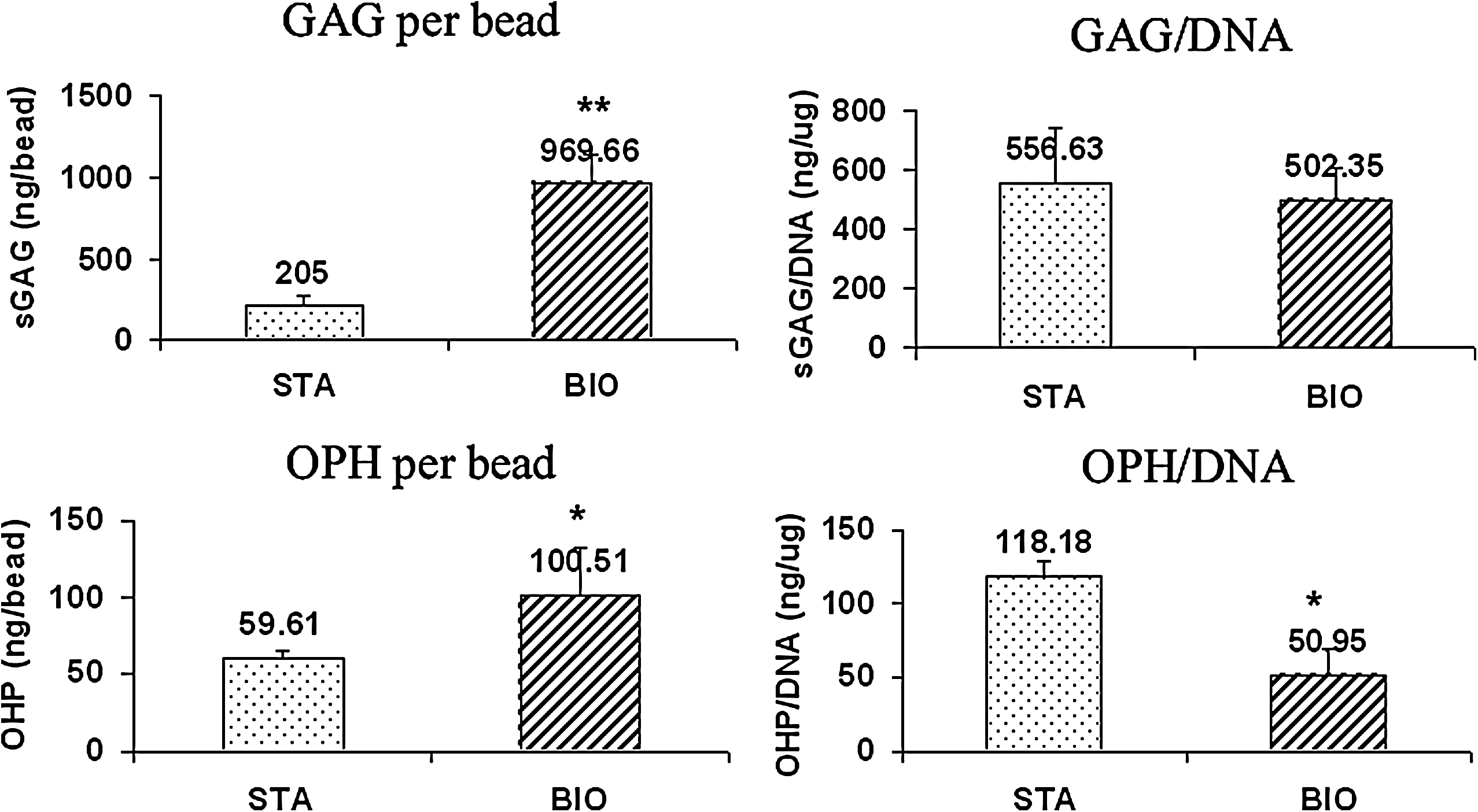
Biochemistry analysis of cell-associated glycosaminoglycan (GAG) and hydroxyproline (OHP) of human AF cells in alginate beads. AF cells mixed with alginate were cultured under either rotating bioreactor (BIO) or static (STA) condition. The contents of DNA, GAG, and OHP were measured by biochemistry assays at 3 weeks. GAG and OHP contents per bead were upregulated in rotating bioreactor culture. However, GAG and OHP contents normalized with DNA amounts under static condition were similar to and larger than those in bioreactor culture, respectively. n=4, *p<0.05, **p<0.01.
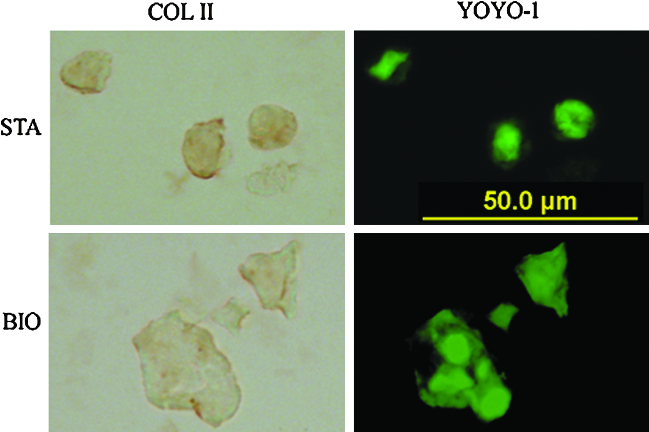
Immunostaining study of type II collagen in human AF cells within alginate beads. After 3-week incubation under either rotating bioreactor (BIO) or static (STA) condition, AF cells were fixed and immunostained with a commercial type II collagen kit. The signals were developed with 3,3′-diaminobenzidine and photographed on an Olympus BX51 microscope. AF cell nucleus was counterstained with YOYO-1. Scale bar=50 μm. Color images available online at www.liebertonline.com/tea

Immunostaining study of aggrecan in human AF cells within alginate beads. After 3-week incubation under either rotating bioreactor (BIO) or static (STA) condition, AF cells were fixed and immunostained with an aggrecan specific antibody. The signals were developed with 3,3′-diaminobenzidine and photographed on an Olympus BX51 microscope. AF cell nucleus was counter stained with YOYO-1. Scale bar=50 μm. Color images available online at www.liebertonline.com/tea
Gene expression of extracellular matrix proteins and their modulators
To elucidate the reason for the increased extracellular matrix production, we analyzed ECM genes and their regulating genes by real-time reverse transcription–polymerase chain reaction. We found that the expression of aggrecan, collagen type I, and type II was elevated significantly under rotating culture at both 1 and 3 weeks, although the increase rate for each gene declined at 3 weeks (Fig. 5A, B). Accordingly, mRNA of matrix-degrading enzymes MMP-3 (Fig. 5C) and ADAMTS-5 (Fig. 5D) was remarkably decreased under rotating bioreactor compared to static culture. C-P4H, a major regulator for collagen synthesis, was also investigated. The expression of C-P4H α(II) was remarkably increased in the rotating bioreactor culture, whereas the expression of C-P4H α(I) remained the same after 3 week-culture (Fig. 5E). Moreover, expression of the pro-inflammatory cytokine IL-1β and its receptor IL-1R was significantly suppressed under rotating culture (Fig. 5F), as well as the hypoxia-regulated genes HIF-1α, HIF-2α, and Glut-1 mRNAs (Fig. 5G).

The gene expression profile of human AF cells in rotating and static cultures. AF cell–alginate constructs were incubated under either rotating bioreactor (BIO) or static (STA) condition up to 3 weeks. Total RNA was extracted. The gene expression was quantified by real-time reverse transcription–polymerase chain reaction and data were normalized to 18s.
Rotating bioreactor promotes AF cell proliferation
To test whether the rotating bioreactor culture has effects on cell proliferation, we measured the DNA contents and the cell proliferation marker PCNA of AF cells in the rotating and static culture conditions. The AF cells were stained with YOYO-1, a nucleic acid staining dye, which binds to DNA. Under the rotating culture, the AF cells tended to grow in cluster (Fig. 6A), and the cell cluster increased as the culture time extended. The same phenomena were not observed in AF cells under static condition. A Hoechst 33258-based fluorimetric quantitative method was used to determine cellular DNA contents. As shown in Figure 6B, cellular DNA contents were markedly increased by fourfolds (n=4, p<0.01) in rotating condition compared with static one. Consistently, the expression of PCNA gene was dramatically increased by fivefolds (n=4, p<0.01) in rotating culture at 1 week compared to static culture, but this increase was diminished at 3 weeks (Fig. 6C).

The proliferation of AF cells in alginate beads was promoted in rotating bioreactor culture.
Discussion
The propagation of cells is one of the essential steps of AF tissue engineering for the treatment of disc degeneration in the future. In the present study, we provided the evidence that the proliferation of AF cells was greatly enhanced in rotating bioreactor culture system. Further, the anabolism and catabolism processes of matrix proteins were well balanced in this system.
Monolayer culture of cells is an important resource for many research applications in vitro. However, it is well known that the cell phenotype might be changed during cell propagation, which is manifested by the extracellular matrix proteins change in monolayer.47–49 Recently, we and others reported that the AF cells have the capability of differentiation into different lineages in monolayer culture.43,50,51 Therefore, a 3D culture is required for AF cell propagation and cell phenotype maintenance. In the current study, we chose alginate hydrogel beads as scaffolds based on following considerations: (1) alginate culture is a mature 3D system for chondocytes.22,33 Our AF cells were isolated from the inner part of human AF tissues that have chondrocyte-like cell phenotype, whose major extracelluar matrix is type II collagen and aggrecan. 52 (2) Alginate has been extensively used in biomedical research and is easily available. (3) The fabrication of cell–alginate beads is a simple and conventional laboratory technique, based on the noncovalent cross-linking with calcium. (4) The cells on alginate beads can be easily released by EDTA.
The metabolism of disc cells depends on the surrounding environments. Herein, we showed that rotating bioreactor culture can promote matrix production and decrease degradation (Figs. 2 and 5). The mRNA levels of aggrecan, type I, and type II collagen were significantly elevated (Fig. 5A, B), whereas the matrix-degrading enzymes MMP-3 and ADAMTS-5 were considerably repressed in AF cells under rotating bioreactor culture compared to static culture (Fig. 5C, D). We also examined the expression of another type of enzyme, C-P4H, to further elucidate the post-translational modification of matrix proteins. C-P4H plays a central role in collagen biosynthesis by catalyzing hydroxylation of proline residues, which contribute to the thermal stabilization of the collagen triple helices at body temperature. 53 In humans, there are three types of C-P4Hs, C-P4H-I, -II, and -III that are categorized by their different catalytic α subunits, α(I)–α(III). C-P4H-II is the main form in chondrocytes and is likely to play an important role in cartilage development. 52 In the current study, we reported for the first time that C-P4Hs were expressed in AF cells (Fig. 5E), and the expression of C-P4H α(II) but not C-P4H α(I) was increased in the AF cells under rotating culture condition. This result implied that C-P4H-II may participate in the regulation of collagen biosynthesis in rotating bioreactor culture.
In avascular tissue such as articular cartilage and intervertebral disc, the transcript factors HIF, including HIF-1 and HIF-2, represent components key to maintain the proper cellular functions under hypoxic condition and have been demonstrated to play a crucial role in moderating the production of extracellular matrix. 54 In addition, Glut-1 is a HIF-1 target gene involved in glycolysis and increase of Glut-1 expression was regarded as an important cellular adaptation to hypoxic stress. 55 Further, gene expression of both HIF and Glut-1 were recently reported in normal and degenerate human intervertebral disc. 56 As mentioned above, the bioreactor allows an environmental atmosphere of relative higher oxygen concentration and lower hypoxia level around and through the alginate constructs compared to a static condition. 38 This raises a question that whether or not hypoxia-related gene expression is differently regulated between human AF cells cultured under these two conditions. Indeed, our data showed that several genes in response to hypoxia, including HIF-1α, HIF-2α, and Glut-1, were transcriptionally downregulated under bioreactor culture (Fig. 5F). Since the involvement of pro-inflammatory cytokines such as IL-1 and TNF is well recognized in the regulation of matrix production in disc cells, 57 we examined gene expression of IL-1β and its receptor IL-1R. Both genes were inhibited in the presence of bioreactor (Fig. 5G). Treatment of IL-1β was reported to upregulate the expression of HIF-1α in human gingival and synovial fibroblasts, and on the other hand hypoxia was able to significantly induce IL-1β expression in human articular chondrocytes.58,59 Interestingly, Gelse et al. demonstrated that in chondrocyte cultures, stability of HIF-1 by dimethyloxaloylglycine under hypoxia was able to cause a decrease in expression of type II collagen, and they attributed such an effect to increased expression of IL-1β and IL-6. 59 This might be also applied in interpreting simultaneous downregulation of HIF and upregulation of collagen expression in our present experiment. Therefore, we suggest that hypoxia-related pathway and/or IL-1 signaling would be probably involved in the regulation of matrix metabolism in human AF cells under bioreactor culture. However, such hypotheses need to be further clarified. Of course, variation of energy metabolism caused by oxygen concentration and mechanical force generated by the dynamic fluid flow may also play some/critical roles and should be seriously taken into consideration in the future study.36,37
At protein level, GAG and collagen contents per bead were significantly increased in the rotating culture system compared to static one. To our surprise, GAG and collagen levels normalized to DNA contents in rotating bioreactor culture were similar to or lower than in static culture (Fig. 2). These observations were also confirmed by our immunohistochemical analysis, which showed less intensive staining of cell-associated COL II and aggrecan in bioreactor culture (Figs. 3 and 4). This might be explained by different rates of matrix molecules transportation after their biosynthesis under different culture conditions. Studies showed that matrix molecules could be assigned into three parts after production by AF cells in alginate beads: the cell-associated matrix (CM) in the construct, the further removed matrix in the construct, and the matrix in the culture medium.45,60 Since the RWVB bioreactor rotates at a vertical axis, the cell–alginate constructs are able to remain suspended in a constantly moving body of nutrient medium and always subjected to a fresh medium. As shown in Figure 7, matrix molecules would probably move faster from the cell-associate part to the other two parts in the bioreactor than under static culture, resulting in a less residence of matrix in each cell. In our study model, only the first part, that is, the CM was detected, leading to achieve less GAG and OPH per DNA (reflecting CM of each cell) but more GAG and OPH per bead (reflecting the CM of the whole cell within a bead) under bioreactor. Therefore, it should be pointed out that failure to examine all three proportions of matrix is an obvious limitation of this study.

Scheme showing possible matrix compartments of the cell–alginate bead culture under static and bioreactor condition. CM, cell-associated matrix in the construct; FRM, further removed matrix in the construct; MCM, matrix in culture medium. The amount of matrix in each compartment is roughly reflected by the size of the circle.
The native AF tissue is currently limited and not enough for most tissue engineering applications. A critical question concerning cell expansion is whether these cells could be propagated in in vitro culture. According to previous report, a bioreactor culture is capable of enhancing proliferation of articular chondrocytes in hydrogel constructs.45,60 In our present study, the AF cells showed aggregated growing pattern under the rotating culture, and the aggregates increased in size over the culture time (Fig. 6A). In contrast, the AF cells under static culture had a sparse separated cell pattern. This data suggested that AF cells proliferated much faster under the rotating culture, and it was supported by the DNA content measurement and PCNA gene expression analysis. Cellular DNA content under bioreactor was about fivefolds of its static counterpart at the end of culture (Fig. 6B). PCNA mRNA level in bioreactor culture was higher than static culture (Fig. 6C). However, it is noticeable that the ratio of PCNA mRNA level in bioreactor vs. static culture decreased remarkably over the culture period (Fig. 6C). These data led us to the notion that, as a result of the increased proliferating capacity during bioreactor culture (evidenced by the increased PCNA mRNA at the first week), more cells (represented by increased DNA contents) could be achieved and retained in alginate constructs, even if cell proliferating ability gradually became not significant between two groups at the end of culture. Thus far, these results implied that 1 week of bioreactor culture would be better than 3 weeks, to keep cells in a relatively high proliferating state.
In summary, this study provided in vitro evidence showing that the matrix production and cell proliferation of human AF cells could be significantly enhanced under rotating bioreactor culture, and the model may potentially be an effective technique for cell amplification in AF tissue engineering.
Footnotes
Acknowledgments
The work was supported by AO Foundation (F-07-97L) and Scoliosis Research Society.
Disclosure Statement
No competing financial interests exist.