
Abstract
We have earlier shown that a peptide derived from the bone morphogenetic protein-9 (pBMP-9) stimulates mouse preosteoblasts MC3T3-E1 differentiation in vitro. Here, we evaluated the effects of two delivery systems (DSs) for pBMP-9, one based on collagen and the other on chitosan. The release kinetics of BMP-9 (used as control) and pBMP-9 from these DSs were first determined in vitro by using enzyme-linked immunosorbent assay and high performance liquid chromatography assays, respectively. Micro-computerized tomography and histological analysis were then performed to study in vivo the ectopic ossification induced by both DSs containing these molecules in C57BL/6 mouse quadriceps. We found that collagen DS released in vitro about 35% of its BMP-9 within 1 h, whereas chitosan DS released 80%. The pBMP-9 was released from both DSs more slowly for up to 10 days. These release kinetics seemed to fit the Korsmeyer–Peppas model. Only chitosan DS containing BMP-9 induced strong bone formation in all mice quadriceps within 24 days. All mice quadriceps treated by pBMP-9 trapped in this DS also favored bone structures that started to mineralize. However, pBMP-9 in collagen DS failed to promote ectopic ossification within 24 days in vivo. This study highlights the importance to optimize carrier, thus improving the efficiency of pBMP-9 in vivo.
Introduction
Only BMP-2/-4/-6/-7/-9 induce complete bone morphogenesis and are found in both normal human intramembranous and endochondral bones.3,7 Recombinant human BMP-2 is the BMP most frequently used in present-day clinical bone healing studies.
8
However, recombinant adenoviruses expressing BMP-6 and BMP-9 induced more robust and mature ossification in the quadriceps of athymic mice than did viruses that express clinically approved recombinant BMP-2 or BMP-7 proteins.
9
However, recombinant BMPs, which are produced in Escherichia coli or Chinese hamster ovarian (CHO) cells, are expensive.
10
Since the BMP-2, -6, -7, and -9 have wrist epitopes that bind to type I receptors and knuckle epitopes which bind to type II receptors,11–17
less expensive osteogenic peptides derived from them have been developed.18–23
Saito et al. observed that incubating mouse multipotent mesenchymal C3H10T1/2 cells for 3 days with the synthetic peptide KIPKA
We have, therefore, developed a 23-residue synthetic peptide
The development of an efficient system for delivering growth factors is of utmost importance, as their actions are often impaired in vivo due to their short half-life and local clearance.26,27 To prevent their breakdown and increase their concentration at the bone repair site, BMPs and their peptides are, therefore, combined with biocompatible carriers.28,29 These implantable carriers, made from natural polymers such as collagen or chitosan, or synthetic materials such as poly(lactide-co-glycolide), act as a stable structural support for cell colonization, proliferation, and differentiation.27–32 Delivery systems (DSs) containing growth factors can be used in a tissue engineering approach to favor cell differentiation in vitro. 33 DSs that contain BMP-2 or BMP-7 entrapped in a type I bovine collagen are currently approved by the Food and Drug Administration (FDA) for clinical use in orthopedic applications in the United States. These include healing bone injuries by mediating spinal fusion and fracture healing.34,35 We have successfully incorporated pBMP-9 into a type I collagen gel, and used this preparation to increase the ALP activity in mouse preosteoblasts MC3T3-E1. 18 Several recent studies have also successfully used chitosan matrix to deliver from 1 to 150 μg of BMP-2 and promote bone formation in vivo.36,37 This study, therefore, describes the in vitro and in vivo effects of two DSs for BMP-9 and pBMP-9, one based on type I collagen and the other based on chitosan. We first characterized and modeled the release of BMP-9 and pBMP-9 from both DSs without cells. Then, we evaluated the osteoinductive potential of pBMP-9 within type I collagen and chitosan DSs by using a C57BL/6 mouse quadriceps model. Since 75–100 μg of synthetic peptides derived from the knuckle epitope of BMP-2 can induce bone formation in rats muscles,22,38 we used 100 μg pBMP-9 for in vivo experiments. BMP-9 was also used as a control at about 6 μg, as some micrograms of BMP-2 trapped in collagen or chitosan DS seem to be able to induce bone formation in vivo.36,39
Materials and Methods
Materials
Recombinant carrier-free human BMP-9, synthesized in CHO cells, was purchased from R&D Systems. The 2.4 kDa peptide pBMP-9 derived from BMP-9 was synthesized by Celtek Peptides with a final purity of at least 98% and was dissolved in ultrapure water (pH 6.3). 18
DS preparation
The DSs were prepared under aseptic conditions in a laminar flow hood. Type I collagen was extracted from rat tail tendon at 3 mg/mL as previously described. 18 The final collagen suspension (1.5 mg/mL) was adjusted to pH 7.4 with 18.75% (v/v) 1N NaOH immediately before incorporating the BMPs or pBMP-9. Medium-molecular-weight chitosan (75%–85% deacetylated; Sigma) was sterilized by exposure to UV radiation overnight, autoclaved for 1 h, suspended in water (45 mg/mL), and adjusted to pH 4.7 with 5N HCl. This stock suspension was gently stirred on a magnetic mixer overnight at room temperature. The chitosan stock suspension kept at room temperature was diluted with water to 15.2 mg/mL before each experiment. All cohesive viscoelastic preparations were homogenized with or without BMPs or pBMP-9 by using a micropipette just before gelification.
BMP-9 release
Two hundred microliters of the type I collagen or chitosan DSs containing 169.2 ng BMP-9 were placed in 24-well low-adhesion plates (Costar®; Fisher Scientific) containing cell culture inserts with porous (0.4 μm) polyethylene terephthalate membranes (Costar) and left to gel. Minimum essential medium (MEM) alpha medium (α-MEM; Gibco®) without ascorbic acid was then added both outside the insert (1000 μL) and inside it (1000 μL), and the plates were incubated at 37°C. About 169.2 ng BMP-9 diluted in 2 mL α-MEM was also used as a control to verify its stability over time. Samples (150 μL) of medium were taken from outside the insert at 7.5, 15, 30, and 60 min and replaced with fresh α-MEM. In another experiment, samples were taken after incubation for 3 and 6 days. Each sample was transferred to a 0.5 mL protein low-binding tube (Eppendorf ) containing 300 μL α-MEM and analyzed by enzyme-linked immunosorbent assay (ELISA). Briefly, 96-well ELISA plates (Falcon, Fisher Scientific) were coated overnight with 100 μL antibodies against BMP-9 (0.75 μg/mL; R&D Systems) diluted in 0.1M carbonate buffer pH 9 and then blocked by incubation with 0.5% bovine serum albumin (BSA) in phosphate-buffered saline (PBS) plus 0.1% Tween® 20 (Sigma) for 1 h. Diluted samples (100 μL) were placed in the wells and incubated for 1 h at 37°C. The wells were then washed thrice with PBS containing 0.1% Tween 20 and incubated with 100 μL/well biotinylated antibodies against BMP-9 (0.8 μg/mL diluted in PBS/0.1% Tween 20/0.1% BSA; R&D Systems) for 45 min at 37°C. The wells were then washed and filled with 100 μL/well ImmunoPure® streptavidin-horseradish peroxidase conjugated solution (0.25 μg/mL diluted in PBS/0.1% Tween 20/0.1% BSA; Thermo Scientific), and the plate was incubated for 30 min. The horseradish peroxidase activity was revealed with o-phenylenediamine dihydrochloride substrate (Fast™; Sigma). Finally, 100 μL 1N HCl was placed in each well to stop the reaction, and the plate was spectrophotometrically read at 492 and 540 nm for correction by using a microplate reader (Synergy™ HT; BioTek). Sample concentrations were calculated from a standard curve generated by using BMP-9 concentrations of 0 to 50 ng/mL.
pBMP-9 release
Two hundred microliters of the type I collagen or chitosan DSs containing 10,000 ng pBMP-9 were placed in 24-well low adhesion plates, and PBS was added outside the insert (1000 μL) and inside it (1000 μL). The plates were incubated at 37°C. Samples (500 μL) of medium were taken from outside the insert at 1 h, 6 h, 1 and 2 days, and each lost volume was replaced with fresh PBS. In another experiment, samples were taken after incubation for 3, 6, 7, and 10 days and replaced with fresh PBS as just described. Each sample was placed in a 0.5 mL protein low-binding tube containing 350 μL PBS and mixed with 0.5 μL trifluoroacetic acid (EMD Chemical). The samples were then placed in glass tubes. Aliquots (20 μL) were analyzed by using a ProStar analytical high-performance liquid chromatography (HPLC) system (Model 335, Varian, Agilent Technology) equipped with a UV detector set at 220 nm and a 5 μm reversed phase column (Luna® C18(2), Phenomenex®). Samples were eluted with the mobile phase, 0.1% trifluoroacetic acid in acetonitrile (A), and 0.1% trifluoroacetic acid in H2O (B), at 1.0 mL/min. Data were analyzed by using the Galaxie Chromatography Data System v 1.9.301.220. Sample concentrations were calculated from a standard curve generated by using pBMP-9 concentrations of 0 to 10,000 ng/mL.
Release kinetics
We analyzed the kinetics of BMP-9 and pBMP-9 release from collagen and chitosan DSs by using four models.40,41 Zero-order kinetics were estimated as the cumulative percentage of drug released in unit time (Eq. 1); first order kinetics as the log of drug remaining against time (Eq. 2); the Higuchi kinetics were estimated from the cumulative percentage of drug released plotted against the square root of time (Eq. 3); and the Korsmeyer–Peppas kinetics were estimated from a plot of the log cumulative percentage of drug released against log time (Eq. 4):41–44
where Mt(%) and M∞(%) are percentages of drug released at time t and at the final time, respectively; k1, k2, k3, and k4 represent the rate constants of zero order, first order, Higuchi, and Korsmeyer–Peppas models, respectively; and n is an exponent that characterizes the mechanism of release.
Bone formation in vivo
The in vivo experiments were performed on twelve 12-week-old male C57BL/6 mice (Charles River). For each experimental condition, four mice were used. All procedures involving animals met the standards set out in “Guide for the Care and Use of Laboratory Animals, Institute for Laboratory Animal Research, 1996”; and the experiments were approved by the Institutional Animal Care and Use Committee of Université de Sherbrooke (Protocol no. 141-07B). DSs with or without BMP-9 or pBMP-9 were prepared just before each injection and kept on ice. The mice were anesthetized by isoflurane inhalation (induction 2.5% and maintained at 1.5%) via a face mask, and the area of injection was shaved and disinfected. Fifty microliters of each DS with or without 6.35 μg BMP-9 or 100 μg pBMP-9 were injected into the quadriceps of the mice by using an insulin syringe with a needle of 25G5/8 (Becton Dickinson, Fisher Scientific). The collagen DS was injected into the left quadriceps muscle, and the chitosan DS was injected into the right muscle. The mice were allowed to move freely and eat after the procedure. They were euthanized by CO2 inhalation 24 days after implantation. The harvested quadriceps were dissected from soft tissue and fixed overnight in formalin. Ectopic bone formation was assessed by using a SkyScan 1072 micro-computerized tomography (microCT) scanner at the Centre for Bone and Periodontal Research. Images were acquired at 45 kV and 222 μA with a 0.9° rotation between frames with lower and upper gray value thresholds of 60 and 255, respectively. Two-dimensional images were used to generate three-dimensional reconstructions and to calculate the muscle and calcified volume in cubic millimeters.
Histology
The specimens used for microCT analysis were then embedded in paraffin. Sections were cut and stained with hematoxylin and eosin (H&E) or von Kossa and toluidine blue. Images were captured at the Material Characterization Centre of the Université de Sherbrooke by using a Leica DM RXP microscope equipped with a 20× objective and a Leica DC300 CCD camera (Leica Microsystems) and analyzed with Leica IM50 version 1.20 software. Sections were also immunolabeled by using specific antibodies directed against desmin by the Laboratory of Pathology at Centre Hospitalier de l'Université de Sherbrooke. Images were captured by using an Olympus microscope equipped with a 40× objective and an Olympus Q color 5 CCD camera.
Statistical analysis
Results are expressed as means±SEM in all experiments. Statistical computations were performed with GraphPad Instat® 3.00 software (GraphPad Software Inc.). The Tukey–Kramer multiple comparison test (ANOVA) was used. Values were considered significantly different if p<0.05.
Results
BMP-9 release
The ELISA assay developed to measure BMP-9 in vitro was linear for BMP-9 concentrations of 0 to 25 ng/mL in α-MEM at 37°C (R 2 =99.9%; Fig. 1A). We first verified that 169.2 ng BMP-9 in solution without a DS was entirely detected within 1 h, which suggested that this cytokine underwent no great conformational change at a short time. However, incubation of BMP-9 without DS showed that only a residual amount (about 2%) was still detected after incubation for 1 day, and none was detected after 3 days. Therefore, we evaluated within 1 h the release of 169.2 ng BMP-9 from a type I collagen DS and a chitosan DS in 2 mL α-MEM. Results showed that the type I collagen DS released 35% of the initial BMP-9 after incubation for 1 h, whereas the chitosan DS released 80% (Fig. 1B). A control performed at 6 days of incubation revealed that only 5% of BMP-9 was still detected for both DSs (data not shown). The release kinetics of BMP-9 was studied with four different models (Fig. 1C–F). The profile of BMP-9 release from the collagen DS fitted the Korsmeyer–Peppas model best (R 2 =90.3%), followed by the Higuchi model (R 2 =86.7%) (Fig. 1E, F). It fitted the zero- (R 2 =76.4%) and first-order (R 2 =78.9%) models less well (Fig. 1C, D). The release of BMP-9 from the chitosan DS fitted all tested models rather well (R 2 ≥97.9%) (Fig. 1C–F). The diffusion exponent n in the Korsmeyer–Peppas model was 0.38 for BMP-9 released from the collagen DS and 0.64 for its release from the chitosan DS. These results suggested that BMP-9 was released faster from the chitosan DS in comparison to collagen DS.
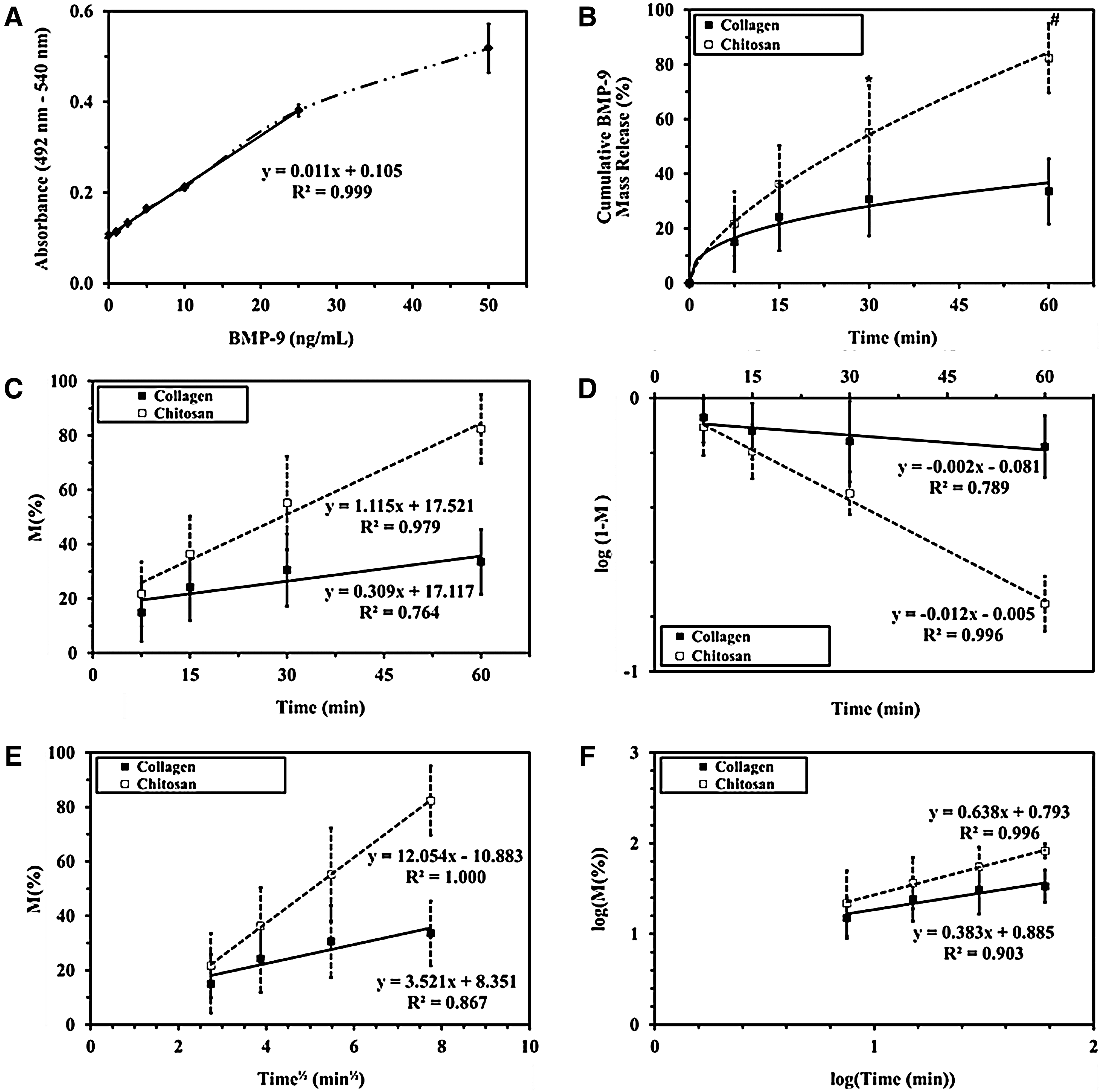
Quantification of BMP-9 by enzyme-linked immunosorbent assay.
pBMP-9 release
HPLC has been used to detect peptides derived from BMP-2.45,46 We, therefore, developed an HPLC assay to quantify the release of 10,000 ng pBMP-9 from the two DSs into 2 mL PBS at 37°C. The peak areas gave a linear response for pBMP-9 concentrations from 0 to 10,000 ng/mL (R 2 =99.9%; Fig. 2A). We used high concentrations of pBMP-9, because the HPLC detection limit is 500 ng/mL. Both DSs released about 15% of the pBMP-9 after incubation for 6 h (Fig. 2B). The release kinetics of pBMP-9 was also studied with four different models (Fig. 2C–F). The pBMP-9 was slowly released from the DSs until it reached a plateau after 10 days (75% for type I collagen DS and 45% for chitosan DS). The profiles of pBMP-9 release from both DSs fitted the Korsmeyer–Peppas model best (R 2 =91.3% in collagen, R 2 =86.9% in chitosan; Fig. 2F), followed by the Higuchi model (R 2 =83.6% in collagen, R 2 =71.3% in chitosan; Fig. 2E). The zero- (R 2 =66.7% in collagen, R 2 =53.9% in chitosan) and first-order (R 2 =78.7% in collagen, R 2 =55.7% in chitosan) models gave the poorest fit (Fig. 2C, D). The factor n in the Korsmeyer–Peppas model was 0.35 for the collagen DS and 0.45 for the chitosan DS. These results suggested that pBMP-9 was released faster from the collagen DS in comparison to chitosan DS.
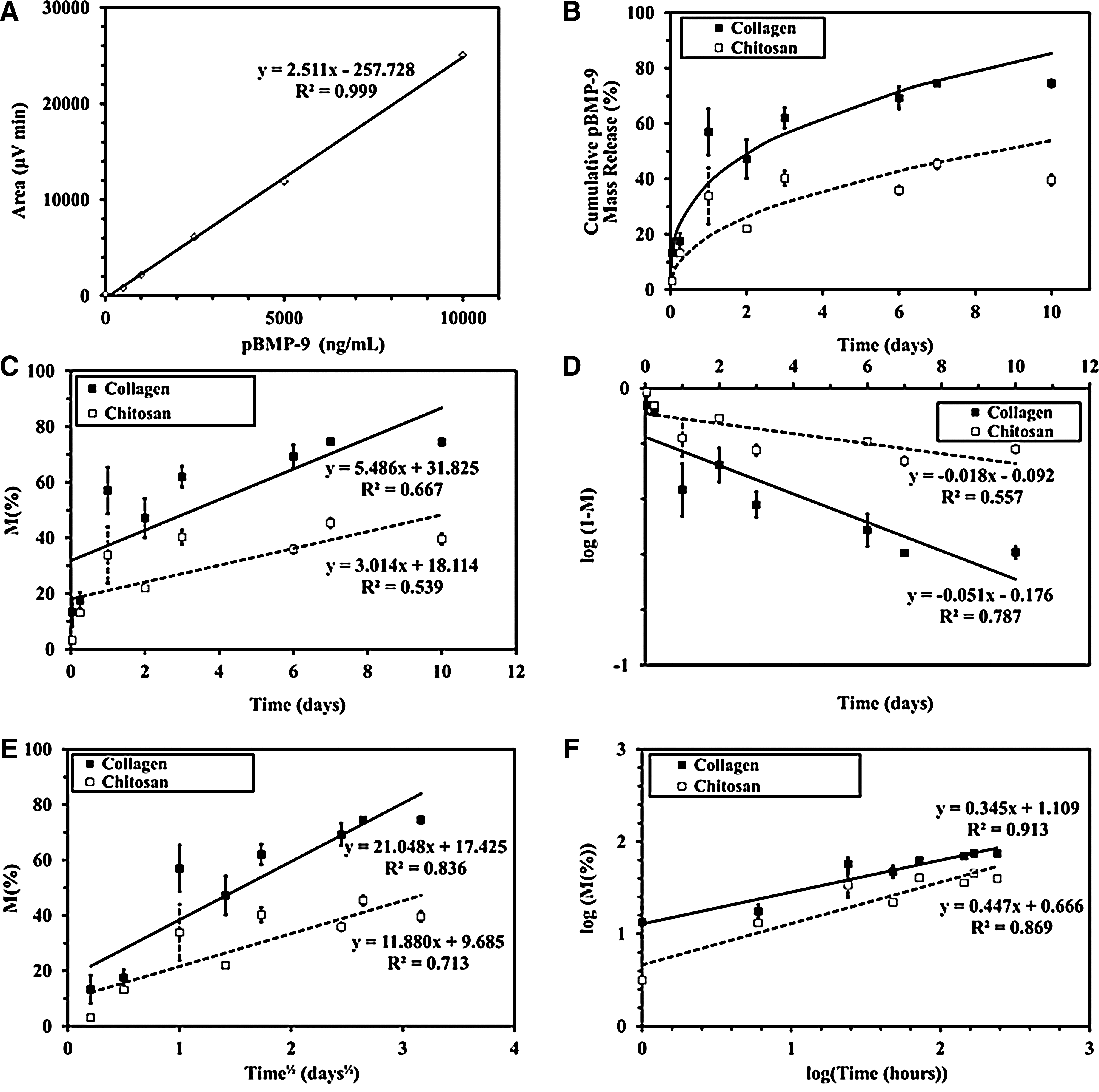
Quantification of pBMP-9 by high performance liquid chromatography.
Bone formation in vivo
To evaluate its osteoinductive property, pBMP-9 trapped into both collagen and chitosan DSs was injected into mouse quadriceps. Osteogenic BMP-9 was also used as a control. After 24 days, bone formation in quadriceps was analyzed by using microCT scans. Our results revealed that BMP-9 in the collagen DS increased mineralization in one mouse (Fig. 3), but had little or no action in the others, which were the same as the control. All the mice treated with type I collagen DS with or without pBMP-9 have a similar response with some diffuse mineralization areas (Fig. 3).

Micro-computerized tomography analysis of C57BL/6 mouse quadriceps muscle 24 days after being injected with type I collagen DS (left leg) or chitosan DS (right leg) with or without 6.35 μg BMP-9 or 100 μg pBMP-9. Three-dimensional images were obtained from an experiment performed in duplicate. The right frame was acquired after a 90° rotation of the left frame for each experimental condition. Another independent experiment gave similar results. Color images available online at www.liebertonline.com/tea
By contrast, BMP-9 in the chitosan DS induced mineralization in the quadriceps of all the mice tested. In addition, pBMP-9 in chitosan DS induced slightly more mineralization than the chitosan carrier alone. The calcification volume quantification revealed that BMP-9 in the chitosan DS produced a strong mineralization varying widely from one mouse to another. Calcification/muscle ratio was 2.1 to 11.4 times that of the chitosan control (0.11%±0.03%). Further, pBMP-9 in the chitosan DS induce mild mineralization in muscles that was 1.4 to 2.1-fold greater than that produced by the chitosan control.
Since the microCT analyses suggested that the mouse quadriceps treated with pBMP-9 underwent some mineralization, we histologically checked for bone formation by staining paraffin tissue sections with H&E or von Kossa and toluidine blue (Fig. 4A). There was no bone formation in any of the muscles that received an injection of type I collagen DS alone (control). However, BMP-9 in type I collagen DS induced a large bone formation in only one quadriceps muscle (Figs. 3 and 4A). Polarized light confirmed the presence of lamellar bone in this muscle (data not shown). H&E staining showed many osteoblasts lining woven bone. The von Kossa staining clearly showed mineralization of bone containing osteocytes that surrounded bone marrow with foci of hematopoiesis and adipose tissue. However, quadriceps injected with a type I collagen DS containing pBMP-9 produced no bone formation. H&E staining showed that the chitosan DS induced more inflammatory response and fibrous tissue formation than did type I collagen DS in all our experimental conditions. However, one quadriceps injected with chitosan DS alone was mildly calcified (data not shown). In fact, von Kossa staining showed that BMP-9 in the chitosan DS induced mineralization in all samples. This treatment also promoted muscle degeneration and the formation of cartilaginous tissue (arrow, Fig. 4A). The pBMP-9 in the chitosan DS also induced some mineralization and concentric structures characteristic of bone tissue in all samples. These structures were only starting to become calcified, as the von Kossa staining was weak and confined to their periphery. To verify the formation of bone tissue, paraffin tissue sections of quadriceps treated by BMP-9 or pBMP-9 in chitosan DS were also immunolabeled by antibodies directed against desmin, a muscle specific intermediate filament protein (Fig. 4B). This staining confirmed that BMP-9 in chitosan DS induced a severe disruption and degeneration of muscle with the formation of cartilaginous tissue. In addition, quadriceps treated by pBMP-9 in chitosan DS clearly revealed the formation of lamellar bone surrounded by degenerative muscle cells.
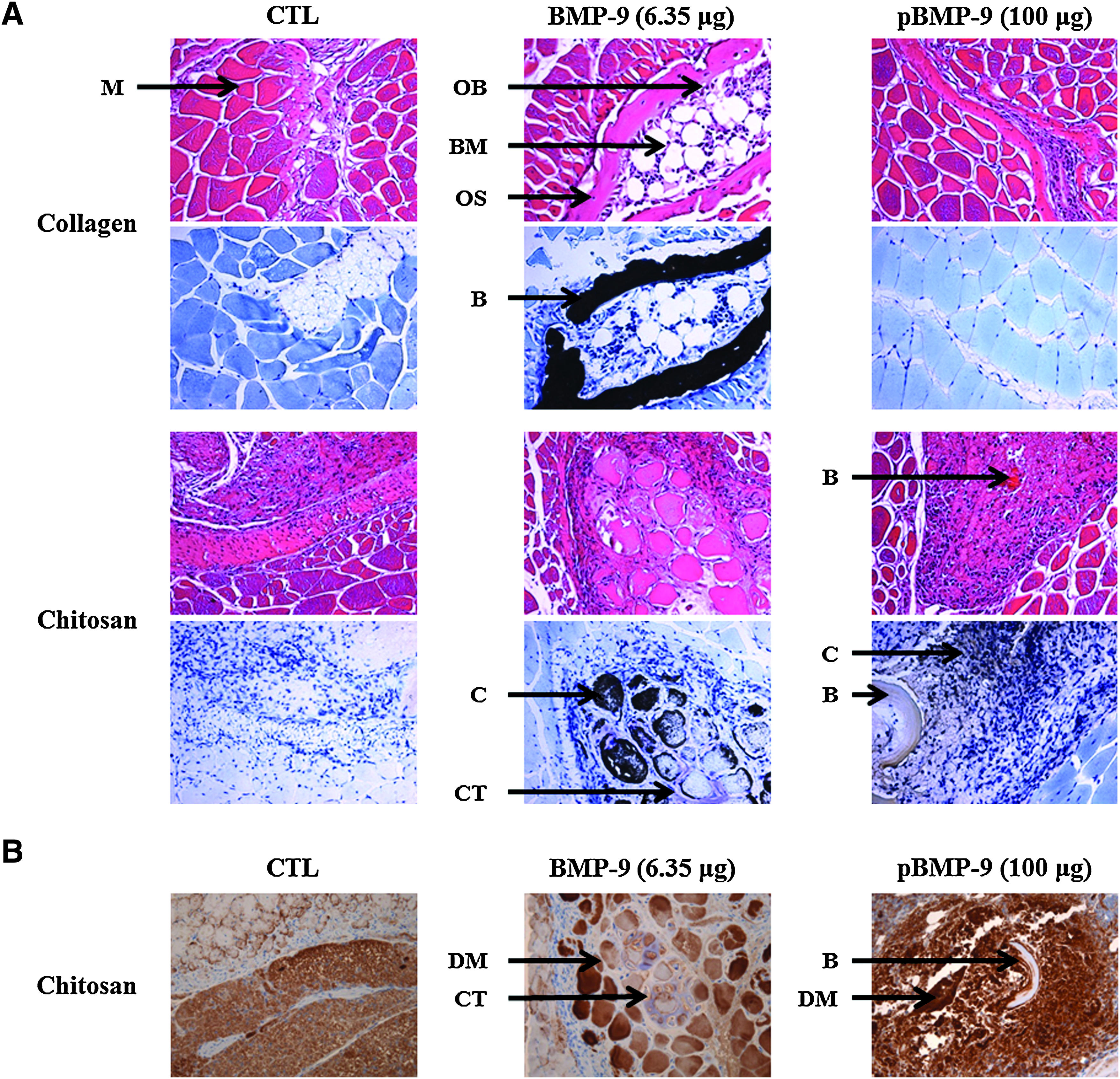
Histological analysis of C57BL/6 mouse quadriceps 24 days after treatment with a type I collagen DS or chitosan DS containing 6.35 μg BMP-9 or 100 μg pBMP-9. Samples from the center of each quadriceps were embedded in paraffin and fixed before
Discussion
BMPs are important modulators of many biological events, including bone formation and repair. 3 The BMPs bound to the extracellular matrix, which regulates their distribution, activation, and presentation to cells. 47 Since type I collagen is the most abundant component of the extracellular bone matrix synthesized by osteoblasts, most DSs have used this weakly immunogenic protein to repair bone defects.48,49 Type I collagen is a biocompatible, biodegradable, malleable, and easily handled carrier and received FDA approval for many clinical applications.35,48 On the other hand, chitosan is a cationic linear polysaccharide derived from chitin, which is made up of glucosamine and N-acetyl glucosamine. 50 This natural biopolymer binds negatively charged molecules such as proteoglycans and glycosaminoglycans through electrostatic interactions, which also bind many growth factors. 51 Thus, both natural polymers type I collagen and chitosan can be used as DSs containing BMPs and peptides derived from them.18,27,36–38,45,52 For example, 150 μg BMP-2 trapped in chitosan can induce ectopic bone formation in rat quadriceps. 37 In addition, we have previously demonstrated that a peptide derived from the knuckle epitope of human BMP-9 (pBMP-9) stimulates mouse preosteoblasts MC3T3-E1 differentiation to mature osteoblasts, just as do BMP-2 and BMP-9.18,24 This peptide in a fast gelling, stable type I collagen gel increased the ALP activity of MC3T3-E1 cells. 18 A recent study also demonstrated that these cells seeded into 100 ng BMP-6-loaded chitosan scaffold induced higher ALP and OC levels than the cells in unloaded chitosan. 53
We first analyzed the stability of the BMP-9 without DS. Interestingly, in our experimental model in serum-free medium, ELISA assays revealed that the BMP-9 did not present a strong conformation change within 1 h. However, only 2% of the BMP-9 was detected after 24 h. The delivery of BMP-9 and its derived peptide by the type I collagen and chitosan DSs was, therefore, determined within 1 h. The release profile of a growth factor from a DS in vitro is important for checking the reproducibility of the DS properties. 52 Our results showed that the type I collagen DS released about 35% of its 3.84 nM BMP-9 loaded after incubation for 1 h, whereas the chitosan DS released 80%. Using 3.84 nM BMP-2, we have previously demonstrated that our type I collagen gel releases 4% of the incorporated BMP-2 after incubation for 1 h. 18 The difference between retentions of BMP-2 and BMP-9 in the collagen DS may be due to the difference of about 15% in their molecular weights. In addition, BMP-2 and BMP-9 are members of different BMP subgroups with notable changes in acidic and basic residues of their receptor binding regions.3,11,14 The retention of active BMPs in natural polymers such as collagen or chitosan may also depend on the pH, the anion concentration, the chemical cross-linking, the carrier nature, and the physical configuration of the carrier.27,37,52 BMPs and pBMP-9 are basic molecules with isoelectric points (pI) of about 7.7 to 9.0.18,54 Since pI of collagen is below pH 7, most of the interactions between BMPs or pBMP-9 and collagen are electrostatic under physiological conditions. 48 The higher pI of 9.0, the low molecular weight of pBMP-9 along with a higher dose may explain why it is released more slowly from both DSs than is BMP-9, which is released in a short burst. 18 Bessa et al. recently studied silk fibroin microparticles as a DS for BMP-2 and BMP-9. Release of both 0.5 and 5 μg BMP-2 and BMP-9 from this DS was biphasic with an initial burst that lasted 2 days and a slower release for up to 2 weeks. These kinetic profiles were best explained by the Korsmeyer–Peppas model with a release exponent varying between 0.30 and 0.39 in all experimental conditions. 40 Since the power-law equation of the Korsmeyer–Peppas model can be viewed as the superposition of Fickian diffusion-controlled (n=0.5) and swelling-controlled Case II transport (n=1), these in vitro releases of both BMP-2 and BMP-9 are likely to be controlled by diffusion mechanism.40,55,56 Here, we also found that Korsmeyer–Peppas kinetics seem to provide the best model for the release of BMP-9 and pBMP-9 from type I collagen and chitosan DSs. Further experiments are required to confirm the release kinetics of both BMP-9 and pBMP-9 at various concentrations.57,58 However, the release of BMP-9 from collagen and the release of pBMP-9 from both DS seem to be diffusion controlled (n from 0.35 to 0.45), whereas the release of BMP-9 from chitosan (n=0.64) seems to be done by anomalous transport where diffusion and swelling transport are coupled.55,56
In addition, we characterized the release kinetics of these growth factors in vitro without cells and serum. The collagen matrix should be degraded more quickly in the presence of preosteoblasts, as these cells secrete matrix metalloproteinases (MMPs) that can degrade collagen. We have previously observed that MMP-2 is the major MMP involved in our type I collagen DS model and that pBMP-9 generated less MMP-2 than did BMP-2. 18 This degradation of the carrier might then affect the kinetics of growth factor release.
Indeed, in vivo experiments did not reveal a strong bone tissue formation in muscles of mice treated with type I collagen DS with or without BMP-9 or pBMP-9. However, we verified the bioactivity of both molecules released from collagen DS in vitro. Indeed, equimolar concentrations (3.84 nM) of BMP-9 or its derived peptide released from this DS induce expression of the genes encoding Runx2, Osx, and the late differentiation marker OC in mouse preosteoblasts MC3T3-E1 in serum-free medium after 6 days (data not shown). This concentration is about 1000 times lower than that used in previous in vitro studies with peptides derived from other BMPs, such as the P4 derived from the knuckle epitope of BMP-2.22,59 Polystyrene coated with P4 (310 μg/cm2) increased the ALP activity in mouse multipotent mesenchymal C3H10T1/2 cells within 3 days. 22 By contrast, Kloesch et al. detected no increase in ALP activity or the transcripts of ALP, Runx2, Osx, and OC in mouse myoblast C2C12 cells incubated with P4 peptide (50 μg/mL) for 5 days. 59 So, the collagen DS containing BMP-9 or pBMP-9 can be used in a tissue engineering approach to favor cell differentiation in vitro.
We verified the osteogenic potential of pBMP-9 and BMP-9 trapped in chitosan DS in vivo in mouse quadriceps. The first report to describe an in vivo effect of BMP-9 found that only a high dose of 25 μg BMP-9 induced ectopic bone formation in 10 days. 60 However, we find that 6.35 μg BMP-9 induced the mineralization of quadriceps in 24 days in all mice when chitosan was used as a carrier. By comparing collagen sponge and hybrid nanofiber mesh/alginate DSs, Boerckel et al. have recently shown that the effective dose of BMP-2 to repair rat bone defect can vary with the DS. 39
Further, histological analysis suggested that BMP-9 trapped in chitosan DS may induce endochondral ossification since we observed cartilaginous tissue. The chitosan DS induced a greater inflammatory response and more fibrous tissue formation than did type I collagen DS, which might explain the greater bone formation by the BMP-9 in chitosan DS. Inflammatory cells play an important role in bone formation as well as bone resorption.61,62 Schett has recently described the effect of inflammatory tissue on bone turnover depending on its cytokine composition. 63 We also recently found that BMP-9 can only induce ectopic bone formation in damaged quadriceps of C57BL/6 mice, whereas BMP-2 can promote it in skeletal muscle regardless of its state. 64 We also observed that the chitosan carrier alone induces some mineralization in the quadriceps of one mouse. Our microCT scans revealed no strong mineralization of quadriceps treated with chitosan DS containing 100 μg pBMP-9. However, microCT does not detect nonmineralized bone tissue that is radiolucent, whereas it can be assessed by histology. 37 Our histological analyses clearly demonstrated that mineralization had begun and that lamellar bone had started to form in muscles treated with pBMP-9 in chitosan DS.
Several DSs containing peptides derived from the knuckle epitope of human BMP-2 have also been studied in vivo. A covalently cross-linked alginate gel containing 75 μg of the P4 peptide promoted the ectopic mineralization of Wistar rat calf muscles after 3 weeks. 22 By contrast, Kloesch et al. observed no ectopic bone formation in the back muscles of Sprague-Dawley rats treated with collagen gel loaded with 50 μg P4 after 4 weeks. 59 They also observed no in vitro effects of P4 in a mouse model. 59 Further, a porous collagen scaffold containing 100 μg of the synthetic peptide P24 S[PO4]KIPKASSVPTELSAISTLYLDDD induced only diffusely distributed small woven bone when injected into the back muscles of Wistar rats at 3 weeks.20,38 These controversial results highlight the importance to get a better understanding of the parameters influencing the efficiency of peptides derived from growth factors both in vitro and in vivo.
Conclusion
To conclude, we have shown that Korsmeyer–Peppas kinetics seem to best describe the release of BMP-9 or its derived peptide, pBMP-9, from type I collagen and chitosan DSs in vitro. Type I collagen DS with BMP-9 (except in one mouse) or pBMP-9 cannot induce a strong bone formation in vivo. By contrast, we find that BMP-9 trapped in chitosan DS induces ectopic ossification in all mouse quadriceps within 24 days. pBMP-9 trapped in chitosan DS also induced bone tissue that starts to become calcified. Thus, we evaluate for the first time the osteoinductive potential of a peptide derived from the knuckle epitope of BMP-9 in vivo. However, further work is needed to determine the optimal dose of pBMP-9 as well as the most appropriate carrier to obtain a strong bone formation in vivo.
Footnotes
Acknowledgments
The authors thank Dr. Owen Parkes for editing the English text, Isabelle Arsenault for technical assistance with the HPLC, and Renée Bernatchez and Yongjun Xiao for microCT and histology analysis at the Centre for Bone and Periodontal Research. Dr. Sophie Roux and Dr. Manuela Pelmus contributed to helpful discussion of the histology data. The authors also thank Prof. Bernard Marcos for his advice on release kinetics models. This research was supported through National Science and Engineering Research of Canada (NSERC) program. E.B. was supported by a Fonds de la recherche en santé du Québec (FRSQ) fellowship. E.L. received a Canadian Institute of Health and Research (CIHR) and Fondation pour la Recherche et l'Enseignement en Orthopédie de Sherbrooke (FREOS) scholarship.
Disclosure Statement
No competing financial interests exist.