




Abstract
Degeneration of the nucleus pulposus (NP) has been implicated as a major cause of low back pain. Tissue engineering strategies using marrow-derived stromal cells (MSCs) have been used to develop cartilaginous tissue constructs, which may serve as viable NP replacements. Supplementation with growth factors, such as transforming growth factor-beta 3 (TGF-β3), has been shown to enhance the differentiation of MSCs and promote functional tissue development of such constructs. A potential candidate material that may be useful as a scaffold for NP tissue engineering is carboxymethylcellulose (CMC), a biocompatible, cost-effective derivative of cellulose. Photocrosslinked CMC hydrogels have been shown to support NP cell viability and promote phenotypic matrix deposition capable of maintaining mechanical properties when cultured in serum-free, chemically defined medium (CDM) supplemented with TGF-β3. However, MSCs have not been characterized using this hydrogel system. In this study, human MSCs (hMSCs) were encapsulated in photocrosslinked CMC hydrogels and cultured in CDM with and without TGF-β3 to determine the effect of the growth factor on the differentiation of hMSCs toward an NP-like phenotype. Constructs were evaluated for matrix elaboration and functional properties consistent with native NP tissue. CDM supplemented with TGF-β3 resulted in significantly higher glycosaminoglycan content (762.69±220.79 ng/mg wet weight) and type II collagen (COL II) content (6.25±1.64 ng/mg wet weight) at day 21 compared with untreated samples. Immunohistochemical analyses revealed uniform, pericellular, and interterritorial staining for chondroitin sulfate proteoglycan and COL II in growth factor-supplemented constructs compared with faint, strictly pericellular staining in untreated constructs at 21 days. Consistent with matrix deposition, mechanical properties of hydrogels treated with TGF-β3 increased over time and exhibited the highest peak stress in stress-relaxation (σpk=1.489±0.389 kPa) at day 21 among all groups. Taken together, these results demonstrate that hMSCs encapsulated in photocrosslinked CMC hydrogels supplemented with TGF-β3 are capable of elaborating functional extracellular matrix consistent with the NP phenotype. Such MSC-laden hydrogels may have application in NP replacement therapies.
Introduction
Tissue engineering strategies may provide a viable NP replacement therapy when compared with current surgical procedures for alleviating back pain and for restoring both the structural and mechanical function of the IVD. 7 Hydrogels have been proposed as ideal scaffolds for NP replacement because of their similarity in mechanical properties to the native tissue. 8 Currently, intradiscal replacements are under investigation, which employ synthetic, nondegradable hydrogel materials such as polyvinyl alcohol and hydrolyzed polyacrylonitrile that do not allow for incorporation of viable cells during gel formation to aid in the regeneration process.9,10 Alginate and agarose are the most widely utilized biomaterials for NP tissue engineering applications that can be used to successfully encapsulate cells.11–16 However, drawbacks of alginate include limited long-term stability, and the inability of agarose to degrade limits its application. Raw alginate has also been shown to stimulate an immune response in vivo in mice and requires additional processing to remove impurities for biomedical applications. 17 Several animal-derived natural materials such as collagen, hyaluronic acid, chitosan, and chondroitin sulfate have also been used to engineer cartilaginous tissue.18–27 Nevertheless, these may require extensive purification procedures and present the risk of animal or bacterial-derived byproducts, thus motivating the investigation of alternative natural biomaterials. A potential candidate material for NP tissue replacement is carboxymethylcellulose (CMC), a water-soluble polysaccharide derivative of cellulose, the primary structural component of plant cell walls. 28 CMC is a biocompatible, environmentally friendly, low-cost, FDA-approved material that is commercially available in high-purity forms, making it attractive for use in tissue engineering. 29 At physiologic pH, the carboxylic acid of the carboxymethyl group is deprotonated, resulting in a negatively charged polymer network, similar to that provided by the glycosaminoglycans (GAGs) in the extracellular matrix (ECM) of cartilaginous tissues, such as the NP. 30
The maturation of engineered constructs may be influenced by several variables, including growth factor supplementation.14,15 For example, recent work has shown that transforming growth factor-beta 3 (TGF-β3), a member of the TGF-β superfamily known to modulate cell proliferation, differentiation, and expression of ECM components, 31 enhances the development of cartilaginous tissue constructs and promotes chondrogenic differentiation of clinically relevant cell sources, including marrow-derived stromal cells (MSCs).15,32,33 This is of particular interest, because MSCs are being investigated as a cell source for NP tissue engineering. Human MSCs (hMSCs) eliminate drawbacks associated with autogenous disc cells, including limited supply, donor site morbidity, and limited regenerative capacity.34–36 Several reports have indicated that MSCs can undergo chondrogenic differentiation under a variety of culture conditions,14,15,33,37 and the similarity between chondrocytes and NP cells suggests that MSCs may be able to adopt an NP-like phenotype.25,36,38 In fact, studies have shown that hMSCs can undergo differentiation toward a phenotype consistent with that of the NP in vitro.39–42
Recently, photocrosslinked CMC has been shown to produce stable hydrogels, which support NP cell viability and promote phenotypic matrix deposition capable of maintaining mechanical properties in vitro when cultured in serum-free, chemically defined media (CDM) supplemented with TGF-β3. 30 However, this scaffold system has not yet been evaluated with hMSCs. Therefore, the objective of this study was to examine the effects of TGF-β3 supplementation on ECM elaboration and the development of functional properties in hMSC-laden CMC constructs cultured in serum-free CDM. We hypothesized that the CMC hydrogel culture system would support the differentiation of hMSCs toward an NP cellular phenotype when supplemented with TGF-β3, as indicated by an increase in characteristic matrix deposition and concomitant improvement in functional material properties.
Materials and Methods
Macromer synthesis
Methacrylated CMC was synthesized through esterification of hydroxyl groups based on previously described protocols. 43 Briefly, a 20-fold excess of methacrylic anhydride (Sigma, St. Louis, MO) was reacted with a 1 wt% solution of 250-kDa CMC (Sigma) in RNAse/DNAse-free water over 24 h at 4°C at a pH of 8.0. The methacrylated CMC solution was purified via dialysis for 96 h against RNAse/DNAse-free water to remove excess, unreacted methacrylic anhydride. The purified methacrylated CMC was recovered by lyophilization and stored at −20°C. The degree of methacrylation was confirmed using 1H-NMR (360 MHz, DMX360; Bruker, Madison, WI) after acid hydrolysis of the purified methacrylated CMC. 43 Molar percentage of methacrylation was determined by the relative integrations of methacrylate proton peaks (methylene, δ=6.2 and 5.8 ppm; and the methyl peak, δ=2.0 ppm) to carbohydrate protons.
Primary cell isolation
hMSCs were isolated from the bone marrow of a 37-year-old man (AllCells, Emeryville, CA) using enriched density centrifugation. Briefly, an hMSC Enrichment Cocktail (StemCell Technologies, Vancouver, BC, Canada) 44 was added to the bone marrow and incubated for 20 min to allow for unwanted cells and red blood cells to be crosslinked via tetrameric antibody complexes. The sample was then layered over a volume of Ficoll-Paque PLUS (StemCell Technologies) 45 and centrifuged for 25 min at 1200 rpm to separate the enriched cell layer of hMSCs from the unwanted and red blood cells. The enriched cell layer was removed from the sample at the Ficoll-Paque PLUS:plasma interface. These primary cells were then cultured in tissue culture flasks using MSC growth medium containing low-glucose Dulbecco's modified Eagle's medium (DMEM; Invitrogen, Carlsbad, CA), 10% fetal bovine serum (FBS; Hyclone, Logan, UT), 100 U/mL penicillin, and 100 μg/mL streptomycin (Invitrogen) and were designated as passage 0. Cells were subcultured four times to obtain the necessary number of cells, and passage 4 cells were used for hydrogel encapsulation.
Cell encapsulation in photocrosslinked hydrogels
Before dissolution, lyophilized methacrylated CMC was sterilized by a 30-min exposure to germicidal UV light. The sterilized product was then dissolved to 2.2% in filter-sterilized 0.05% photoinitiator, 2-methyl-1-[4-(hydroxyethoxy)phenyl]-2-methyl-1-propanone (Irgacure 2959, I2959; Ciba Specialty Chemicals, Basel, Switzerland), in sterile Dulbecco's phosphate-buffered saline (DPBS) at 4°C. Passage 4 hMSCs were resuspended in a small volume of 0.05% I2959 and then homogenously mixed with dissolved methacrylated CMC at 20×106 cells/mL for a final macromer concentration of 2%. The solution was then cast in a custom-made glass casting device and exposed to long-wave UV light (EIKO, Shawnee, KS; peak 368 nm, 1.2 W) for 10 min to produce covalently crosslinked hydrogel disks of 5 mm diameter×2 mm thickness.30,43 Each hydrogel was incubated in 1.5 mL of MSC growth medium, consisting of low-glucose DMEM (Invitrogen) with 10% FBS (Hyclone), 100 U/mL penicillin, and 100 μg/mL streptomycin (Invitrogen), at 37°C under 5% CO2. At day 1, the growth medium was fully exchanged with CDM. CDM was comprised of high-glucose DMEM with 1% insulin–transferrin–selenium+universal culture supplement (BD Biosciences, San Jose, CA), 100 U/mL penicillin, 100 μg/mL streptomycin, 40 μg/mL
Swelling ratio
The equilibrium weight swelling ratio, Qw, was calculated at day 7 (n=3) and day 21 (n=4). Constructs were weighed to determine the wet weight (Ws), lyophilized, and then weighed again to measure dry weight (Wd). Qw was calculated using the following equation:
Histology/immunohistochemistry
Constructs were fixed for 45 min in acid formalin/ethanol at room temperature, dehydrated in a graded series of ethanol, and embedded in paraffin. 46 Samples were sectioned at a thickness of 8 μm using a Thermo Scientific Microm Rotary Microtome (Model HC 325; Walldorf, Germany). Hematoxylin and eosin staining was conducted to visualize cellular distribution throughout the hydrogel. ECM localization was determined at 7 and 21 days by Alcian Blue staining of sulfated GAGs. In addition, immunohistochemical analyses were performed on days 7 and 21 constructs according to previous studies.16,30 Briefly, monoclonal antibodies to COL II (1:3 dilution in blocking solution, composed of 10% horse serum diluted in DPBS) and chondroitin sulfate proteoglycan (CSPG; 1:100 dilution in blocking solution, consisting of 10% goat serum diluted in DPBS; Sigma) were used. A peroxidase-based system (Vectastain Elite ABC, Vector Labs, Burlingame, CA) and 3,3′-diaminobenzidine (Vector Labs) as the chromogen were employed to visualize ECM localization. Samples were viewed with a Zeiss Axio Imager Z1 (Carl Zeiss, Inc., Thornwood, NY) optical microscope and images were captured using AxioVision software (Carl Zeiss, Inc.).
Biochemistry
After lyophilization, total protein and DNA were extracted at day 7 (n=3) and day 21 (n=4) by pepsin digestion based on previous studies.16,30 Briefly, lyophilized samples were homogenized and treated with pepsin (Sigma) in 0.05 N acetic acid (1.9 mg/mL) for 48 h at 4°C. Afterward, pepsin was neutralized by the addition of 10× Tris-buffered saline. Cell-free hydrogels (n=2) were maintained for all groups to serve as negative controls. Total DNA content was measured using the PicoGreen DNA assay 47 (Molecular Probes, Eugene, OR) with calf thymus DNA (Sigma) as the standard. 16 Samples were analyzed at 480 nm excitation and 520 nm emission using a BioTek Instruments microplate reader (Synergy 4, Winooski, VT).
Total sulfated GAG content was quantified at day 7 (n=3) and day 21 (n=4) using the 1,9-dimethylmethylene blue (DMMB) assay. 48 The DMMB dye was reduced to pH 1.5 to minimize the formation of CMC carboxyl group–DMMB dye complexes 49 and absorbance was determined at 595 nm using a shark-derived chondroitin-6 sulfate standard curve (Sigma). 30
COL II production was quantified at day 7 (n=3) and day 21 (n=4) via an indirect enzyme-linked immunosorbent assay using monoclonal antibodies (II-II6B3, Developmental Studies Hybridoma Bank, University of Iowa, Iowa, IA) based on previous protocols.16,30 Protein values for each sample were determined using a standard curve generated from human COL II (Chondrex, Inc., Redmond, WA). Absorbance was measured at 450 nm. DNA, GAG, and COL II content are presented normalized to wet weight.
Mechanical testing
Unconfined compression testing was conducted on CMC hydrogels at days 7 and 21 (n=5) using a custom-built apparatus as previously described.16,30 Briefly, the unconfined compression testing protocol was comprised of a creep test followed by a multiramp stress-relaxation test. The creep test consisted of 1 g tare load at a ramp velocity of 10 μm/s for 1800 s until equilibrium was reached (equilibrium criteria: <10 μm change in 10 min). Equilibrium creep strain (ɛeq; at ∼1800 s) in the axial direction was determined by measuring the change in specimen thickness divided by the initial, unloaded thickness. After creep, a multiramp stress-relaxation test was done on the samples, consisting of three 5% strain ramps, each followed by a 2000-s relaxation period (equilibrium criteria: <0.5 g change in 10 min). Peak stress (σpk) was measured at the third ramp, corresponding to 15% strain. Equilibrium stress was calculated at each ramp using surface area measurements plotted against the applied strain. An average equilibrium Young's modulus (Ey) was determined from the slope of the stress versus strain curves of each sample.
Statistical analysis
A two-way ANOVA was used to determine the effects of time and TGF-β3 on Qw (n=3 or 4), mechanical properties (n=5), and biochemical content (n=3 or 4). A Tukey's post hoc test was performed on the two-factor interaction. Significance was set at p<0.05. Data represent the mean±standard deviation. All statistical analyses were performed using JMP software (SAS Institute, Cary, NC).
Results
CMC was methacrylated at a 4.12% modification, as verified by 1H-NMR (data not shown). Constructs were isolated at days 7 and 21 to determine the swelling ratio, mechanical properties, and biochemical content. Stereomicrograph images of the treated (CDM+) and untreated (CDM) constructs after isolation indicated that the CDM+ constructs were more opaque compared with CDM samples (Fig. 1). There was no significant difference in Qw between CDM and CDM+ samples at day 7. At day 21, the Qw of CDM hydrogels was significantly higher (51.27±6.38) than all other groups (Fig. 2).
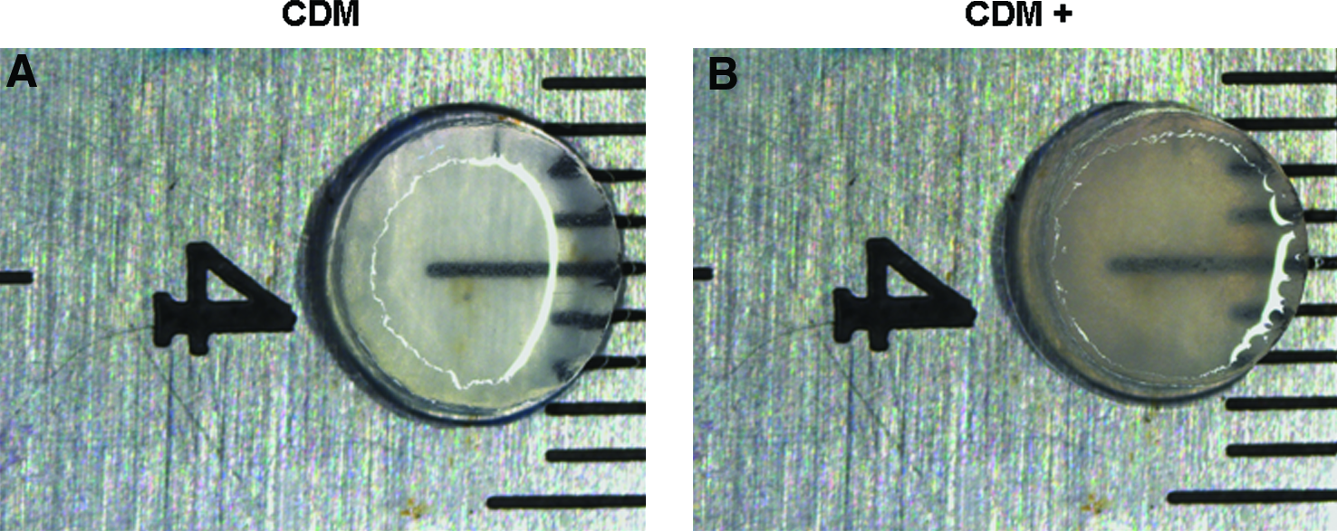
Stereomicrographs of hMSC-laden CMC hydrogels cultured in CDM
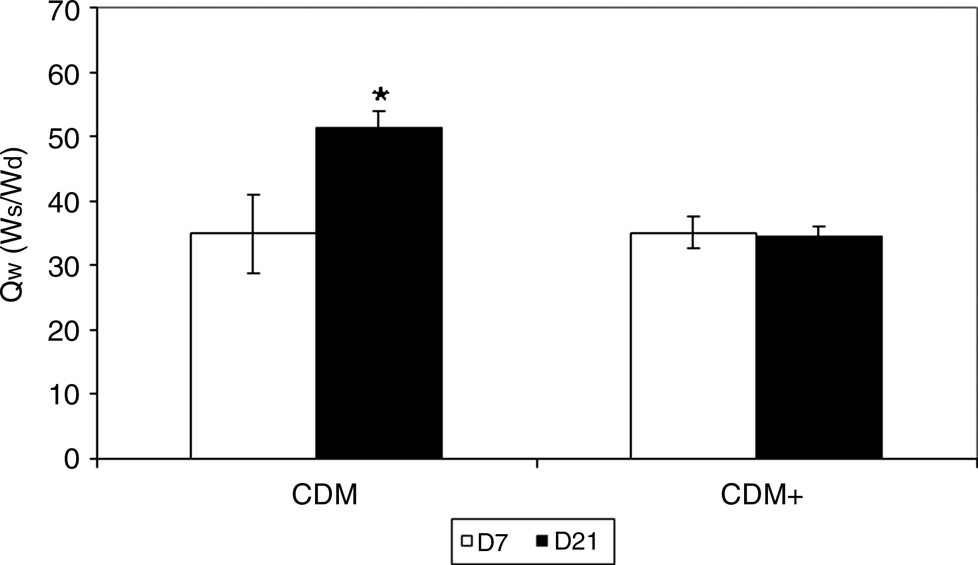
Equilibrium weight swelling ratio (Qw) of hMSC-laden CMC constructs cultured in CDM and CDM+ media at day 7 (open bar) and day 21 (solid bar). *Significantly different compared with all other groups (n=3–4).
COL II and GAG content at day 7 were similar between CDM and CDM+ groups, whereas the content of both COL II and GAGs in treated samples (CDM+) at day 21 was significantly greater than all other groups. DNA content was significantly greater at day 21 versus day 7 across both the treated and untreated groups; however, no significant differences were seen between CDM and CDM+ hydrogels at each time point (Table 1).
Data represent the mean±SD.
Significantly different from all other groups.
Significantly different from grouped day 7 samples (n=3–4).
SD, standard deviation; GAG, glycosaminoglycan; Coll II, type II collagen; CDM, chemically defined medium.
Mechanical properties at days 7 and 21 were analyzed by unconfined compression. There were no significant differences in σpk and ɛeq between treated and untreated constructs at day 7; however, treated constructs (CDM+) at day 21 exhibited the highest σpk (1.489±0.389 kPa) and the lowest ɛeq (0.15±0.02), both of which were significantly different from all other groups (Table 2). In addition, untreated constructs (CDM) at day 21 exhibited the highest ɛeq (0.338±0.063) among all groups. Although Ey increased with TGF-β3 treatment over time, the increase was not significantly greater and there were no significant differences between treatment groups at day 7 or 21.
Data represent the mean±SD.
Significantly different from all other groups (n=5).
Histological and immunohistochemical analyses were performed on the constructs at days 7 and 21. At 7 days, TGF-β3–treated constructs (CDM+) exhibited Alcian Blue and CSPG staining that was localized both pericellularly and in the interterritorial space, whereas COL II elaboration was detected as intense pericellular deposits (data not shown). Untreated controls (CDM) only displayed faint pericellular staining (data not shown). After 21 days, CDM+ specimens stained more intensely with Alcian Blue (Fig. 3B), CSPG (Fig. 3D), and collagen II (Fig. 3F) than at 7 days and in comparison to CDM hydrogels (Fig. 3A, C, E). In addition, CDM+ constructs exhibited dense pericellular and largely uniform interterritorial staining patterns, whereas CDM staining was more faint and strictly pericellular.
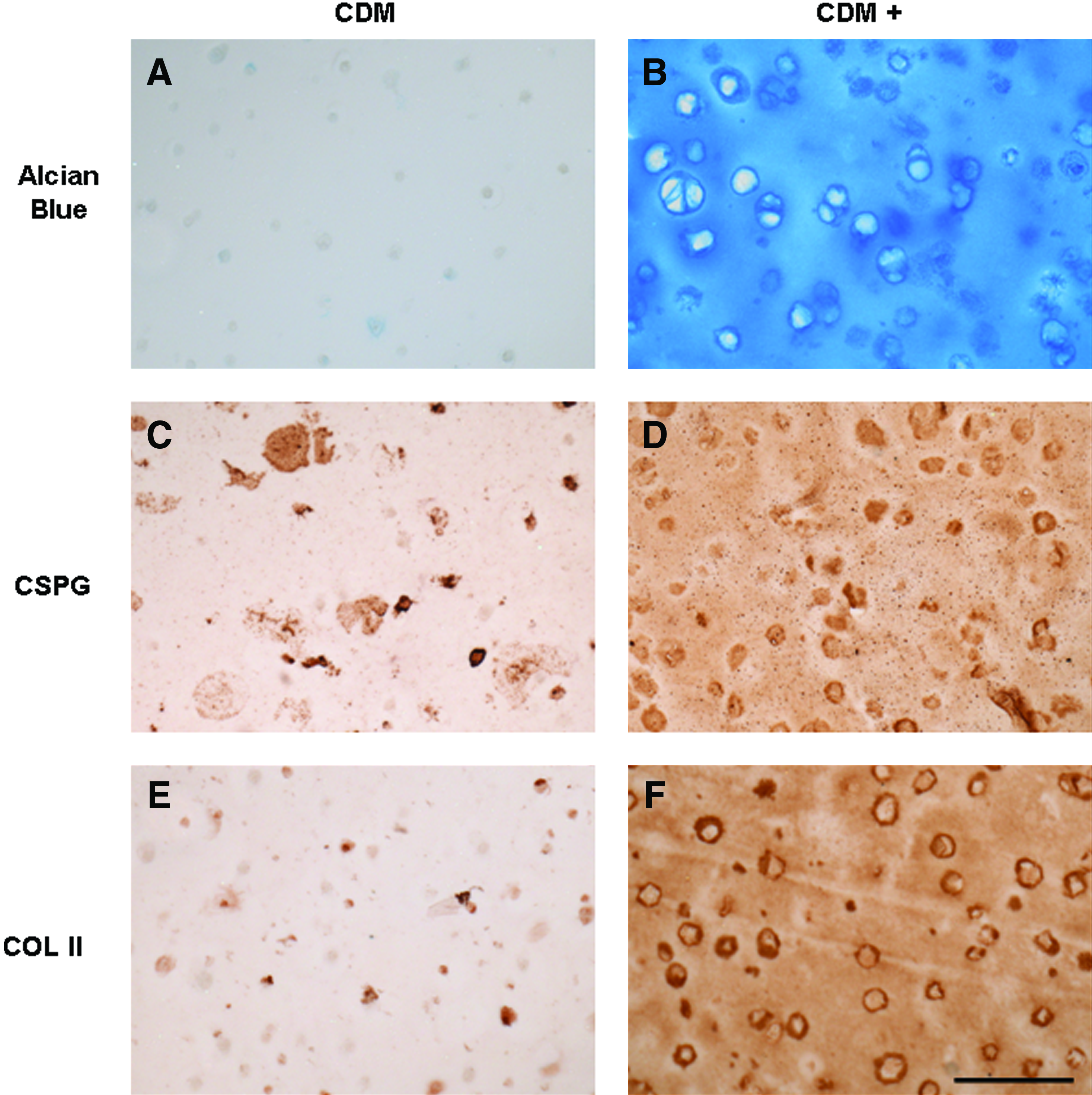
Histological and immunohistochemical analyses of hMSC-laden CMC constructs at day 21. Alcian Blue staining of sulfated GAGs for CDM
Discussion
This study is the first to investigate the effects of TGF-β3 on the response of hMSCs encapsulated in photocrosslinked CMC hydrogels cultured in chemically defined, serum-free medium. Consistent with the hypothesis of this study, it was demonstrated that serum-free, CDM supplemented with TGF-β3 resulted in increased GAG and COL II accumulation and enhanced functional material properties. These findings underscore the importance of growth factors in promoting the differentiation of hMSCs toward a chondrogenic phenotype for the development of engineered constructs.
CMC is a well-established derivative of cellulose, which is rendered water-soluble through the introduction of carboxymethyl groups along the polymer backbone. Photocrosslinked CMC gels form stable networks that undergo controlled degradation by hydrolytic scission of interchain ester crosslinks between the methacrylated CMC chains under physiologic conditions. This provides void space for the accumulation of secreted matrix macromolecules while maintaining sufficient structural integrity. 43 The functional properties and cellular response to the CMC constructs used in this study are comparable to those of other materials employed for similar applications, such as hyaluronic acid and collagen.21,22,50,51 Moreover, photocrosslinked CMC hydrogels provide additional advantages of a low cost, nonanimal or bacterial-derived starting material. A particular benefit is that the hydrogels are not susceptible to matrix metalloprotease catalysis, as CMC is only degraded by cellulase, an enzyme that is not found in humans. The CMC properties are expected to be maintained in the hypoxic, low-glucose, and acidic environment of the native human NP.52,53 Although hydrogel permeability was not assessed in this study, it is anticipated to be sufficient for effective oxygen and nutrient transport in vivo given the increase in cell proliferation and matrix elaboration measured in vitro. However, the acidic environment of the NP may result in accelerated hydrolytic degradation of the CMC interchain ester crosslinks.
Previously, CMC hydrogels were shown to support NP cell viability over several weeks in vitro.30,43 In this study, DNA content was significantly higher after 3 weeks for the control and TGF-β3–treated groups, confirming that CMC hydrogels are capable of supporting hMSC cell proliferation over time. The addition of TGF-β3 resulted in enhanced matrix deposition compared with untreated constructs, in support of our hypothesis. The Qw for the untreated constructs significantly increased over time, whereas the swelling ratio of TGF-β3–treated constructs was unchanged over the course of the study. This was due to an increase in the dry weight of the CDM+ constructs from day 7 to 21 (∼11%), in contrast to the CDM samples, which displayed a decrease in dry weight (∼24%) over the same time period. These findings are indicative of an increase in matrix accumulation in CDM+ specimens compared with the CDM constructs, which was confirmed by the increased opacity of the treated hydrogels. Although both groups underwent hydrolytic degradation of the CMC interchain crosslinks, allowing for greater water uptake and a concomitant increase in wet weight, the increased GAG and COL II content in the CDM+ group enabled the swelling ratio of the treated constructs to be maintained.
The enhancement of matrix deposition in TGF-β3–treated hydrogels was further verified by the mechanical properties of these constructs when tested in unconfined compression. The CDM+ hydrogels at day 21 exhibited the lowest axial deformation in creep, whereas CDM gels at day 21 resulted in the highest deformation, indicating a weakening of the constructs due to lack of matrix accumulation and CMC hydrolysis. CDM+ constructs also displayed the highest σpk among all groups at day 21, demonstrating a greater transient mechanical response. Nevertheless, TGF-β3 treatment did not give rise to increased equilibrium mechanical functionality, as Ey was not significantly different from control gels at either time point, but was within the range reported for native NP tissue (5–6 kPa). 54
The distinct effects of growth factor supplementation were also evident when evaluating specific ECM macromolecule accumulation. TGF-β3–treated constructs resulted in extensive Alcian Blue, CSPG, and COL II staining in photocrosslinked CMC hydrogels at days 7 and 21, characteristic of cartilaginous tissues, such as the NP. Alcian Blue staining indicated the presence of sulfated GAGs, whereas CSPG staining and quantification via the DMMB assay (which uses a chondroitin sulfate standard) specifically measured the accumulation of chondroitin sulfate. CSPG is an important factor to monitor, because it is a major component of aggrecan, which is the most abundant proteoglycan found in the IVD. 4 The Alcian Blue and CSPG staining were supported by a significant increase in GAG content in CDM+ constructs at day 21 compared with all other groups. There was no quantifiable COL II present in untreated samples at either time point, whereas TGF-β3–treated constructs displayed significant accumulation at day 21. Similar findings have been reported using TGF-β3 to stimulate functional matrix elaboration in chondrocyte-laden hydrogels.14,15
There were potential limitations to this study. Although matrix accumulation was significantly greater overall in TGF-β3–treated constructs, the GAG and COL II content did not reach the levels seen with bovine NP cells encapsulated in CMC hydrogels. 30 In addition, the 27:1 ratio of GAGs to collagen reported in native NP was much lower than the ratio seen in this study. 55 These differences could be due to cell source and study duration, as hMSCs will require a greater length of time to differentiate and produce characteristic matrix molecules, specifically collagen, whereas NP cells only need to maintain their phenotype over time. Although the equilibrium Young's modulus (Ey) in unconfined compression of these constructs was similar to that seen in human NP tissue, the peak stress and swelling behavior were not analogous to the native NP.30,54 Several previous investigations using hMSC-laden hydrogels observed improved functional properties after at least 28 days with stiffer starting materials than those employed here.15,32 Future studies will evaluate the long-term response of hMSCs encapsulated in CMC hydrogels of varying initial stiffness to determine the effect on functional tissue development. These long-term studies will also confirm whether the cellular arrangement in the constructs can create a structure that is comparable to native NP tissue. In addition, more specific NP cell markers such as SNAP25, KRT18, and FOXF1 56 will be assessed to further validate the differentiation of hMSCs toward an NP-like phenotype, as opposed to that of other cartilaginous tissues. For clinical translation, in vivo studies will need to be performed to verify biocompatibility (i.e., subcutaneous pouch model) 57 as well as functional efficacy using relevant IVD injury models.58–60 Nevertheless, a recent study confirmed the biocompatibility of acellular photocrosslinked methylcellulose hydrogels, suggesting that these CMC constructs may elicit a minimal inflammatory response in vivo. 61
Taken together, this study suggests that photocrosslinked CMC hydrogels can support functional cartilaginous ECM assembly consistent with an NP-like phenotype by hMSCs cultured in serum-free, CDM supplemented with TGF-β3. This hydrogel system may eventually have application in intradiscal NP replacement therapy.
Footnotes
Acknowledgments
This work was supported by an Early Career Translational Research Award from the Wallace H. Coulter Foundation and the National Science Foundation CAREER Award CBET 0747968 (to S.B.N.). The II-II6B3 monoclonal antibody developed by T. Linsenmayer was obtained from the Developmental Studies Hybridoma Bank developed under the auspicious of the NICHD and maintained by The University of Iowa, Department of Biological Sciences, Iowa City, IA. The authors also thank Dr. Sihong Wang for the generous gift of hMSCs and Dr. Anna T. Reza for helpful discussions.
Disclosure Statement
No competing financial interests exist.