
Abstract
Biodegradable gelatin sponges incorporating various amounts of magnesium calcium phosphate (MCP) were introduced and the in vitro osteogenic differentiation of rat bone marrow mesenchymal stem cells (MSCs) in the sponges was investigated. The MCP was added to the gelatin sponges at 0, 25, 50, 75, and 90 wt%. The pore sizes of the gelatin sponges ranged from 143 to 154.3 μm in diameter and the porosity percentage was 34.3–50.1%. The compression modulus of the sponges and the resistance to the volume change significantly increased with increases in the amount of MCP. When seeded into the sponges by an agitating method, MSCs were distributed throughout the sponges. Following the incubation of MSCs in the gelatin sponges, a significantly higher cellular proliferation and alkaline phosphatase activity was observed in the gelatin sponges incorporating higher MCP contents. On the other hand, the osteocalcin content of MSCs seeded in the gelatin sponges incorporating no or low MCP showed a significantly higher levels in comparison with the MSCs seeded in the gelatins incorporating high MCP. These findings indicate that the MCP incorporation maintained the pore size and porosity percentage of the gelatin sponges and enabled the sponge to achieve mechanical reinforcement as well as promoting MSC proliferation and osteogenic differentiation.
Introduction
Gelatin is one of the materials extensively used in biomedical fields. It is a biodegradable polymer that can be easily chemically modified and has been applied to pharmaceutical, medical, and food usage. We have prepared biodegradable hydrogels from different types of gelatin for the controlled release of various growth factors. 5 We also prepared gelatin sponges with or without tricalcium phosphate as a scaffold for bone regeneration to demonstrate that it is suitable for cell culture. 6
Magnesium is the fourth most abundant cation in the human body and is naturally found in bone. 7 It is reported that magnesium is highly involved in bone formation and promotes cell attachment or spreading on other surfaces. 8 From the chemical view point, the presence of magnesium ions can reduce the crystallinity of calcium phosphate and increase the water solubility of phosphates. 9
Different cells have been used to test the biological functions of scaffolds, among them, mesenchymal stem cells (MSCs) are clinically popular in regenerative medicine because they can be readily isolated from the bone marrow. 10 It is well recognized that MSCs have an inherent potential to differentiate into cell lineages of various types. 11 During the osteogenic differentiation of MSCs, it is known that cells differentiate into osteoprogenitors with a limited self-renewal capacity, then to preosteoblasts with limited proliferation, and finally mature into osteoblasts that secrete osteoid. 12
The objective of this study was to evaluate the biological behavior of MSCs in gelatin sponges incorporating various amounts of magnesium calcium phosphate (MCP) and compare this with those without MCP. We also examine the physical and mechanical properties of the sponges.
Materials and Methods
Materials
Gelatin samples with an isoelectric point of 9.0 were kindly supplied by Nitta Gelatin Co. Calcium dihydrogen phosphate and magnesium oxide (90% and 99%, respectively) were obtained from Nacalai Tesque Ltd. and used without further purification. Culture media were obtained from Invitrogen Corporation (Carlsbad). Other chemicals were obtained from Wako Pure Chemical Industries.
Preparation of gelatin sponges incorporating MCP
Gelatin sponges containing different amounts of MCP were prepared by the dehydrothermal crosslinking of gelatin. Briefly, calcium dihydrogen phosphate and magnesium oxide at a molar ratio of 2:1 13 were mixed with 3 wt% of gelatin aqueous solution at different weight percentages of 0, 25, 50, 75, and 90 wt%. The mixed solution was agitated at 5,000 rpm for 3 min by using a homogenizer (ED-12; Nihonseiki Co.). The resulting foamy solution was cast into a polypropylene dish of 138×138 cm2 and 5 mm depth and then immediately frozen at −80°C. Finally, the sponges were freeze dried and dehydrothermally crosslinked at 140°C for 96 h.
Physical characterization of gelatin sponges incorporating MCP
The inner structure of sponges was viewed under a scanning electron microscope (SEM, S2380N; HITACHI), after sputter coating with gold/palladium. The porosity percentage and the mean diameter of the pores were determined by using ImageJ Version 1.43, Wayne Rasband, National Institute of Health.
The compression moduli of the freeze-dried gelatin sponges, with or without MCP incorporation (5×5×5 mm3), were measured by a mechanical apparatus (AG-5000B; Shimadzu) at a rate of 1 mm/min. A stress–strain curve was obtained and the compression moduli of samples were calculated from the initial slope of the load-deformation curve. Four sponges of each MCP concentration were used to calculate the average value and the standard deviation of the mean.
MSC preparation and culture
MSCs were isolated from the bone shaft of femurs of 3-week-old male Fisher 344 rats according to the technique reported by Lennon et al. 14 Briefly, both the ends of the femurs were cut away from the epiphysis and the bone marrow was flushed out by a syringe (21-gauge needle) with 1 mL of alpha-minimum essential medium supplemented with 15 vol.% fetal calf serum (FCS) and 50 IU/mL penicillin and streptomycin. The cell suspension (5 mL) was placed into T-75 culture flasks (SUMILON; Sumitomo Bakelite Co., Ltd.). The medium was changed every 3–4 days during culture. When the cells became subconfluent, they were detached by 0.25 wt% of trypsin, −0.02 wt% of ethylenediaminetetraacetic acid, and subcultured. Cells of the third passage at subconfluence were used for all the experiments.
MSC seeding into gelatin sponges incorporating MCP and culture
Gelatin sponges with or without MCP incorporation were cut into cylinders of 8-mm diameter and 1.25±0.25 mm using a biopsy punch (Kai Industries Co. Ltd.). MSCs were homogenously seeded into the cylindrical sponges by the agitated seeding method as it has been demonstrated that this method is effective in seeding cells homogeneously throughout three-dimensional porous scaffolds.15,16 Briefly, 50 μL of cell suspension (5×10⁵ cells) was dropped on the sponges that had been placed into the wells of a 48-multiwell tissue culture plate (IWAKI Glass Co. Ltd.) and agitated on an orbital shaker (ORBITAL SHAKER; Bellco Glass, Inc.) at 180 rpm for 1 h. Then, 1 mL of culture medium was added and the shaking was continued for a further 5 h.
The cell-seeded sponges were placed into 6-well multiwell tissue culture plates (3815-012; IWAKI Glass Co. Ltd.). Each sponge was incubated in Dulbecco's modified Eagle medium supplemented with 15 vol.% FCS, 10 nM dexamethasone, 50 μg/mL ascorbic acid, and 10 mM β-glycerophosphate (osteogenic differentiation medium) at 37°C in a 5% CO2–95% air atmosphere. The medium was changed and collected twice a week. The number of sponges used for each experimental group was 2 to 3.
SEM observation of MSCs cultured in gelatin sponges incorporating MCP
The gelatin sponges cultured with cells for 6 h were fixed with 2.5 wt% glutaraldehyde solution in 1× phosphate-buffered saline solution (PBS, pH 7.4). After PBS rinsing and subsequent dehydration with ethanol aqueous solutions, the dehydrated samples were immersed in t-butanol and dried with a critical point dryer (ES-2030; HITACHI). After sputter coating with gold/palladium, the samples were viewed on SEM.
Volume changes of the gelatin sponges incorporating MCP during culture
The change in the volume of gelatin sponges with or without MCP incorporation was determined by taking serial photographs of the scaffolds in the presence of a reference scale attached to the outside surface of the bottom of the culture dish, during the culture period. Image J software was used to calculate surface area of the scaffold and the reference scale. By correlating the size of the scaffold to the reference scale, it was possible to determine the dimensional change in the scaffold during the culture period. The photographs were taken with a digital camera (Cyber shot, DSC-F707; Sony).
Evaluation of cell behavior after incubation in gelatin sponges incorporating MCP
The number of MSCs attached to the gelatin sponges with or without MCP incorporation was determined by the fluorometric quantification of cellular DNA according to the method reported by Rao et al. 17 Briefly, the cell-seeded sponges were lysed in 500 μL of 30 mM sodium citrate–buffered saline solution (SSC) (pH 7.4) containing 0.2 mg/mL sodium dodecylsulfate by using a tissue lyser (Retsch Qiagen 85210 Tissue Lyser), for 10 min at 20 Hz, and then the samples were incubated at 37°C for 1 h. The cell lysate was centrifuged at 14,000 rpm and 4°C for 5 min to separate the cell lysate from the sponge remnants. The cell lysate (100 μL) was mixed with a dye solution (100 μL; 30 mM SSC, 1 μg/mL Hoechst 33258 dye) and the fluorescent intensity of the mixed solution was measured in a fluorescence spectrometer (F-2000; HITACHI) at excitation and emission wavelengths of 355 and 460 nm, respectively. The calibration curve between the DNA and cell number was prepared by using cells of known numbers.
As a measure of MSC osteogenic differentiation, the alkaline phosphatase (ALP) activity and osteocalcin content were determined. The ALP of cells was determined by using the conventional p-nitrophenylphosphate method, 18 while the osteocalcin content of the cells was determined by the enzyme-linked immunosorbent assay (ELISA) method. Briefly, MSCs cultured in the sponges for different time periods were mixed with 1 mL of 40 vol.% formic acid for more than 12 h to decalcify them using a mixer (CM-1000; Eyela Co. Ltd.). After the decalcified samples were centrifuged, the supernatant of the cell extraction was applied to gel filtration on a SephadexTM G-25 column (PD-10; Amersham Pharmacia Biotech AB). The resulting solution was freeze dried, redissolved in double distilled water (DDW), and subjected to an osteocalcin rat ELISA (Rat osteocalcin ELISA system; Biomedical Technologies Inc.).
Evaluation of the magnesium concentration in culture medium
The collected cultured media were freeze dried. The freeze-dried powder was then redissolved with DDW in a similar volume to that of the culture medium originally collected to ensure that all the samples had the same volume. The magnesium was determined by using the xylidyle blue method (Magnesium B reagent Kit; Wako Pure Chemical Industries) and standardized by the culture medium of the 0 wt% scaffolds. Briefly, 5 μL of culture medium was mixed with 750 μL of color reagent. Absorbance was measured at 520 nm.
Statistical analysis
All the data were analyzed by one-way analysis of variance with Tukey's test to compare significance, and statistical significance was accepted at p<0.05. Graph Pad Prism 5 software was used to conduct the statistical analysis. Experimental results were expressed as the mean+standard deviation.
Results
Characterization of gelatin sponges incorporating MCP
Figure 1 shows scanning electron micrographs of gelatin sponges with or without MCP incorporation. Irrespective of the MCP content, a similar infrastructure was observed, where the MCP appeared to be incorporated uniformly into the matrix of the gelatin sponges and deposited within the wall of every sponge. All the sponges had an interconnected porous structure with the pore size ranging from 143 to 154 μm, while the porosity percentages were around 34.3–50.1% (Table 1). The compression modulus of the sponges increased significantly with increases in the amount of MCP incorporated. For example, the compression modulus of the gelatin sponges containing 75 and 90 wt% MCP was significantly different from those incorporating lower amounts of MCP (Fig. 2).
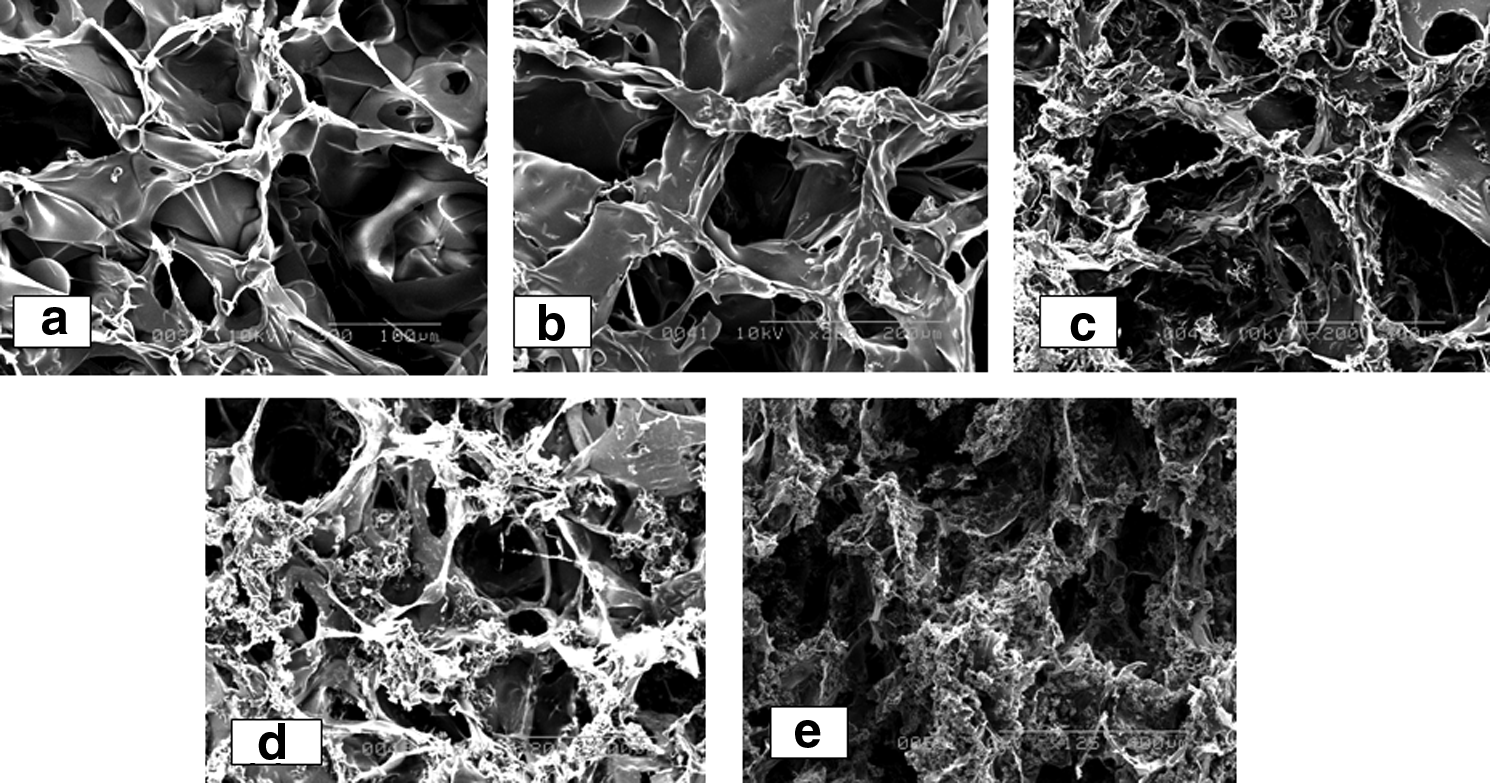
Scanning electron micrographs of gelatin sponges incorporating 0

Compression moduli of gelatin sponges incorporating different MCP contents. *p<0.05; significant against the compression modulus of gelatin sponges incorporating 0 wt% of MCP. †p<0.05; significant against the compression modulus of gelatin sponges incorporating 25 wt% of MCP. ‡p<0.05; significant against the compression modulus of gelatin sponges incorporating 50 wt% of MCP. §p<0.05; significant against the compression modulus of gelatin sponges incorporating 75 wt% of MCP.
There is no significant difference among groups.
MCP, magnesium calcium phosphate.
The gelatin sponges containing MCP showed an initial burst of magnesium followed by a steady-release profile. On the other hand, the gelatin sponges incorporating 90 wt% MCP showed a significantly higher release profile over the whole culture period in comparison with the other sponges (Fig. 3).

Magnesium concentration in the medium after culturing with gelatin sponges incorporating 25 (⋄), 50 (Δ), 75 (▪), and 90 (♦) of MCP content. †p<0.05; significant against magnesium concentration of the medium of gelatin sponges incorporating 25 wt% of MCP. ‡p<0.05; significant against magnesium concentration of the medium of gelatin sponges incorporating 50 wt% of MCP. §p<0.05; significant against magnesium concentration of the medium of gelatin sponges incorporating 75 wt% of MCP.
Attachment and proliferation of MSCs in gelatin sponges incorporating MCP
Figure 4 shows SEM photographs of gelatin sponges incorporating MCP 6 h after MSC seeding. The cells attached to all types of sponges and they were distributed throughout the sponges. No difference in the shape of the attached MSCs was observed among sponges.

Scanning electron micrographs of MSCs attaching to gelatin sponges incorporating 0
Volume change of gelatin sponges incorporating MCP
Figure 5 shows the percent change in the volume of the gelatin sponges incorporating MCP. The dimensional stability of gelatin sponges incorporating MCP seemed to increase in proportion to the amount of MCP. The gelatin sponges incorporating 90 wt% MCP showed the highest dimensional stability; about 88.7% of the initial size was maintained even after 4 weeks of culture. On the other hand, the gelatin sponges containing 0 wt% of MCP showed the least dimensional stability.
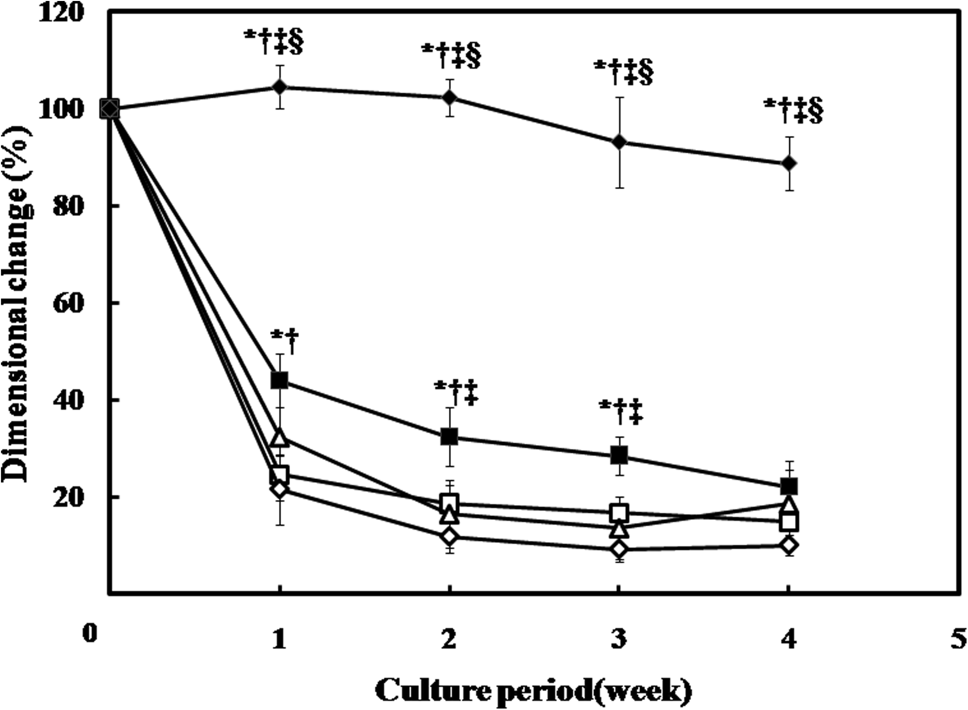
Percent change in the volume of the gelatin sponges incorporating 0 (□), 25 (⋄), 50 (Δ), 75 (▪), and 90 wt% (♦) of MCP contents. *p<0.05; significant against the volume change of gelatin sponges incorporating 0 wt% of MCP. †p<0.05; significant against the volume change of gelatin sponges incorporating 25 wt% of MCP. ‡p<0.05; significant against the volume change of gelatin sponges incorporating 50 wt% of MCP. §p<0.05; significant against the volume change of gelatin sponges incorporating 75 wt% of MCP.
MSC proliferation and osteogenic differentiation in gelatin sponges incorporating MCP
Figure 6 shows the number of MSCs proliferated in the gelatin sponges with or without MCP incorporation. In the first week of culture, no significant difference was seen in the cell number among different types of sponges. On the contrary, 2 weeks later, the gelatin sponges incorporating 90 wt% MCP showed a significant increase in the cell number compared with the other sponges. In the third and fourth week of culture, the cell number significantly increased for the sponges incorporating 75 and 90 wt% MCP in comparison with others. The cells seeded in the gelatin sponges incorporating 50 wt% MCP showed a significant difference only in the fourth week of culture compared with those seeded in gelatin sponges incorporating lower amounts of MCP.

Time course of MSC proliferation in gelatin sponges incorporating 0 (□), 25 (⋄), 50 (Δ), 75 (▪), and 90 (♦) of MCP content. *p<0.05; significant against MSC number in the gelatin sponge incorporating 0 wt% of MCP. †p<0.05; significant against MSC number in the gelatin sponge incorporating 25 wt% of MCP. ‡p<0.05; significant against MSC number in the gelatin sponge incorporating 50 wt% of MCP. §p<0.05; significant against MSC number in the gelatin sponge incorporating 75 wt% of MCP.
Figure 7 shows the time course of the ALP activity of MSCs after culture in gelatin sponges with or without MCP. In the second week of culture, the ALP activity of the MSCs cultured in the gelatin sponges incorporating 90 wt% MCP was significantly higher than that of the other gelatin sponges. In the third and fourth week of culture, the MSCs cultured in the gelatin sponges incorporating 50, 75, and 90 wt% MCP showed a significantly higher ALP activity in comparison with the other groups.

Time course of the ALP activity of MSCs in the gelatin sponge incorporating 0 (□), 25 (⋄), 50 (Δ), 75 (▪), and 90 (♦) of MCP content. *p<0.05; significant against the ALP activity of the cells in gelatin sponges incorporating 0 wt% of MCP. †p<0.05; significant against the ALP activity of the cells in gelatin sponges incorporating 25 wt% of MCP. ‡p<0.05; significant against the ALP activity of the cells in gelatin sponges incorporating 50 wt% of MCP. §p<0.05; significant against the ALP activity of the cells in gelatin sponges incorporating 75 wt% of MCP. ALP, alkaline phosphatase.
The osteocalcin content of MSCs cultured in the gelatin sponges containing 0 or 25 wt% of MCP was significantly higher when compared with those of the gelatin sponges containing 75 and 90 wt% of MCP, in the fourth week of culture (Fig. 8).

Osteocalcin content of the MSCs in the gelatin sponge containing different MCP contents at 2 weeks (black column) and 4 weeks (white column). §p<0.05; significant against the osteocalcin content of the cells in the gelatin sponges incorporating 75 wt% of MCP. ¤p<0.05; significant against the osteocalcin content of the cells in the gelatin sponges incorporating 90 wt% MCP.
Discussion
This study investigated the proliferation and osteogenic differentiation of MSCs in gelatin sponges incorporating different amounts of MCP or without MCP incorporation. Incorporation of MCP enabled the gelatin sponges to mechanically reinforce and promote the proliferation and osteogenic differentiation of MSCs. The gelatin sponges incorporating MCP could be prepared by an easy, one-step procedure with the advantage of creating a porous structure for cell-based bone regeneration. In other words, the simple foaming of a gelatin solution permitted the formation of gelatin sponges, while the addition of MCP improved the physical, mechanical, and biological properties of the sponges. The sponges also showed slow magnesium release, which may contribute to bone regeneration in vivo. 7 In addition, the gelatin used in the sponge fabrication, I.P.9 gelatin, is a known carrier for bone morphogenetic protein-2, 19 an important osteoinductive and ectopic bone-forming protein. 20
There is growing consensus that the physical properties of materials, such as topography, geometry, porosity, and stiffness, can be used to direct biological outcomes in a manner similar to traditional approaches involving chemistry or biomolecules. 21
The addition of MCP to the gelatin sponges did not affect their porosity percentage and pore size because the MCP was added during, not after, the sponge fabrication. In other words, the MCP was mixed during the gelatin foaming procedure with the end result of no difference in pore size and porosity percentage among groups. On the contrary, the compression modulus increased with the addition of MCP (Fig. 2), which is of great value for the regeneration of hard tissue such as bone. 4 The cells cultured in the gelatin sponges with or without MCP incorporation did not show a different morphology (Fig. 4), indicating that MCP has no negative effect on cell morphology.
When MSCs were cultured in the gelatin sponges incorporating MCP, the number of MSCs proliferating in the sponges increased with increases in the amount of MCP. For example, there was an eightfold increase in the MSC number cultured in the gelatin sponges incorporating 90 wt% MCP compared with those of MCP-free sponges (Fig. 6). This can be explained in terms of pore space in the sponges. As shown in Figure 5, although the initial sponge size and porosity were almost the same for all the groups, they were deformed during the culture period, and since the gelatin sponges with higher amounts of MCP incorporated showed less shrinkage, there was more room for the cells in these sponges to grow and multiply, with more surface area exposed to oxygen and nutrients. 6 Another possible reason is the dissolution of magnesium and calcium ions, which can stimulate cellular proliferation.22,23
The ALP activity of MSCs in the sponges increased with increases in the amount of MCP; for example, the ALP activity in the gelatin sponges incorporating 90 wt% MCP showed a threefold increase in the second week of culture. This could be due to the presence of calcium and phosphate with magnesium, which is known to facilitate cell differentiation.24–29 In addition, the differentiation of cells was maximized when the modulus of the scaffold increased to match that of bone tissue. 30 Another possible reason is the change in surface topography, as seen in Figure 4, where 90 wt% gelatin sponges shown a rough microscopic morphology in comparison with 0 wt%. Rough surfaces have been showed to enhance osteogenic differentiation in comparison with smooth surfaces. 31
Osteocalcin, a late osteo-differentiation marker, showed a higher amount per cell in the gelatin scaffold incorporating low or no MCP. This could be explained by the fact that the cells in the gelatin sponges containing a high MCP content are at the early stage of osteogenic differentiation, which is characterized by a high proliferation index and ALP activity. Meanwhile, the cells in the low or without MCP gelatin sponges are at the late osteogenic differentiation stage, which is characterized by low cell proliferation, low ALP, and high osteocalcin levels.32,33 The dimensional shrinkage, which limits cellular proliferation and enhances contact differentiation of MSCs, could be a possible explanation for the high osteocalcin levels in the gelatin sponges with low MCP content. On the other hand, a high magnesium concentration has been known to have an inhibitory action on late osteogenic differentiation by increasing the expression of the anticalcification protein osteopontin with upregulation of the calcification inhibitor, matrix Gla protein. 34
In summary, the addition of MCP did not affect the pore size and porosity percentage adversely. Moreover, it improved the dimensional stability and compression modulus of the gelatin scaffolds, which in turn along with the slow magnesium release and surface roughness promoted MSC proliferation and osteogenic differentiation.
Conclusion
Novel scaffolds based on gelatin and MCP were introduced. The addition of MCP to the gelatin sponges during preparation did not reduce the pore size and porosity percentage of the scaffold. Furthermore, a significant increase in dimensional stability and compression modulus was identified in the sponges containing high amount of MCP.
The MSCs seeded into the gelatin sponges containing MCP showed consistency in morphology with those without MCP content. In addition, the MSC proliferation and osteogenic differentiation benefited greatly by the increase in dimensional stability, hardness, roughness, and magnesium release in the gelatin sponges containing MCP.
Footnotes
Disclosure Statement
No competing financial interests exist.