
Abstract
The aim of the study was to investigate the use of a hyaluronic acid/polycaprolactone material for meniscal tissue engineering and to evaluate the tissue regeneration after the augmentation of the implant with expanded autologous chondrocytes. Eighteen skeletally mature sheep were treated. The animals were divided into three groups: cell-free scaffold, scaffold seeded with autologous chondrocytes, and meniscectomy alone. The implant was sutured to the capsule and to the meniscal ligament. At a 12-month gross assessment, histology and histomorphometry were used to assess the meniscus implant, knee joint, and osteoarthritis development. All implants showed excellent capsular ingrowth at the periphery. The implant gross assessment showed significant differences between cell-seeded and cell-free groups (p=0.011). The histological analysis indicated a cellular colonization throughout the implanted constructs. Avascular cartilaginous tissue formation was significantly more frequent in the cell-seeded constructs. Joint gross assessment showed that sheep treated with scaffold implantation achieved a significant higher score than those underwent meniscectomy (p<0.0005), and the Osteoarthritis Research Society International score showed that osteoarthritic changes were significantly less in the cell-seeded group than in the meniscectomy group (p=0.047), even though results were not significantly superior to those of the cell-free scaffold. Seeding of the scaffold with autologous chondrocytes increases its tissue regeneration capacity, providing a better fibrocartilaginous tissue formation. The study suggests the potential of the novel hyaluronic acid/polycaprolactone scaffold for total meniscal substitution, although this approach has to be further improved before being applied into clinical practice.
Introduction
Lesions of the meniscus are frequently observed in orthopedic practice, with a mean annual incidence of 60 to 70 per 100,000 knee injuries. 3 Damage or loss of meniscal tissue leads to pain and knee dysfunction, making a meniscectomy often inevitable despite its well-known deleterious effects. In fact, even a partial meniscectomy causes abnormal cartilage stresses, leading to the development of osteoarthritic changes of the tibia and femur at long term. 4 Therefore, the possibility of preserving the meniscal structure should be always considered, especially in the young active patients.
Many devices have been developed to fix the loose segment or the tear onto the main body of the meniscus: other than the traditional suturing procedures, various devices, such as anchors, screw, and staples, have been proposed for the fixation of the damaged fragment. 5 Unfortunately, the limited blood supply, together with the low healing potential of this tissue, does not allow often a biological repair to be achieved 6 . To increase the low-standard biological healing process, vascular access channels and synovial abrasions have been used for enhancing the vascularization. 7 However, despite these attempts to facilitate repair and improve healing, many repairs continue to fail. 8
In the last years, thanks to the continuous development in regenerative research field, tissue engineering is emerging as a possible solution for meniscal regeneration.1–3,9–12 In a previous study, tissue integration between the original meniscus and a new scaffold made of hyaluronic acid esters (HA, HYAFF®, Fidia Advanced Biopolymers) and poly-ɛ-caprolactone (PCL) for total meniscal substitution was observed with tissue formation, cellular infiltration, and vascularization. 11 In a recent study 12 we applied a tissue-engineering approach, using cells seeded onto this new resorbable biomaterial for total meniscal substitution in sheep. Different cell sources have been analyzed in vitro to find the most suitable source for cell augmentation of tissue-engineered meniscus. Autologous articular chondrocytes were chosen, as we have demonstrated that, in vitro, they were better than either meniscal cells or synovial or fat pad cells in their capacity to develop into a tissue with a relevant cell phenotype and matrix production, generating tissues with cellular and matrix phenotypes, including glycosaminoglycan (GAG) expression, similar to those of both the inner and outer meniscus regions. 13 The choice was also influenced by the fact that articular cartilage harvesting is less damaging to the joint with respect to healthy meniscal tissue harvesting: most of the patients suitable for this procedure have been previously meniscectomized with very few meniscal tissue leftover. Finally, because the surgical technique for total meniscal replacement has also not been highly investigated and is not consistent among authors,9,14,15 the secondary aim of that study 12 was to evaluate two different surgical scaffold implantation techniques: suture to the capsule and to the meniscal ligament, with or without transtibial fixation of the horns. The results obtained supported the idea that HA-PCL scaffold used alone has the potential for total meniscal substitution, as it is tolerated immunologically and induces tissue ingrowth. There was no evidence of a significant synovitis or lymphoplasmacytic infiltrate, but a prominent foreign-body giant cell response to the implant, presumably a part of the physiological resorption process,16,17 was documented. Seeding of the scaffolds with autologous articular chondrocytes provided some benefits; in fact, although no significant difference was shown between cell-seeded and cell-free implants at 4-month follow-up macroscopically, the histological analysis demonstrated deposition of a cartilaginous matrix only in cell-seeded implants. With regard to the surgical techniques analyzed, a higher incidence of tears and implant extrusions, other than worse implant and joint appearances, were observed in cases where trans-tibial, rigid fixation of the horns was utilized. It is likely that too rigid horn fixation in this weight-bearing, large-animal model resulted in overly high mechanical stresses on the implant, leading to a higher incidence of graft tears. 12
Therefore, the previous short-term study showed the potential of this HA-PCL scaffold and allowed us to determine the more favorable implantation procedure, and we concluded that longer term animal studies were necessary to fully evaluate the role of cell augmentation in promoting fibrocartilaginous tissue regeneration in complete meniscal substitution. 12 The aim of our present study at 12-month follow-up was to evaluate if the observed benefits at 4-month follow-up become more evident at longer observation, leading to better results and meniscal tissue regeneration. Gross assessment, histology, and histomorphometric scores were used to assess the meniscus implant, knee joint, and osteoarthritis development.
Materials and Methods
Study design
All procedures were approved by the Scientific and Ethics Committees of the experimentation center and by the Minister of Health. The study was performed by strictly following the National Laws on animal experiments (Law by Decree 116/92).
Eighteen skeletally mature female adult sheep (Bergamasca–Massese, 70±5 kg b.w.) were acquired from authorized farms, and kept in quarantine for at least 7 days before use. Under general anesthesia, animals underwent medial meniscectomy of the right stifle joint and were divided into three groups. The cell-seeded group (n=6) received a total medial meniscal replacement by the implantation of a scaffold seeded with autologous chondrocytes, whereas in the cell-free group (n=6), the meniscus was replaced by a cell-free scaffold. In the cell-free group, the left stifle joints served as nonoperated joints for comparison. In the cell-seeded group, the cartilage biopsy was performed from the left stifle joint (from non-weight-bearing areas of the trochlea) to obtain the chondrocyte culture. The medial meniscus was left untouched, and these joints were used as controls (it is not a real sham control, since it differs not only for the meniscus treatment but also for the dissection and suture of the medial collateral ligament [MCL], other than the biopsy, but it was useful to see the impact of the arthrotomy in a healthy joint). In the empty group (n=6), medial meniscectomy was performed using the same surgical technique without insertion of a meniscus device. Antibiotics (cefalosporin, 1 g/day for 4 days) and analgesics (metamizole sodium 40 mg/kg/day for 2 days) were administrated postoperatively.
All sheep received an external plaster (Scotch/Soft Cast, 3M Health Care) to limit motion at the operated limb for 5 days, as described by Dorotka et al. 18 Twelve months after surgery, the sheep were euthanized under general anesthesia by endovenous administration of Tanax (Hoechst). X-rays were routinely performed in every animal both at implantation surgery time and at the end of experiment after euthanasia.
Surgical procedure
The MCL cut was preparatory to meniscectomy and thus was performed in all right stifle joints. The MCL was cut slightly above the joint line to achieve a good exposure of the posterior aspect of the joint. The meniscus was circumferentially dissected from the capsule; the posterior horn of the meniscus was exposed by flexion, external rotation, and valgus stress of the joint, and the meniscus was detached at its posterior and anterior horns. The implant was sutured to the capsule at the level of the original meniscal rim, the anterior and posterior horns of the implant being fixed to the meniscal ligaments. 12 The capsule was closed. The MCL was reconstructed with 4 sutures, using resorbable sutures. The skin was closed by standard surgical techniques.
Biomaterial and implant
Porous scaffolds preparation
The scaffold was a porous blend of PCL and HYAFF, a class of hyaluronan-derived polymers obtained by a coupling reaction (Fidia Advanced Biopolymers), with a pore size of 200 to 300 μm. The meniscus-like composite devices were produced by a lamination technique, using molds that were designed according to original sheep menisci. About 294 mg/mL of PCL pellets (Mw=65000, Aldrich) was dissolved in a tetrahydrofuran (THF)/dimethyl sulfoxide (DMSO) 80/20 w/w solution, by stirring the components at 45°. A mixture of salts consisting of 93.9% w/w NaCl (with granulometry 315–400 μm), 3.5% w/w NaHCO3 (with granulometry 140–315 μm), and 2.6% w/w citric acid (with granulometry inferior to 200 μm) was prepared. Successively, the PCL solution and HYAFF 11 p75 HE powder (supported by FAB, AbanoTerme) at a ratio of PCL/HYAFF 70/30 w/w were mixed with salts, until a homogeneous paste was obtained. The ratio between the polymers and salts was 1/10. The paste obtained was then poured in a meniscus-shaped teflon mold containing a polylactic acid (PLA) net. The meniscus device was augmented with circumferential PLA fibers. PLA fibers were arranged in the mold, and the remaining paste was used as a matrix to fill it. The samples obtained were left in air for 24 h and immersed in bi-distilled water for 1 day. Subsequently, they were immersed in bi-distilled water at 40°C for 5 h to remove the solvents. The porous scaffolds were then left in bi-distilled water for a further 5 days, changing the water every day for complete removal of salts and any residual solvents. The samples were removed from molds and immersed in an ethanol/water 70/30 w/w solution, followed by placement in an Edwards lyophilisator to remove the water. The porous scaffolds obtained were immersed in an 8 mg/mL hyaluronic acid solution and lyophilized again. The structures were finally sterilized through γ radiation (2.5 Mrad) (Patent PCT/EP2005/056792, WO/2006/064025: “A biocompatible material and a prosthetic device made thereof for the replacement, repair and regeneration of meniscus”). The length of the final implants was 32 mm, the diameter 22 mm, and the height at the periphery 6–8 mm. The biomechanical analysis of implants was performed in shear and dynamic in compression. 19 The in vivo degradation time can be estimated on the basis of the behavior of the individual components. PCL degrades slowly by hydrolytic chain scission of the ester linkage, 20 losing its in vivo molecular weight slowly (up to 12 months, depending on the polymer composition), and its mechanical stability much earlier (between 12 and 16 weeks).21,22 HYAFF-11 degrades by de-esterification in 3 to 4 months in vivo. 16
Cell-scaffold construct preparation
Cartilage was harvested from the non-weight-bearing areas of the trochlea. The cartilage biopsies were put into sterile 50-mL tubes with a nutrient medium and sent to FAB (Fidia Advanced Biopolymers) for the cell-seeding procedure. Chondrocytes were isolated by sequential enzymatic digestions: 30 min with 0.1% hyaluronidase (Sigma), 1 h with 0.5% pronase (Sigma), and 1 h with 0.2% collagenase (Sigma) at 37°C. The chondrocytes were then centrifuged for 10 min at 1800 rpm, and the pellet was washed twice with the Dulbecco's Modified Eagle Medium with 10% fetal calf serum (FCS, Sigma). The cells were cultured under conventional monolayer culture conditions at 37°C in 5% CO2. The medium was changed every 3 to 4 days. Once sufficient cells were available, usually after the third to fourth passage, chondrocytes were dynamically seeded onto the meniscus prototype at a density of 25×106 cells/cm3 (i.e., 40×106cells/scaffold) in a mixed flask (50 rpm) in a volume of 100 mL of medium. The medium was removed 24 h after seeding and replaced by a complete medium supplemented with 10 μg/mL insulin, 0.1 mM ascorbic acid, and 1 ng/mL transforming growth factor ß1. Media were changed twice a week thereafter. After 14 days of spinner flask culture, prototypes were ready for in vivo implantation. Characterization of the cell-based grafts before implantation was previously reported. 12
Evaluation
Specimen preparation and gross inspection
At sacrifice, the joints were assessed clinically for swelling and stability. Under sterile conditions, the right and the left joints were opened, a swab for microbiological analysis and a synovial aspirate were taken, and the presence of effusion or popliteal cysts was documented. Synovial fluid was analyzed for the presence of inflammatory cells.
The joint was resected in toto, and the restoration of the MCL was inspected. A Gross Evaluation of Meniscus Implant score was used to assess the implant outcome. 11 This includes nine different categories: implant integration, implant position, horn position, implant shape, presence of tears, implant surface, size, tissue quality, and condition of the synovia. The specimens were photographed, and then the implant was dissected from the tibia. Joint status was assessed using the Gross Assessment of Joint Changes score published by Jackson et al., 17 which scores 12 different areas of the joint, including the femoral and tibial cartilage, the patella, its femoral groove, the menisci, and the femoral junction.
Histological evaluation
The meniscal implants, synovial samples, tibial plateau, and femoral condyles were analyzed by histology. The meniscal implants were cut into an anterior and posterior part at the level of the MCL. Consecutive 3-mm cross-section slices were cut to produce blocks, allowing histological sections that showed inner and outer regions, superior and inferior surfaces of the potentially regenerated meniscus, and attachment to the joint capsule. Tissue blocks were fixed in 10% neutral buffered formalin, decalcified when necessary, and processed into paraffin wax. Paraffin sections were cut at 3–4 μm and stained with (i) hematoxylin and eosin for assessment of cellular features and (ii) alcoholic toluidine blue for assessment of the GAG content.
Blocks of synovia were assessed for the presence of synovial cell hyperplasia/hypertrophy and an inflammatory cell infiltrate. Blocks of meniscal implant were assessed for the presence of residual scaffold, cellularity, foreign-body response, inflammatory cell (lymphocytes, plasma cells, and neutrophils) infiltrate, blood vessel ingrowth, fibrous and cartilaginous matrix, and integration of the implant with the joint capsule. Histological features were scored as being either present or absent.
Femoral condyles and tibial plateau, including articular cartilage and subchondral bone, were scored by the Osteoarthritis Research Society International (OARSI) cartilage OA histopathology grading system. 23 Sections were blindly evaluated by an experienced osteoarticular pathologist.
Statistical analysis
All continuous data were expressed in terms of mean and standard deviation of the mean. Grouping variables were expressed as frequency and percentage. The Student t-test or the Mann-Whitney test (two groups) and the Kruskal-Wallis test (more than two groups) were performed to test differences between and among groups' means. The Jonckheere-Terpstra test for ordinal groups was performed to investigate the trend among ordinal groups. Due to the small samples, all evaluations were carried out by the Exact Method. For all tests, p<0.05 was considered significant. A statistical analysis was carried out by means of the Statistical Package for the Social Sciences (SPSS) software version 15.0 (SPSS Inc.).
Results
Clinical assessment
All animals tolerated surgery well and survived the post-surgical period. All joints were stable. No differences in gait and joint stability were found between left not-operated limbs and cartilage-harvested limbs (controls). No signs of infection or allergic reactions were detected.
All sheep showed soft-tissue swelling on the medial aspect of the operated joints. A mild effusion in the form of a clear synovial fluid was detected in 11 joints (4 cell-free, 2 cell-seeded, 4 meniscectomy group, and 1 control). One sheep in the cell-free group presented signs of synovitis with synovial hypertrophy. Comparison of postoperative and preoperative radiographs revealed no signs of fracture.
Implant gross assessment
The results are summarized in Table 1. The statistically significant difference was observed between the cell-seeded (mean score: 23.2) and cell-free (mean score: 15.8) groups (p=0.011).
Figure 1 illustrates macroscopic appearance of meniscal implant in different groups. Implants showed capsular ingrowth at the periphery. The implant was dislocated in three cases (all cell-free). A slight extrusion of the implants at the periphery and wrinkling in the posterior region was frequently observed; only 2 cases from the cell-seeded group did not present any signs of extrusion. Both horns were anatomically positioned and firmly attached in all cases in the cell-seeded group, and only in 1 case in the cell-free group. The menisci implants had maintained the meniscus-like shape in all cases of the cell-seeded group, and in four cases of the cell-free group implants were out of shape. Three implants (all cell-free) had a full substance tear. Complete integrity of the implant was observed in four implants of only the cell-seeded group. Also better regenerated tissue quality was observed in the cell-seeded group. Normal synovia was observed in the majority of the cases. In 1 case, (cell-free) synovial hypertrophy was noted.
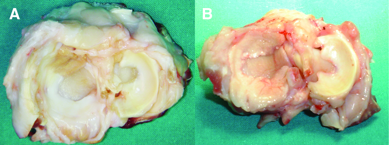
Macroscopic photographs of meniscal implant in the cell-seeded group
Joint gross assessment
The statistical analysis showed different results among groups. Joint degeneration in the nonoperated joints (mean score: 0.8) and in controls (mean score: 1.5) was negligible. Sheep treated with scaffold implantation (with or without cells) achieved significantly better scores than those that underwent the only meniscectomy (p<0.0005), even if they did not achieve the level of the sheep that underwent arthrotomy and biopsy or were not operated at all (p<0.0005). Further analysis, performed to assess differences in the outcome obtained in the treatment groups, failed to show any difference between cell-free and cell-seeded scaffolds (Fig. 2). A better chondroprotection was observed in the two sheep where menisci were not extruded, both part of the cell-seeded group. The subchondral bone was exposed in 3 cases of the cell-free group, in 2 sheep of the cell-seeded group, and in 6 cases in the meniscectomy group (Fig. 3).

The Joint Gross Assessment Data showed negligible joint degeneration in the nonoperated joints (no op) and in controls. Sheep treated with cell-free and cell-seeded scaffold implantation achieved significantly better scores than those that underwent only meniscectomy (***p<0.0005), but worse with respect to sheep in which only arthrotomy (control) was performed or were not operated at all (***p<0.0005). No difference between cell-free and cell-seeded scaffolds was found.
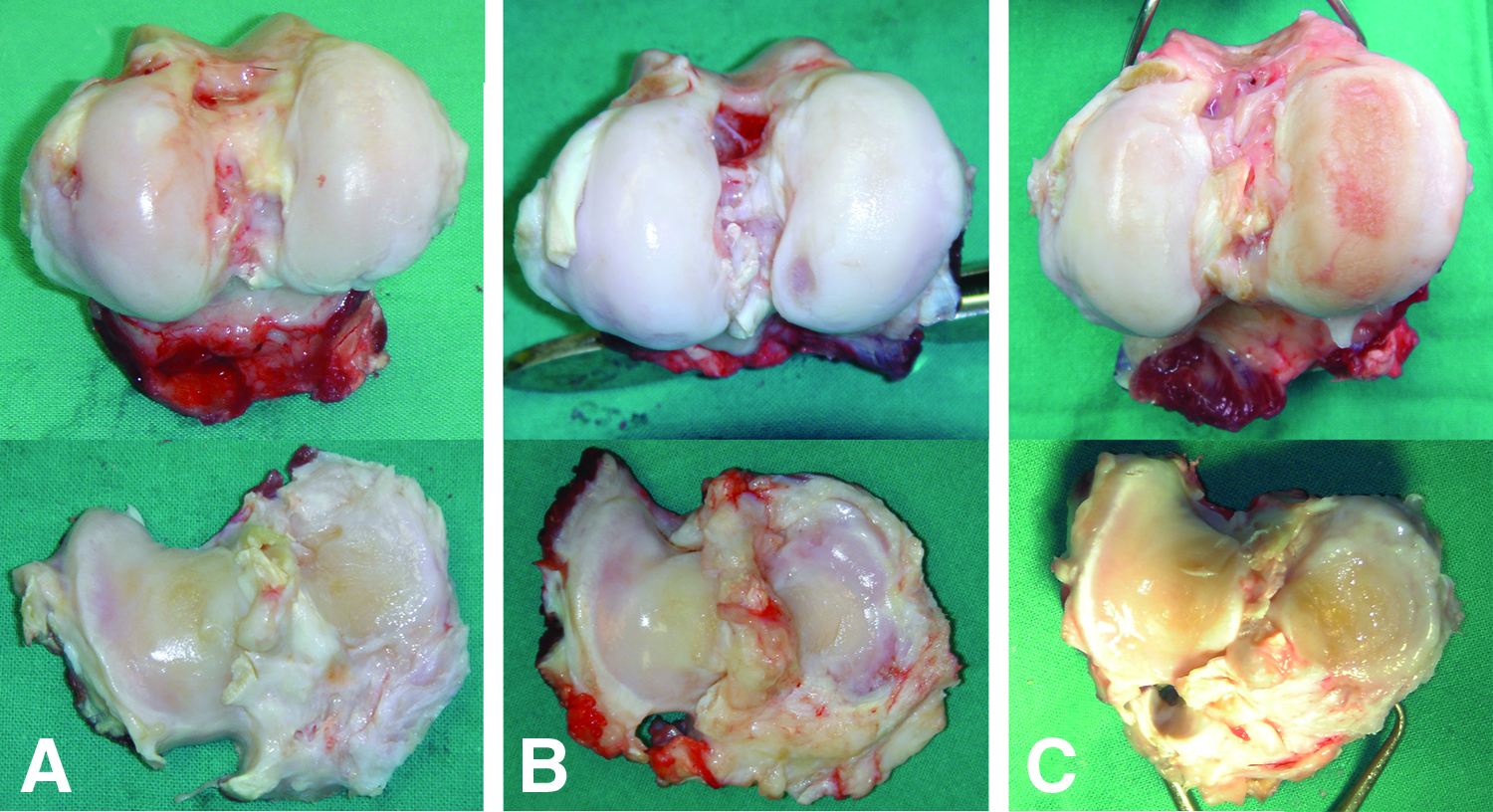
Macroscopic photographs of operated joints: femoral condyle (at the top) and tibial plateau (at the bottom) aspects. The Joint Gross assessment showed lower joint degeneration in the cell-seeded
Histological evaluation
Osteoarthritis was evident histologically in all joints. This was most severe in the meniscectomy-alone group with an OARSI score of 12.0±3.0. In contrast, the OA changes were less severe in the cell-seeded group with an OARSI score of 8.7±2.3 (p=0.047). The OA score of the cell-free group was also less being 9.7±3.5, which was not significantly different to the meniscectomy-alone or the cell-seeded groups. There was no evidence of a significant inflammatory cell infiltrate in the synovia of joints, which contained either cell-seeded or cell-free implants. The results of the histological assessment of the meniscal implants are summarized in Table 2.
Results showed the persistence of scaffold residuals together with a consequent foreign-body reaction in both the cell-seeded and cell-free implant groups, although in two cell-seeded cases, the foreign-body reaction was weak, and giant cells were evident only at periphery of the implant close in continuity with the scaffold residuals.
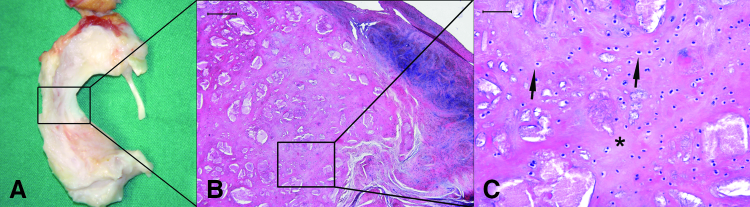
Moreover, as far as the scaffold degradation was concerned, in five cases (4 cell-seeded and 1 cell-free), the scaffold appeared completely resorbed at the implant tip (Fig. 4).

Histological images of two cell-seeded implants showing the scaffold degradation at the implant tips. (S) Scaffold residuals and (R) regenerated avascular tissue. Hematoxilin/eosin staining, 4× magnification, scale bar=500 μm. Color images available online at www.liebertpub.com/tea
Implants had a well-organized and tight integration with the surrounding capsule and presented an excellent cell colonization throughout the implants: only one cell-seeded case presented areas of hypocellularity. In all groups, new vessel formation was recorded, although not homogeneously distributed: neoangiogenesis was observed near the residual scaffold materials, but where the scaffold was degraded, the regenerated meniscal tissue did not have any blood supply.
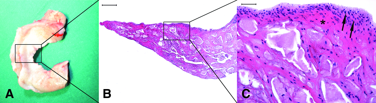
The main differences observed between the cell-seeded and cell-free groups were related to cartilage metaplasia and fibrosis (Figs. 5 and 6). New cartilaginous matrix deposition in the central region of the implant was observed in three cell-free cases, among which, two cases presented cartilaginous staining also at the tip and along the implant surface. In the cell-seeded group, cartilage metaplasia was noted in five cases at the tip and in the inner implant region. Otherwise, cartilage features appeared different between groups: in the cell-seeded group, cells had a spherical and chondroid appearance and were enclosed in a quite amorphous and hyaline-like matrix, whereas in the cell-free group, cells exhibited a fibroblast morphology with elongated nuclei and were surrounded by a fibrous and GAG-rich tissue. An inflammatory infiltrate was also noted: it consisted mainly of innate and nonspecific immune system responders (lymphocytes and neutrophils). The inflammatory response was present in a similar manner in both groups and was confined in areas with scaffold residuals.
Images of a cell-seeded implant.
Images of a cell-free implant.
Discussion
In the current study, we have investigated the feasibility of using autologous chondrocytes seeded on a new resorbable HA-PCL biomaterial for total meniscal substitution in a sheep model. Our findings confirmed the usefulness of cells, showing significant results of the cell-seeded group with respect to the cell-free group at the implant gross assessment and at histology. Joint gross assessment as well as the OARSI grading score documented the role of meniscal implantation in reducing joint articular degeneration. The histological assessment suggests that cell seeding of implants may result in a better outcome than when scaffolds are inserted without preloading of cells. Moreover, a better chondroprotection was observed in the two sheep where menisci were not extruded, both part of the cell-seeded group, even though a higher number of cases are needed to confirm the statistical significance of this finding. The implants were well tolerated immunologically by the sheep. Histology showed the presence of a residual scaffolding material coupled with a foreign-body reaction in both cell-free and cell-seeded groups. Residual materials consisting mainly of polymeric components had, as expected, a slow degradation rate that caused the foreign-body reaction. However, all implants showed excellent integration with surrounding tissues, connective tissue formation, and new vessel ingrowth. Cartilage metaplasia was achieved in 5 cases of the cell-seeded group versus three cases of the cell-free group. In the remaining cases, fibrous tissue interposition, made of dense and well-arranged collagen deposition, was noted. In particular, the tissue ingrowth consisted of a fibrovascular connective tissue with a foreign-body response in the areas where the implant material was still present. In areas where the scaffold was resorbed, the regenerated tissue consisted of an avascular tissue as expected in normal menisci. Collagen appeared as a fine fibrillar network with orientation, and cells showed a chondroid morphology in the cell-seeded group and a fibroblast aspect in the cell-free group. Compared with the 4-month results, 12 the histological analysis revealed an improvement concerning the implant colonization, integration, and cartilage metaplasia. In both groups, the implants were fully colonized and had a tight integration with the surrounding capsule; more numerous foci of cartilaginous differentiation in the central regions were recorded in the 12-month samples in comparison with the 4-month samples (cell-seeded group: 5 vs. 1 cases; cell-free group: 3 vs. 2 cases). At the implant tip, the newly formed tissue was composed of an avascular hyaline-like connective in the cell-seeded group and of a fibrous connective in the cell-free group. The observed differences between groups in the regenerated tissue could be attributed to the presence of cells on implants and to their phenotype of differentiated articular chondrocytes. Moreover, the 4-month results on OA progression have shown that the meniscal construct engineered with the use of cells was able to delay the degenerative processes, whereas at 12 months showed to be not sufficient to significantly influence on the final joint alteration in this experimental model. Whether similar outcome would be encountered in the human clinical setting is difficult to foresee. In fact, despite cast immobilization, full weight bearing cannot be completely avoided in a sheep model, 24 whereas controlled rehabilitation with limited weight bearing and the range of motion in humans may allow for a better tissue regeneration and joint preservation.
Another important aspect that has to be considered is the engineered construct stage of maturation. In fact, the reactivity and susceptibility of the implanted graft appear to be inversely related to the maturity of the engineered construct, particularly in terms of extracellular matrix presence.25,26 In the present study, two cell-seeded implants induced a weaker foreign-body reaction as compared to cell-free implants: it can be hypothesized that the presence of cells and the new matrix might have protect the implant, being responsible of the attenuated body reaction. Moreover, the regenerated meniscus of cell-seeded cases consisted of a quite avascular tissue that might have reduced the macrophage recruitment and might have switched off the inflammatory stimulus. The local environment into which the construct is implanted is critical for its fate on one hand because of the catabolic cytokine pattern 27 that is often present in joints either as pre-existing chronic conditions or as a consequence of surgery itself; on the other hand, because of the fledgling, engineered constructs are more susceptible to degradation than more mature constructs or native tissue.28,29 In our study, the construct maturation was performed by a 14-day in vitro cultivation under dynamic culture conditions. Despite this, the engineered-graft characterization had shown that stirring of the cell suspension in the mixed flask resulted in a cell distribution predominantly at the edges and at the periphery of the scaffold. 12 Thus, a more homogenous and efficient seeding of the inner two-thirds of the scaffold could probably allow a better tissue regeneration. Further studies need to prove if the improvement of the overall mechanical and biological properties of the implant would allow to obtain better results and to delay or prevent joint degeneration, before human studies can be made and this cell-seeded scaffold can be applied in clinical practice.
In case of extensive destruction or a complete loss of the meniscus, meniscal substitution could restore the knee biomechanics and prevent the development of early osteoarthritis: allograft transplantation and synthetic scaffolds implantation are two replacement strategies. In properly selected patients, meniscal allografts based on various preservation techniques (fresh, fresh-frozen, and cryopreserved) have been shown to heal to the capsule and relieve pain. However, their long-term success, durability, safety, and chondroprotective effect are still uncertain.30,31
The possibility to entirely reproduce the meniscus structure and function is highly attractive. Regenerative approaches have been advocated to improve the reparative processes of joint tissues, and various biomaterials have been studied and applied in preclinical and clinical studies. Veth et al. tested carbon fiber for meniscus repair in dogs, with scarce results. 32 Small intestine submucosa was successfully used to repair posterior vascular defects, but not for complete meniscal substitution. 33 A polyvinyl alcohol–hydrogel meniscus showed chondroprotective effects in rabbits, but with unresolved problems, such as durability of the polymer, fixation method, and lack of tissue adherence.34,35 Tienen et al.14,15 studied different meniscus substitutes: despite the promising results in tissue formation obtained with porous polyester urethane polymers, one of the materials (aromatic 4,4,-diphenylmethanediisocynate) is thought to degrade into toxic products, whereas other polymer implants did not show to prevent cartilage degeneration or were not suitable as meniscal substitute because of poor tissue ingrowth and poor mechanical properties. Other experimental studies concerning synthetic devices made of polyurethane, polytetrafluoroethylene, and polyester carbon have yielded controversial results in terms of biocompatibility, material properties, and chondroprotection.33–38
Among the numerous products tested, only two scaffolds have reached so far the clinical setting. Stone et al.39,40 have developed a bioreabsorbable collagen matrix (CMI) (ReGen Biologics, Inc.) that acts as a scaffold to restore the meniscus. In a dog model, the bovine collagen scaffold showed to host-tissue ingrowth and formation of fibrocartilage resembling a normal canine meniscus after 9 and 12 months. 40 Clinical trials subsequently showed encouraging results; however, despite improvements in pain and self-evaluation, the histological and MRI results are controversial, as well as the evidence of chondroprotection.41–44
Recently, a new polyurethane scaffold (Actifit™,Orteq Ltd) has been introduced in clinical practice as an alternative for the CMI. Brophy et al. showed in sheep cadavers that the contact pressures after partial meniscectomy and replacement with polyurethanes were less than the contact pressures after partial meniscectomy only. 45 The polyurethanes are believed to have better material properties to suture to the remaining tissue and to resist the extreme forces within the knee joint, thereby potentially preventing chondral joint damage. Preclinical and clinical preliminary findings are positive, but not sufficient to clearly demonstrate it yet46,47; randomized clinical trials are needed to reveal if this hypothesis would hold.
The current clinical studies only concern partial replacement of the resected meniscus with cell-free scaffolds, and total meniscus substitution remains more difficult. As for the CMI, which is indicated for partial meniscal replacement when the peripheral meniscal rim is still intact, also polyurethanes, initially developed and tested in animal studies for total meniscal replacement, are now being assessed as a scaffold for partial meniscal replacement. 47
Tissue engineering has recently been proposed as a possible solution for meniscal regeneration, aiming to offer better tissue regeneration and to improve the results obtained with the use of scaffolds alone, thus possibly allowing total meniscus replacement. There have, however, been very few animal studies reported in the literature.3,9,48–50 Weinand et al. 3 showed that the use of both autologous and allogenic chondrocytes delivered via a biodegradable Vycril mesh enhanced healing of avascular meniscal lesions. Kang et al. 50 demonstrated the feasibility of regenerating whole-meniscal cartilage in a rabbit total meniscectomy model using allogeneic meniscal cells seeded onto a polymer (plyglycolic-polylactic acid) scaffold. Walsh et al. 48 used a collagenous sponge loaded with mesenchymal stem cells to heal a partial meniscus defect in rabbits and reported that the presence of cells augmented the repair process, but did not prevent degenerative osteoarthritis. Martinek et al. 9 reported better macroscopic and histological results in CMI implants seeded with meniscal fibrochondrocytes in comparison to cell-free implants in sheep. However, the tissue-engineered meniscus was biomechanically unstable and the implant size reduced during the 3-month observation period. Ibarra et al. 49 also reported on a pilot study in sheep, where they used autologous meniscal fibrochondrocytes and a polyglycolic acid polymer. The authors reported that the constructs produced a new tissue with fibroblasts and chondrocytes. The presence of collagen fibers was observed histologically, and the cells produced proteoglycans. The number of animals was small, but the authors demonstrated the proof of principle for the technique.
Finally, in a recent study, 12 we applied a tissue-engineering approach using cells seeded onto a new resorbable biomaterial for total meniscal substitution, showing promising interesting results in sheep at short follow-up (4 months). This study confirms at longer follow-up (12 months) the potential of this hyaluronic-acid–polycaprolactone (HA-PLC) scaffold for total meniscal substitution in a large-animal model and the role of cells for increasing the tissue quality obtainable.
In conclusion, seeding the scaffold with autologous chondrocytes increases its tissue regeneration capacity, providing a better fibrocartilaginous tissue formation, but this approach has to be further improved before being applied into clinical practice.
Footnotes
Acknowledgments
The work was supported by the European Commission 5th Framework Program, project titled Innovative Materials and Technologies for a Bio-Engineered Meniscus Substitute, project n. GRD1-2001-40401, contract n. G5RD-CT-2002-00703.
Disclosure Statement
The author L.A. is one of the inventors of the patented meniscal prosthetic device.