
Abstract
Nanofibers have been applied to tissue engineering scaffolds because fiber diameters are of the same scale as the physical structure of protein fibrils in the native extracellular matrix. In this study, we utilized cell matrix engineering combined with cell sheet matrix and electrospinning technologies. We studied small-diameter vascular grafts in vitro by seeding smooth muscle cells onto electrospun poly(lactide-co-ɛ-caprolactone) (PLCL) scaffolds, culturing and constructing a three-dimensional network. The vascular grafts constructed using cell matrix engineering were similar to the native vessels in their mechanical properties, such as tensile strength, tensile strain, and e-modulus. Also, they had a self-sealing property more improved than GORE-TEX because PLCL has compatible elasticity. Small-diameter vascular grafts constructed using matrix engineering have the potential to be suitable for vascular grafts.
Introduction
Nanostructured materials offer a great potential for tissue engineering, such as controlling the nanofiber diameter, geometry, and mechanical properties of biomaterials. Nanofibers have been recently applied to tissue engineering scaffolds because nanosized fiber diameters are of the same scale as the physical structure of protein fibrils in the native extracellular matrix (ECM).12,13 Cells can attach and organize easily around fibers that have smaller diameters. 14 In addition, the nanofibrous structure has a high surface area-to-volume ratio and this may provide more surface area for cell attachment and protein absorption. Recent studies have shown that electrospun nanofibers of polymers and matrix proteins allow adhesion, proliferation, and organized assembly of cells in vitro.14–17
Native blood vessels consist of three distinct layers: intima, media, and adventitia layers. The intima is composed of a simple epithelium called endothelial cells. The media contains numerous smooth muscle cells (SMCs) and the adventitia layer is made up of several connective tissues. The media layer in blood vessels plays an important role to maintain the vessel structure and to withstand, regulate, and endure blood pressure. Also, blood vessels are a dynamic tissue with high elasticity and strength suited for the pressure and flow of blood.9,11,18 Having the right mechanical properties, conferred mainly by the media layer of blood vessels, is essential in the development of a functional tissue-engineered blood vascular graft.
The nature of native blood vessels requires the artificial blood vascular grafts to possess the right mechanical properties, such as sufficient elasticity and strength. Recently, poly(lactide-co-ɛ-caprolactone) (PLCL) copolymers have been applied as biomaterials for vascular grafts because they have high elastic and biodegradable properties.10,19–22
In this study, we fabricated vascular scaffolds that possessed similar mechanical properties as native vessels and combined several techniques, such as electrospinning (ELSP) and cell matrix. We seeded SMCs on the two-dimensional (2D) thin electrospun scaffold. The seeded SMCs grew and formed a cell layer on the electrospun scaffold. After this process, the firmly attached SMC layer on the electrospun scaffold was rolled around, creating a tubular-shaped vascular graft. The electrospun scaffold supported the SMC layer as a frame to exert appropriately their mechanical properties such as burst of strength. This approach was designated as cell matrix engineering. We generated small-diameter vascular grafts using cell matrix engineering in vitro by seeding SMCs onto the electrospun scaffolds, culturing them, and constructing a tubular-shaped vascular graft. We investigated the feasibility of using an electrospun scaffold with SMCs as a vascular graft by testing the biomechanical properties and cell compatibility in vitro.
Materials and Methods
Scaffold preparation
Copolymerization of PLCL was prepared as previously described. Briefly, PLCL (50:50) was polymerized in a 100 mL glass ampoule containing L-lactide (100 mmol) and ɛ-caprolactone (100 mmol) at 150°C for 24 h in the presence of 1,6-hexanediol (0.5 mmol) and stannous octoate (1 mmol) as catalysts. The ampoule was sealed under vacuum after purging three times with nitrogen at 90°C and heated to 170°C for 24 h with stirring in an oil bath. After the reaction, the obtained polymer was dissolved in chloroform and filtered through a 4.5-μm-pore membrane filter. The polymer was precipitated into an excess of methanol, filtered, and dried under vacuum.
PLCL nanofibrous membranes were fabricated by ELSP. The custom-designed ELSP machine consisted of a KD-200 syringe pump (KD Scientific), a high-voltage power supply (Nano NC), and a rotating mandrel to collect the fibers. The ELSP solution was prepared with 9 wt% in 1,1,1,3,3,3-hexafluoro-2-propanol (99+ %) (HIFP; Sigma-Aldrich). A positive voltage (15 kV) was applied to the polymer solution by the high-voltage power supply. The polymer solution was delivered through a 22-gauge blunt-tip syringe needle at a constant flow rate of 1 mL/h using a syringe pump. The air gap between the syringe tip and the mandrel was 18 cm, and the rotation rate was 500 rpm.
Cell isolation and culture
SMCs were isolated from the descending aortas of male, 3-week-old New Zealand white rabbits. After removing endothelium, adventitia, fat, and connective tissue, the vascular smooth muscle tissues were cut into pieces and incubated in an enzymatic dissociation buffer under agitation for 90 min at 37°C. This buffer consisted of 0.125 mg/mL elastase (Sigma-Aldrich), 1.0 mg/mL collagenase (CLS type I, 204 units/mg; Worthington Biochemical), 0.250 mg/mL soybean trypsin inhibitor, and 2.0 mg/mL crystallized bovine serum albumin (Sigma-Aldrich). Following the complete dissolution of the matrix, the resultant cell suspension was filtered through a 100-μm Nitex filter (Tetko) and centrifuged at 200 g for 5 min. The pellet was resuspended in growth medium consisting of Medium 199 (M199; Welgene) supplemented with 10% v/v fetal bovine serum (Gibco) and 100 units/mL penicillin and 0.1 mg/mL streptomycin (Welgene). Isolated SMCs were cultured in tissue culture flasks under a humidified atmosphere of 5% CO2 and 95% air at 37°C. The culture medium was changed three times a week. SMCs were used at passage 4.
Cell seeding and in vitro culture
Before SMCs were seeded on the electrospun PLCL scaffold, 0.1% collagen type I was coated on the scaffold. SMCs at passage 4 were seeded on the scaffold for 1 day for attachment to occur and then the same process was repeated for the other side of the scaffold. The density of cell seeding was 104 SMCs per 1 cm2 of scaffold (Fig. 1a). After the seeding process, a 2D-shaped scaffold was rolled (Fig. 1b), and then the flask was cultured. The nanofibrous sheet was wrapped around a 4-mm-diameter mandrel and bound by fibrin gel (Green Cross) to maintain the tubular shape (Fig. 1c). The medium was changed three times a week for 4 weeks.
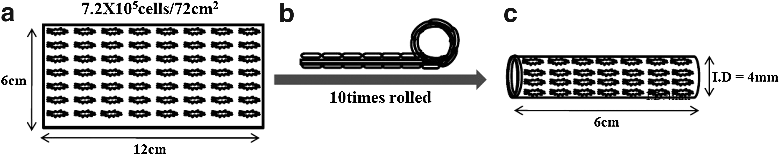
A schematic diagram of the manufacture of small-diameter vascular grafts using cell matrix engineering.
Scanning electron microscopy
The morphology and surface topography of scaffolds and tissue-engineered grafts were observed by scanning electron microscopy (SEM; Hitachi) operated at 15 kV. For the SEM examination, cell-seeded scaffolds were fixed in 4% (v/v) formaldehyde for 24 h and then dehydrated using a graded ethanol series and dried. The specimens were coated with gold using a model IB3 sputter-coater (Eiko).
Tensile properties
A cell-matrix-engineered vascular graft cut from each graft (5 mm×10 mm) was tested for tensile properties. The mechanical properties of the electrospun PLCL fiber sheets were measured by a tensiometry using a model 5567 of instron (TestResources) with a 10-N load cell number at a cross-head speed of 10 mm/min (n=3). Young's modulus, tensile strength, and elongation at break were obtained from the stress–strain curves.
Needle-hole leakage comparison of the vascular grafts
A Gore-TEX vascular graft (W.L. Gore & Associates), an ELSP-PLCL scaffold graft, and 8-week-old rabbit aorta were compared for their self-sealing property. Grafts were tested by an identical system that provided 100 mmHg static water pressure at ambient temperature. A needle puncture was accomplished with a RENAX A.V. Fistula needle set (Sunder Biomedical Tech). Two test cases were chosen: a single puncture and a multiple puncture case. For a multiple puncture case, two punctures were chosen, as it is the minimum puncture. After removal of the needle, leakage through the resultant needle hole was measured for 1 min.
Burst pressure strength
Grafts were mounted in a system custom designed for pressurizing individual grafts to failure. They were pressurized with water, and the pressure was increased by 5 kPa to a maximum of 30 kPa for 5 min and maintained at 30 kPa for 30 min. Then the pressure was gradually increased until the grafts failed.
Cell proliferation and DNA quantification
For the in vitro cell adhesion and proliferation study, cell suspensions (1×10⁴ SMCs/200 μL) were seeded onto ELSP PLCL sheet scaffolds, which were coated with type I collagen (Sigma-Aldrich). Cell proliferation was measured at days 1, 4, and 7 using the WST test (Dojindo Molecular Technologies), which is based on the ability of living cells to reduce a tetrazolium salt into a soluble colored formazan product.
Viable cells in vascular grafts were determined by an AccuPrep genomic DNA extraction kit (Bioneer). Briefly, the scaffolds were freeze-dried for 3 days and 25 mg DNA was collected according to the manufacturer's protocol for DNA quantification. The DNA content was determined by a Nanodrop ND-1000 apparatus (Thermo Fisher Scientific).
Histology
Cell-matrix-engineered vascular grafts were harvested after 1, 2, 3, and 4 weeks of incubation. The grafts were fixed in 4% formalin for 24 h for histologic analysis. The scaffolds were processed in a tissue processor, embedded in paraffin, and sectioned at a thickness of 5 μm. Sections were deparaffinized and stained with hematoxylin and eosin (H&E) to stain the nucleus and cytoplasm. For the immunohistochemical staining, the antibody against smooth muscle α-actin (SMA, 1A4; DAKO) was used to identify SMCs. Also, adhered cells were observed using a model TE2000-U fluorescence microscope (Nikon) by staining with 4,6-diamino-2-phenylindole-dihydrochloride (DAPI; Vector Laboratories).
Statistical analyses
Where applicable, all data are expressed as mean±standard deviation. Student's t-test and single-factor analysis of variance were used for parameter estimation and hypothesis testing, with p<0.05 and p<0.01 considered as being statistically significant (*) and extremely significant (**), respectively.
Results
Scanning electron microscopy
Hexafluoro-2-propanol was used as a solvent to dissolve PLCL. The 9 wt% dissolved solution was electrospun with a pumping rate of 1 mL/h at 15 kV. SEM of representative electrospun PLCL fibers obtained by the cell matrix engineering revealed a small-diameter vascular graft (Fig. 2a). Fiber diameter determined by the image analysis of SEM images (Fig. 2b, c) was 1.05±0.23 μm. Figure 2c shows a cross-sectional SEM image of the vascular graft and the sheet of ELSP with a thickness of 500±40 and 46±4 μm, respectively. SMCs (7.2×10⁵) were seeded on both sides of a scaffold coated by collagen type I. Attachment of SMCs on the scaffolds was confirmed by SEM after 4 weeks (Fig. 2d–f).

Scanning electron microscopic findings of a small-diameter vascular graft using cell matrix engineering.
Tensile properties
Tensile properties were analyzed by three values (i.e., stress at break, strain at break, and Young's modulus; E-modulus). These factors are important for assessing the mechanical properties in the native environment in which blood pressure exists. Small-diameter vascular grafts constructed using cell matrix engineering had similar tensile properties as a rabbit aorta as a native vessel (Fig. 3). The tensile strength values of small-diameter vascular grafts were 1.91±0.56 MPa (0 week), 2.09±0.2 MPa (1 week), 2.38±0.53 MPa (2 weeks), 3.16±0.4 MPa (3 weeks), and 3.23±0.57 MPa (4 weeks) at a strain 135%, 244%, 254%, 263%, and 270%, respectively (Fig. 3a, b). Also, the rabbit aorta and GORE-TEX grafts were analyzed. The tensile strength of the rabbit aorta was 2.61±0.4 MPa at a strain of 86.7% and that of GORE-TEX was 14.03±0.72 MPa at a strain 27.8% (Fig. 3a, b). The E-modulus of PLCL small-diameter vascular grafts was lower than GORE-TEX: 0.85±0.14 MPa (0 week), 0.86±0.2 MPa (1 week), 0.90±0.1 MPa (2 weeks), 0.98±0.2 MPa (3 weeks), and 1.2±0.3 MPa (4 weeks) versus 31.61±4.76 MPa (GORE-TEX), while it was higher than the rabbit aorta, which had an E-modulus value of 0.72±0.1 MPa (Fig. 3c).

Comparison of tensile properties of small-diameter vascular grafts, rabbit aorta, and GORE-TEX (RA: rabbit aorta).
Needle-hole leakage comparison of the vascular grafts
The cell-matrix-engineered vascular graft that was made using the ELSP technology, a GORE-TEX vascular graft, and rabbit aorta were punctured by an A.V. fistula needle at 100 mmHg static water pressure. Leakage of the rabbit aorta was minimal for both puncture conditions: 0.48±0.05 mL/min for one-puncture condition and 1.0±0.03 mL/min for two-puncture condition (Fig. 4). The leakage of the conventional GORE-TEX graft was 32.45±1.8 mL/min for one-puncture condition and 63±9.9 mL/min for the two-puncture condition, while the leakage of the engineered vascular graft was 0.65±0.1 mL/min and 1.18±0.1 mL/min for the one- and two-puncture conditions, respectively.

Needle-hole leakage comparison of a small-diameter vascular graft using cell matrix engineering and GORE-TEX (p<0.01). PLCL, poly(lactide-co-ɛ-caprolactone).
Burst pressure strength
The burst pressure strength test was performed to identify the maximum pressure that the scaffolds could endure before failure, and to determine whether the vascular graft possessed adequate strength to endure the physiologic forces. The burst pressure strength was enhanced with increasing SMC culture time (Fig. 5). The burst pressure strength at weeks 0, 1, 2, 3, and 4 was 604±4, 765±44, 809±44, and 933±22 mmHg, respectively. When the burst strength with a cell-matrix-engineered vascular graft using cell matrix engineering was compared with the GORE-TEX graft, the latter displayed a higher burst strength value at 4 weeks of a small-diameter vascular (1323±383 vs. 933±22 mmHg). The burst strength of the rabbit aorta was the highest value among others at 1647±201 mmHg.
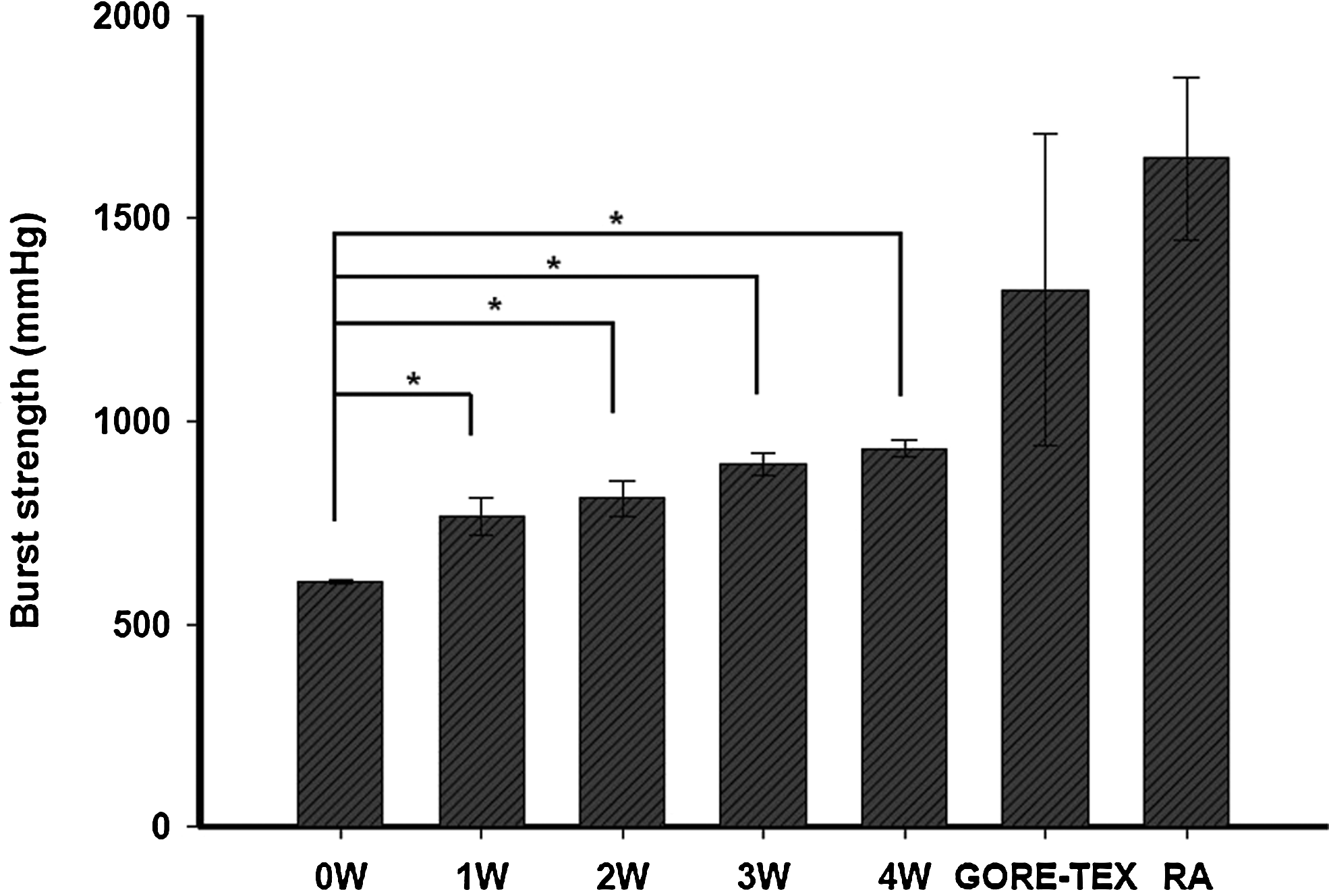
Burst pressure strength of a small-diameter vascular graft using cell matrix engineering and GORE-TEX (*p<0.01).
Cell proliferation and DNA quantification
The cell proliferation for up to 7 days for SMCs cultured on PLCL ELSP scaffolds and tissue cell culture plates (TCPs) is plotted in Figure 6. ELSP scaffolds supported better cell proliferation than TCPs after 3 days. At 7 days, the viability of SMCs on PLCL ELSP scaffold was significantly higher than TCPs. As also seen in Figure 6, the number of viable cells cultured on PLCL ELSP scaffolds was comparable to that cultured on TCPs at 3 and 7 days. The DNA content was measured to determine the growth quantification of SMCs on small-diameter vascular grafts using PLCL cell matrix engineering for up to 4 weeks. As seen from Figure 7, the DNA content increased rapidly at week 1, while only a slight increase was observed after 2 weeks. When the DNA contents were compared with 0 week (1.4±0.1 μg/μL), there were significant differences after 1 week (4.2±0.6, 4.8±0.4, 5.1±0.2, and 5.6±0.3 μg/μL, respectively, at 1, 2, 3, and 4 weeks) (p<0.01).
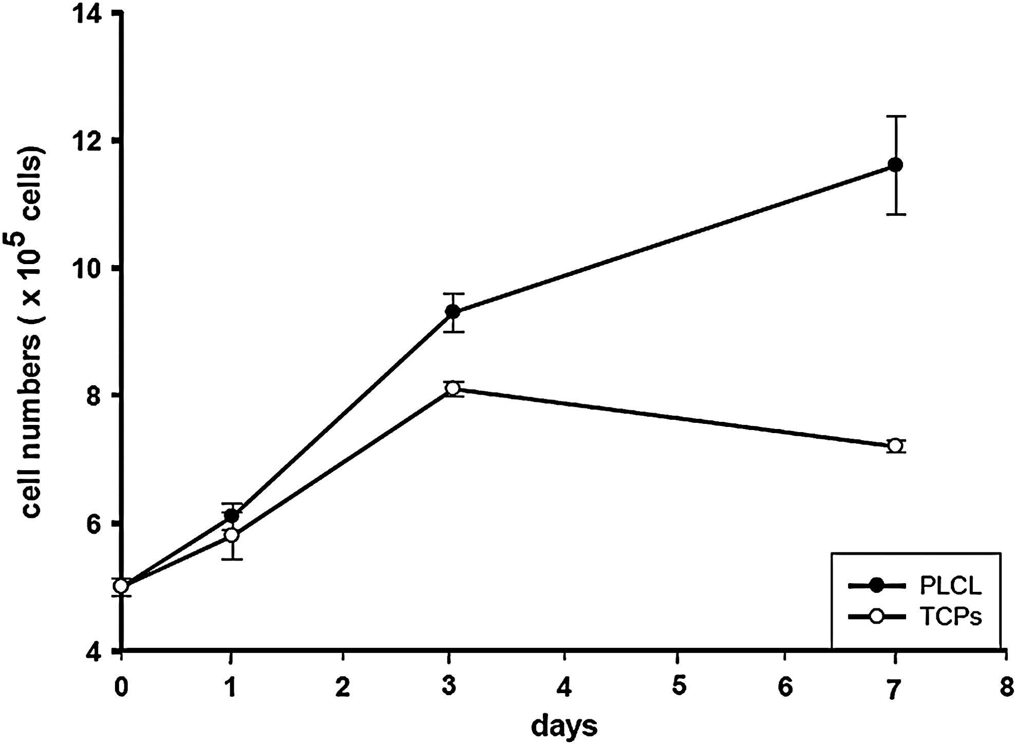
Viability of SMCs cultured on PLCL electrospun scaffold and on TCPS. TCPS, tissue culture polystyrene.

DNA content of SMCs cultured on a small-diameter vascular graft using cell matrix engineering; SMCs were seeded at a density of 1×10⁴ cells/cm2 and cultured for short term (1, 3, and 7 days) (*p<0.01).
Histology
SMC nuclei and cytoplasm within the cell-matrix-engineered vascular grafts were visualized by staining with H&E. SMCs were present on each graft layer (Fig. 8a, b). Immunochemical analysis showed that the formed tissue stained positively for SMA, which indicates SMCs (Fig. 8c, d). As shown in Figure 8c and 8d, SMCs were evident on each layer of the scaffold, indicating SMC regeneration. To examine the distribution of SMCs that adhered to small-diameter, cell-matrix-engineered vascular grafts, DAPI staining was performed. DAPI-stained cells appeared as bright blue spots on both sides of each layer of the vascular grafts (Fig. 8e).
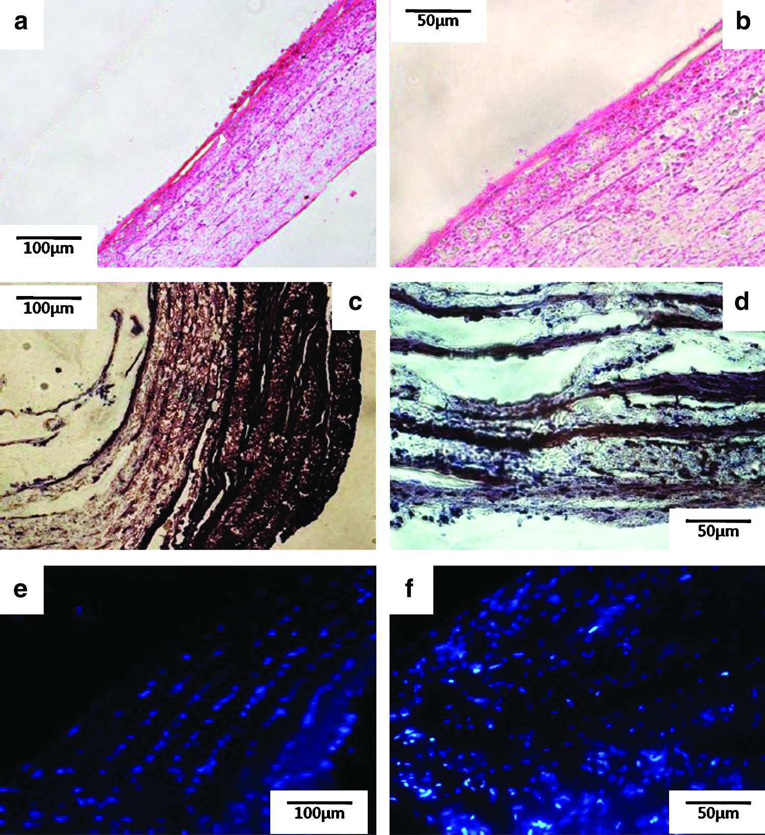
Histological evaluation of a small-diameter vascular graft using cell matrix engineering by H&E staining, SMA, and DAPI staining.
Discussions
Electrospun nanofibrous scaffolds have several advantages, such as an extremely high surface-to-volume ratio, tunable porosity, and malleability to conform to a wide range of sizes and shapes.8,23–25 However, ELSP scaffolds display some limitations as a means of culturing cells. Cells have difficulty infiltrating into the electrospun scaffolds because they usually have nanosized pores.26–29 Also, the cell matrix usually does not have enough mechanical properties to endure blood pressure as the artificial vascular grafts do.30–32 To overcome difficulties associated with the nanostructured scaffold and cell sheet matrix in this study, we investigated the potential of adapting cell matrix engineering based on ELSP scaffold as a fabrication method for vascular scaffolds. In this approach, cell matrix engineering was combined with cell matrix and electrospun scaffold. The 2D electrospun PLCL scaffold could be seeded with more SMCs than the tubular-shaped electrospun vascular grafts, because there are different methods of scaffold preparation. Cells were seeded on both sides of the 2D electrospun scaffold, whose surface area is 144 cm2, compared with the surface area of 15.1 cm2 in cell-seeded area of three-dimensional (3D) electrospun scaffold (common tubular scaffold: inner diameter=4 mm and length=60 mm).
Cell-matrix-engineered small-diameter vascular grafts have been suitably fabricated. The scaffolds have several favorable characteristics for the potential applications for small-diameter vascular grafts, such as 3D-shaped blood-vessel-like structure (Fig. 2a), biomechanical properties, and in vitro biocompatibility. In this study, the small-diameter vascular grafts were manufactured with a length of 6 cm and a thickness of 500±40 μm (Fig. 2c). The surface of a small-diameter vascular graft displayed an ECM-like matrix (Fig. 2b). This could provide a suitable environment to attach and grow SMCs. After 4 weeks, we observed a confluent growth of SMCs on the surface of vascular grafts by SEM (Fig. 2e, f).
The mechanical properties of PLCL cell-matrix-engineered small-diameter vascular grafts are depicted in Figure 3. Comparison of tensile properties between small-diameter vascular grafts, rabbit aorta, and GORE-TEX included tensile strength, tensile strain, and E-modulus. The tensile strength and tensile strain were enhanced with increasing culture time from 0 to 4 weeks in the small-diameter vascular grafts (Fig. 3a, b).
The tensile strength of GORE-TEX was not comparable to the rabbit aorta as a native vessel because the value of GORE-TEX was seven times higher than that of the rabbit aorta. Rather, the tensile strength of small-diameter vascular grafts was comparable to that of the rabbit aorta (Fig. 3a). The tensile strain results echoed those of the tensile strength. GORE-TEX had the lowest tensile strain, indicating that it was stiff in comparison to the rabbit aorta (Fig. 3b). The E-modulus value was closely related to compliance because the inverse of modulus is compliance in elastic materials like polymer.33,34
The E-modulus of small-diameter vascular grafts showed a similar value to that of the rabbit aorta (Fig. 3c). We could expect that the cell-matrix-engineered small-diameter vascular grafts would have similar tensile properties to the rabbit aorta, and this would cause less problems related to a mechanical mismatch to native vessel in vivo.35–37
For some artificial vascular grafts, such as the A.V. Fistula, which is used for dialysis, having a self-sealing property that minimizes blood leakage following piercing is crucial.38–40 Self-sealing property of vascular grafts seems to depend on the graft material elasticity. As seen in Figure 4, the small-diameter vascular grafts showed a superior self-sealing property than the GORE-TEX grafts. This was expected because of the elasticity of PLCL materials.19–22 As shown in Figure 3b, PLCL small-diameter vascular grafts were more elastic than GORE-TEX grafts.
Figure 5 shows that the burst pressure strength increased with SMC culture time. The burst pressure strength at 4 weeks in the PLCL small-diameter vascular scaffold rose to 933±22 mmHg. Although it was less than the burst pressure strength of the GORE-TEX grafts, it could be enough to endure the physiological blood pressure.41–44 The significant improvement of the burst pressure strength of the PLCL small-diameter vascular scaffolds confirmed the analysis of cell proliferation and DNA quantification (Figs. 6 and 7). The burst pressure strength increased about 35%, from 604±4 to 933±22 mmHg, and DNA quantification was risen almost 400%, from 1.4±0.1 to 5.6±0.3 μg/μL, from 0 to 4 weeks (Figs. 5 and 7). The collective data indicate that SMCs on the small-diameter vascular grafts grew and might have conferred glue-like properties between each layer as the culture period increased.
At less than 1 week of culture, the viability of SMCs on the electrospun PLCL scaffolds was better than TCPs (Fig. 6). There are several explanations. In this study, the vascular scaffold was coated by type I collagen to improve cell-seeding efficiency and growth on the scaffold. Collagen, one of the main classes of structural ECM proteins, has excellent cell-binding properties and cell compatibility, which makes it a widely used biomaterial in the tissue engineering field. Previous studies assessed the role of type I collagen, which is the most important component of ECM in terms of amounts, in cell growth, and function. The cell-seeding efficiency was approximately twofold higher in collagen/SMC–incorporated scaffolds than in SMC-seeded scaffolds.19,44,45 Electrospun nanofibers have a structure that is similar to the native ECM, which is composed of nonoscaled protein fibrils, providing SMCs with a more familiar environment to attach and grow on the scaffold.23,24,45–47 H&E staining, SMA, actin, and DAPI staining showed that the SMCs attached and grew on the layers of the small-diameter vascular graft (Fig. 8). This indicates that the electrospun cell matrix scaffold can support long-term cell growth and proliferation.
Conclusion
Nanostructured materials have a tremendous potential for tissue engineering. ELSP technology can be used to generate nanofibrous scaffolds made of synthetic polymers or native matrix molecules.
In this study, we pioneered an approach named cell matrix engineering that combined the electrospun scaffold and the cell matrix. We expected that the viable cell numbers on the ELSP scaffold and mechanical strength properties of the cell matrix could improve by cooperating. Usually, cells have a difficulty infiltrating into the tubular-shaped electrospun vascular grafts because of their nanosized pores. The cell-matrix-engineered blood vascular grafts showed similar mechanical properties as native vessels, such as tensile strength, tensile strain, and E-modulus. Also, they had better self-sealing property than the GORE-TEX grafts because PLCL scaffold has higher elasticity.
We investigated the small-diameter vascular grafts in vitro by seeding SMCs onto the electrospun scaffolds, cultured them, and constructed a 3D vascular structure. We found that the small-diameter vascular grafts constructed using the cell matrix engineering have the potential to be used as artificial vascular grafts in tissue engineering.
Footnotes
Acknowledgment
This study was supported by a grant from the Korea Healthcare Technology R&D Project, Ministry of Health & Welfare (MOHW), Republic of Korea (A110962).
Disclosure Statement
No competing financial interests exist.