
Abstract
While it has been shown that cells respond to topographical cues, most studies of the influence of topography have been restricted to culture substrates with regular, single-scale features, such as grooves. In contrast, in vivo topography is highly complex, irregular, and multiscale. In this work, we demonstrate the use of chemical vapor deposition (CVD) on native tissue to fabricate a precise nonbiological replica of irregular macro-to-microscale biological topography. Specifically, the porcine intestinal basement membrane was decellularized and used as a template to create a silica replica from which tissue was removed to produce a free-standing topographically biomimetic silica film. Preservation of the crypt–villus structure (tens to hundreds of micrometers in scale), which is theorized to influence intestinal cell development and behavior, as well as the porosity of the native tissue membrane (1–5 μM in scale), was demonstrated; however, submicrometer topography appeared to be masked by ball-like structures believed to be a result of the CVD process. CVD process parameters, including reactor pressure and deposition temperature, were explored in efforts to enhance structural and mechanical integrity of the silica replica. A rigid inorganic replica can be used as a template for casting of biocompatible polymeric membranes; thus, this is the first step in fabricating cell culture substrates that precisely mimic their in vivo counterparts in terms of irregular, multiscale topography.
Introduction
These results suggest that culturing cells on biomimetic constructs that replicate complex, irregular in vivo topography as fully as possible may induce a phenotype that resembles that of cells in vivo to a much greater extent than culture on flat, nonmimetic substrates. Intestinal epithelium is an example of a tissue that possesses significant topographical features that could potentially impact cell development and phenotype. The basement membrane of the small intestinal epithelium consists of an intricate invaginated structure with features at many different-length scales. Folds occur at the millimeter-scale to increase the overall surface area. Crypts and villi, structures that are tens to hundreds of micrometers in scale, are located in a close proximity on the surface of these folds. On the surface of the intestinal basement membrane, pores ∼1–5 μm in diameter are found. 15 Finally, at the nanometer scale, a fibrous material in an interwoven pattern provides the ultrastructure of the extracellular matrix (ECM). Culture of intestinal epithelial cells on a biomimetic construct may be useful for a variety of applications such as tissue engineering of intestine for short-bowel syndrome treatment and improved pharmaceutical transport studies.
In terms of replicating the complex structure of the intestinal basement membrane, chemical vapor deposition (CVD) offers the ability to precisely reproduce micro- and nanoscale topography. CVD has a long history in the semiconductor industry as a method for creating thin, conformal coatings on substrates that exhibit a three-dimensional (3D) topography at the micro- and nanoscales.16–18 Many of the biological topographical features of native tissue, and intestinal basement membrane in particular, are micrometers or tens-to-hundreds of micrometers in scale—easily within the scope of CVD process capabilities. CVD is able to provide a conformal coating that is challenging for traditional wet chemical processes, primarily because CVD is a solventless, vapor-phase process, and as such avoids many solvent-related issues, such as wetting, surface tension effects, and fluid flow issues at small-length scales (e.g., capillary flow).
In addition to its extensive use in the semiconductor processing fields, CVD has also been used to deposit biocompatible coatings for a variety of applications, such as coatings for neural probes, cell culture, controlled release of drugs, and biosensors.19–22 Cook et al. demonstrated the replication of the complex micro- and nanoscale structure of a butterfly wing in silica. 23 These conformal silica thin films were deposited using silanes and organosilanes in excess hydrogen peroxide with no need for an external excitation source. This method is attractive as it can occur at near-ambient temperatures (5°C–25°C) and pressures in the Torr range (∼10−3 atmospheres), both of which are benign to a properly prepared intestinal sample.
Here, the use of CVD silica to coat the irregular, complex, multiscale topography of native porcine intestinal basement membrane is described. As the ultimate goal is to develop a rigid mold of tissue from which suitable cell culture materials (e.g., biocompatible polymers) can repeatedly be cast, the tissue is removed, leaving a standalone silica replica of intestinal topography. The limits of replication of topographical scale, as well as sensitivity of the CVD silica properties to process parameters, are presented.
Materials and Methods
Intestinal preparation
Two methods were used to remove the cellular material from the intestinal basement membrane: incubation in ethylenediaminetetraacetic acid (EDTA) and maceration in osmium tetroxide (OsO4). The methods described in the following section were modified from previous reports of these treatments, enabling removal of intestinal cells from basement membrane.24,25
A segment (∼12-cm long, 2.5 cm in diameter) of porcine intestine received from a local abattoir <2 h after slaughter was cut open in the direction of the flow axis, and any bulk material was removed. The tissue was then washed twice with Hank's balanced salt solution (HBSS) and blotted on filter paper to remove mucus. In the first procedure, the cut intestine samples were submersed in 200 mL 0.02% EDTA solution in 0.5 mM Dulbecco's phosphate-buffered saline (PBS) at 37°C in a shaking water bath for 15 min. 24 This procedure was repeated three additional times. The tissue was then rinsed twice with 100 mL HBSS at 37°C and placed into 300 mL 2.5% glutaraldehyde in 0.05 M sodium cacodylate buffer at pH 7.4 and left overnight at 4°C. Samples were then washed with 100 mL HBSS three times, placed in 100 mL 1% OsO4 in PBS for 90 min, rinsed with 100 mL distilled water twice, and dehydrated in a graded ethanol series, as previously described. 25 After the final ethanol wash, samples were critical point dried and then utilized for scanning electron microscopy (SEM) analysis or silica coating. Samples that were imaged via SEM were sputter coated with palladium.
In the second procedure, samples were placed in a mixture of 200 mL 1% glutaraldehyde and 1% paraformaldehyde in 0.1 M PBS (pH 7.3) at 4°C overnight. Samples were then cut into small pieces (∼1 cm2), rinsed in 100 mL PBS three times, and subjected to maceration in 100 mL 0.1% OsO4 buffer in 0.1 M PBS at 20°C for 48 h. Samples were agitated vigorously to remove all cellular material, rinsed three times in 100 mL distilled water, dehydrated in a graded ethanol series, and critical point dried.
CVD of silica
Two different reactors were used in the silica deposition experiments. The first reactor is a custom-built stainless-steel vacuum chamber ∼17 L in volume. Methylsilane (99.9%: Gelest) from a gas cylinder was metered through a series of needle valves. Hydrogen peroxide (30% by weight; Sigma Aldrich) was contained in a temperature-controlled Pyrex® jar and volatilized for flow into the reactor. Peroxide flow was metered through a second needle valve. The two reactant gases mixed in a manifold system before being introduced into the reactor chamber. Methylsilane:peroxide flow ratios were varied between 1:5 and 1:10; reactor pressure was varied between 1800 and 3000 mTorr, and the stage temperature varied between 5°C and 25°C.
The second reactor is a custom-built stainless-steel vacuum chamber ∼3 L in volume. In this setup, the reactants were individually admitted to the reactor directly above the substrate stage, and not premixed in a manifold. The smaller chamber design was chosen, so as to minimize dead zones and wasted volume in the larger chamber, as well as to prevent premature mixing of the reactants. Methylsilane:peroxide flow ratios were again varied between 1:5 and 1:10. The total gas flow rate was varied between 9 to 60 sccm (standard cubic centimeters per minute). Reactor pressure was varied between 1000 and 3000 mTorr, and the stage temperature was again varied between 5°C and 25°C.
Silica film postprocessing
Selected silica films were subjected to a gentle annealing process (3 h at 150°C) in an attempt to further condense the silica network in films that showed evidence of a substantial hydroxyl peak in the Fourier-transform infrared (FTIR).
In addition to the prepared intestinal samples, silica films were also deposited onto 550-μm-thick silicon wafers from Montco Silicon (lot# S4988) for characterization purposes. In both reactors, substrates were placed on the deposition stage, which was maintained via backside cooling controlled by a Polyscience 6000 series chiller. Vacuum was achieved using an Edwards E2M40 rotary vane pump (17 L reactor) or a Leybold D8b (3-L reactor), and chamber pressure was controlled using a butterfly valve connected to an MKS model 252-A exhaust valve controller and an MKS Baratron capacitance manometer. In situ monitoring of the silica thickness was accomplished using a laser interferometric technique, and the films were grown to a thickness of ∼1 μm. Samples collected from reactor 1 are represented in Figures 1, 2a and b, and 3a, while samples collected from reactor 2 are represented in Figures 2c–f, 3b and c, and 4.
Scanning electron microscope image of
Scanning electron microscope images of uncoated
Removal of the intestinal tissue from the silica
Four different solution-based techniques26,27 were explored to remove the biological tissue from the silica coating postdeposition: 5% KOH, 20% NaOH, 2 N NaOH, and household bleach (3%–6% NaOCl). For the removal studies, 1 cm2 of fixed and dried intestinal tissue was placed into 1 mL of each solution and maintained at 60°C.
Variable-angle spectroscopic ellipsometry
Thin film thicknesses were measured using a J.A. Woollam Co., Inc. M-2000 spectroscopic ellipsometer at angles of 65°, 70°, and 75°. The Cauchy equation was used to model the system and determine the thicknesses. 28 Thicknesses were collected in triplicate for each sample, and then averaged to obtain an overall thickness for each deposition.
Fourier-transform infrared spectrometry
The structure of the silica thin films was analyzed using a Perkin Elmer Spectrum GX-2000 FTIR running Spectrum software suite (version 5.3.1). Spectra represent the average of 32 scans between 4000 and 400 cm−1 at a resolution of 4 cm−1. Measurements were done in absorbance mode, and spectra were baseline corrected using Spectrum software, and the thickness was normalized for quantitative comparison.
Energy dispersive X-ray spectroscopy
Surface chemical composition was examined using energy-dispersive X-ray spectroscopy (EDS) with a 15 keV electron beam in situ during SEM imaging.
Results
Intestinal preparation
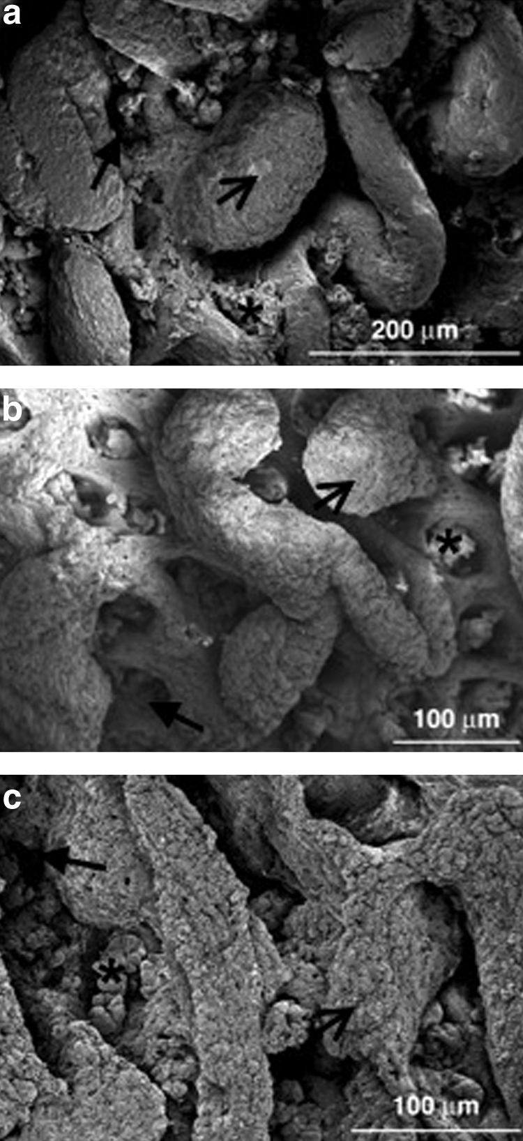
To prepare tissue to withstand low pressure in the CVD chamber, dehydration and aldehyde-based fixation techniques were utilized. As we are interested in the topography of the basement membrane that the cells are resting upon, the ultimate goal in tissue processing is to prepare a fixed, dried sample of intact basement membrane with as much of the bulk waste material, cells, and mucus removed as possible. A piece of intestine was blotted and washed multiple times with EDTA before dehydration and critical point drying. An SEM image of the fixed and dried intestinal sample is shown in Figure 1a. As can be seen, the surface has a complex, multiscale three-dimensional topography that is dominated by the intestinal villi, which are separated by the crypts. Also, some cellular material, and possibly mucus, which was not removed during the initial sample preparation, remains. This is especially evident in the crypts.
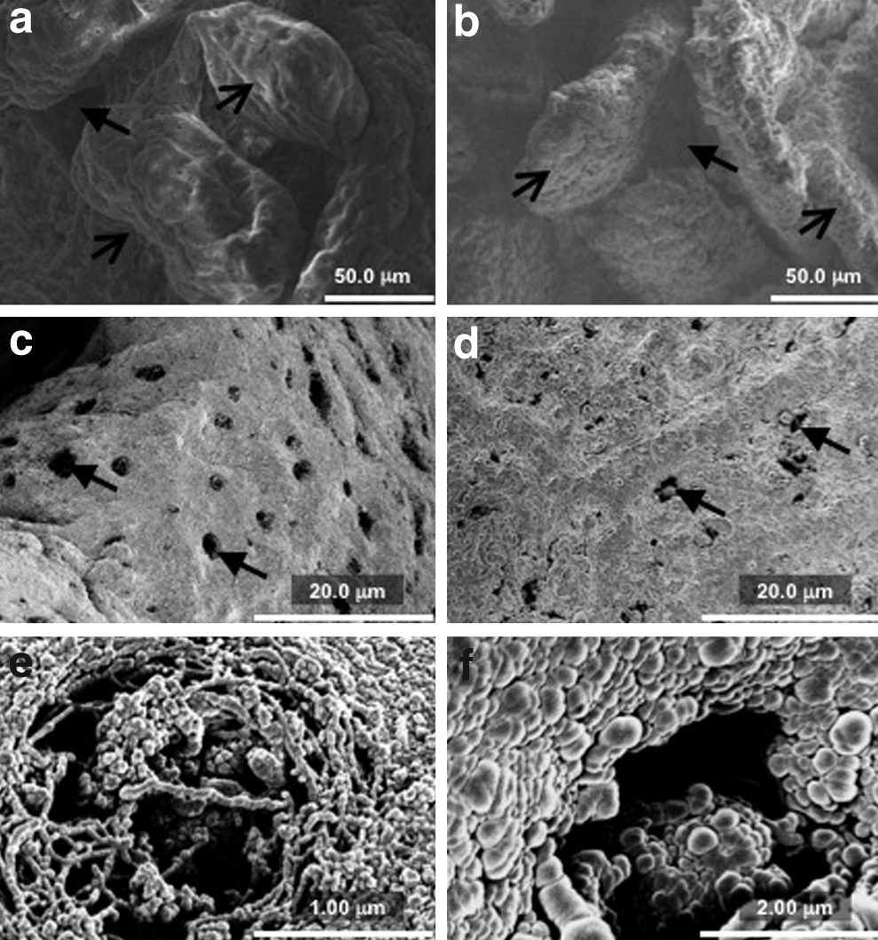
The presence of the cellular debris and likely associated residual epithelial cells was undesirable, as our goal was to replicate the structure of basement membrane upon which cells grow in vivo. Thus, maceration/osmication with OsO425,29 was investigated as an alternative method of removing epithelium and exposing the basement membrane. Figure 2a shows an SEM image of basement membrane that was prepared via this technique. As compared to Figure 1, note the absence of any obvious cellular material on the surface after the osmication treatment.
CVD of silica
To establish CVD process conditions for silica film formation on intestinal tissue, the sensitivity of film formation to process parameters was explored. In the 17-L reactor configuration, methylsilane:hydrogen peroxide flow rate ratios between 1:5 and 1:10 were studied. Total gas flow rates were between 42 and 77 sccm, and the residence time ranged between 29 s and 90 s, depending upon the pressure (1800 or 3000 mTorr, respectively). Table 1 details the results of experiments carried out in the 17-L reactor.
PM, reactant partial pressure; PSAT, reactant saturation pressure at temperature of the substrate.
As the reaction is anticipated to be surface driven, an important parameter to examine is the ratio of the reactant partial pressure (PM) to the saturation pressure at the temperature of the substrate (PSAT). As detailed by Lau and Gleason, at low values of PM/PSAT, the adsorption of the reactant species is limited, and the surface coverage is small. 30 As PM/PSAT trends toward a value of 1, more of the reactant species is adsorbed onto the substrate, until eventually, at a value of 1, the vapor will condense on the surface. For CVD applications, PM/PSAT ratios generally <0.8 are considered desirable, 30 and for codeposition of two species, matching the PM/PSAT values can control the film growth by controlling the relative proportion of the two species on the surface. As a gas at ambient conditions, the PM/PSAT ratio for methylsilane will generally always be low, as it has little incentive to adsorb onto the surface. As a liquid at ambient conditions, the peroxide will have larger, more traditional values of PM/PSAT.
Of the reactions shown in Table 1, two produced conformal silica films, and one produced a powdery coating. We can attempt to explain these results by looking at the relative values of PM/PSAT for methylsilane and peroxide, as this provides an estimate of the relative surface densities of the two reactants. The final column in the table gives the ratio of the PM/PSAT values for peroxide:silane. As can be seen, the deposition regime occurs in a sweet spot in the middle range of this ratio. At either end of the spectrum, the surface concentration of reactants is either too rich or too lean in one of the species to support a reaction. In the mid-range, the surface compositions are adequately matched to allow the reaction to take place and films to form. The 17-L reactor, however, was not optimal for the silica growth studies due to the large volume, creating significant potential for dead zones and recirculation cells, as well as the gas delivery manifold, which premixed the reactants. To mitigate some of these drawbacks, a smaller, 3-L reactor was used to continue the silica deposition studies.
In the 3-L reactor, the methylsilane:hydrogen peroxide ratios between 1:5 and 1:7.5 were examined, with total flow rates between 18 and 60 sccm. This gave residence times of between 11 and 25 s. Major results from the 3-L reactor are presented in Table 2.
In the smaller reactor, we see similar results to the larger reactor in terms of the effect of surface concentration of the reactants. At too low of a peroxide:silane ratio, we see no film formation (in this case, at the low substrate temperature and high PM/PSAT for silane, we actually see condensation), and as the reaction enters the sweet spot, film formation begins to occur. In the small reactor, the limiting high ratio was not observed. From these experiments, it is clear that controlling the relative ratios of the two reactants on the surface of the substrate is key to the formation of a film versus either no deposition or overaggressive deposition resulting in powder formation. The PM/PSAT parameter can be used as a guide to selecting the most desirable deposition conditions as well as precursor selection. Since the ratio can be adjusted by controlling either monomer partial pressure (PM) or the surface saturation pressure (PSAT, as a function of TSTAGE), multiple avenues for optimization are available. Future work should focus on finding a silane or silica precursor more easily matched to the PM/PSAT characteristics of the peroxide.
Once reactor conditions producing viable silica films were established in each reactor configuration, intestinal samples were coated in each chamber. Blank pieces of silicon wafer were placed next to the fixed, dehydrated intestinal samples to provide visual feedback and confirmation of silica growth during the deposition. Figure 1b shows an SEM of an intestinal sample prepared in the same fashion as that in Figure 1a, now coated with ∼1 μm of silica. Note that the cellular debris that was evident in the uncoated sample is still seen in the sample that has been coated with silica. FTIR analysis on the wafer sample is shown in Figure 3a. Spectroscopically, the sample shows a large Si–O absorption in the characteristic location between 1000 and 1200 cm−1, which is indicative of the silica network. Also prominent is a larger –OH absorption in the high wave number region, most likely due to Si–OH bonds created during the deposition step that were not condensed to produce Si–O–Si bonds. This is supported by the presence of the large absorption between 830 and 920 cm−1 that corresponds to Si–OH bonding. 31 A small amount of organic C–H bonding is evident at ∼2800 cm−1, due to retention of the CH3 group in the silane monomer. Also present is Si–H bonding, appearing at 2200 cm−1. The presence of the Si–H bonds, coupled with the large –OH peak, suggests that this silica network is not fully condensed, as we would expect no Si–H bonding and minimal –OH incorporation in the final coating if all Si–H sites were reacted.
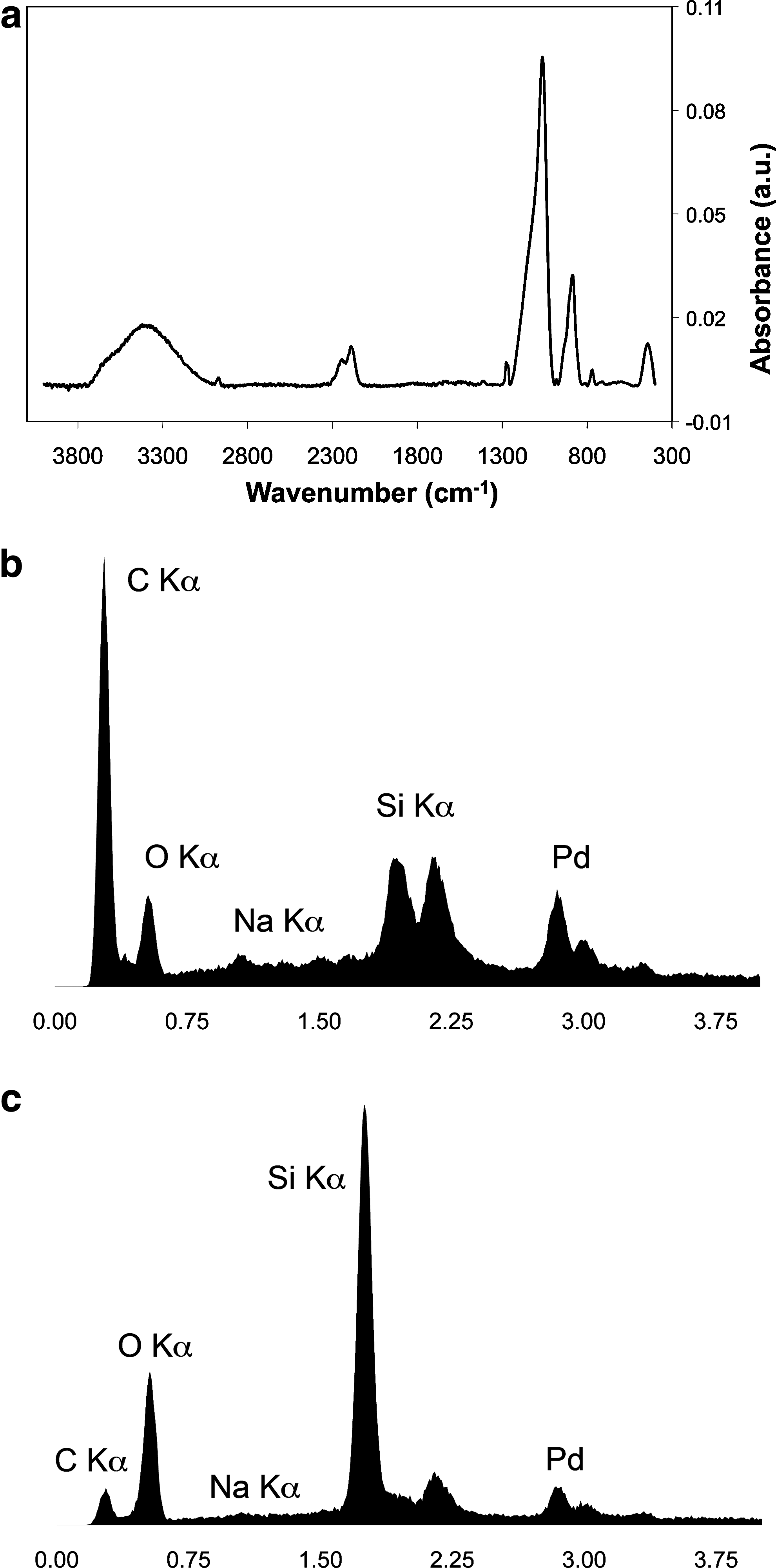
The presence of the silica coating on the biological substrate is confirmed via EDS analysis (Fig. 3b, c). As can be seen, the EDS spectrum of the uncoated intestinal sample is dominated by the carbon peak, indicative of the organic nature of the intestinal sample (Fig. 3b), whereas the coated sample (Fig. 3c) shows a large increase in the Si and O peaks, indicative of the silica network on the sample surface. Further examination shows that the C peak still has a small presence in the EDS spectra of coated samples. It is believed that this can either be attributed to the sampling depth of the electron beam, ∼1 μm, being very similar to the thickness of the silica film deposited, or residual carbon in the silica network from the organosilane precursor.
Multiscale replication of intestinal tissue
Examination of topographical features of uncoated and coated intestinal basement membrane at the macro- and microscale demonstrates the capabilities of CVD to replicate biological features at multiple scales (Fig. 2a–f). The macroscopic folds of intestinal tissue as well as the microscopic crypt–villus topography were clearly conserved during coating with silica (Fig. 2a, b). The pores in the intestinal basement membrane (Fig. 2c, d), which are on the order of 2 μm in diameter, were also successfully replicated. However, examination of the topography at the submicrometer scale reveals a fibrous material that is masked by spherical structures (Fig. 2e, f). These spherical structures appear to result specifically from CVD on a biological material, as silica depositions on silicon wafers resulted in a flat nanoscale surface observed by SEM.
Removal of the intestinal tissue from the silica
After the successful deposition of silica onto the basement membrane substrate, several methods were investigated to remove the biological tissue from the silica coating. Cook et al. used a thermal degradation process for removing a butterfly wing from a silica mold, but at a temperature of 500°C, this resulted in severe deformation and shrinkage of the silica mold itself, with an overall reduction in all dimensions of ∼25%. 23 As this would be unacceptable for our intended application, we investigated tissue dissolution techniques. About 1 cm2 of fixed and dried intestinal tissue was placed in 1 mL of various solutions, including household bleach (3%–6% NaOCl), at 60°C. After 2 h, the sample in the bleach solution had completely dissolved. Samples placed in the 5% KOH and 20% NaOH solutions dissolved after 48 h under soak. The sample placed in 2N NaOH took ∼60 h under soak to completely dissolve. Figure 1c shows an SEM image of the freestanding silica mold after the basement membrane has been removed via the bleach bath method. As can be seen, the complex, 3D topography of the basement membrane has been preserved in the freestanding silica mold, and comparison with Figure 1b shows that the overall dimensional fidelity has been retained. However, this 1-μm-thick silica mold was brittle and delicate, making it difficult to handle while keeping it intact, likely due to thinness, but also possibly to the incomplete condensation of the silica network, which would result in a reduced mechanical strength.
CVD process parameter variation
Additional experiments were conducted in an attempt to fully condense the silica network and improve the integrity of the stand-alone replica. It is hypothesized that a film with more long-chain silica will have greater mechanical stability as well as a more uniform surface topography at the nanoscale.
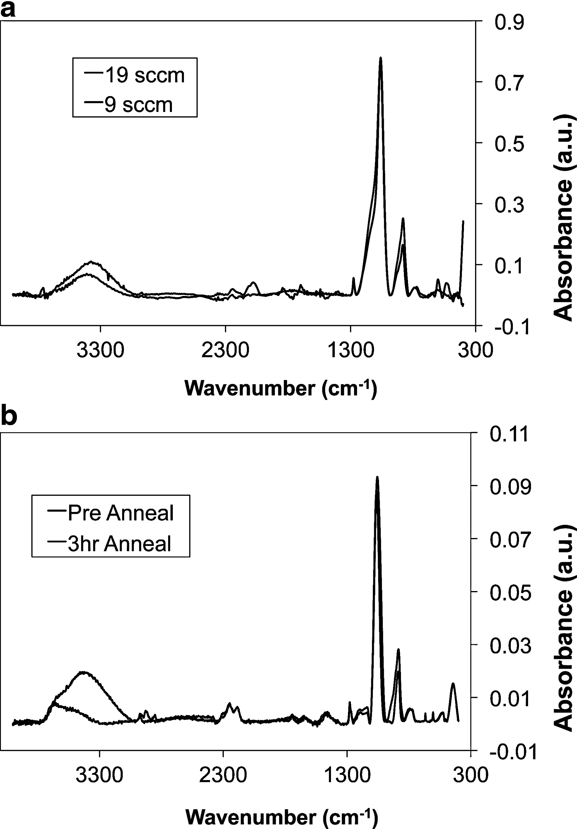
Lower chamber pressure (1000 mTorr), lower reactant ratios (1:4), and lower methylsilane flow rates (1.5 sccm vs. 3 sccm) were explored. Of these changes, only the lower flow rate conditions notably decreased the hydroxyl peak and increased long-chain silica formation observed via FTIR, most notably at a total reactor pressure of 1500 mTorr and reactant ratio of 1:5 (Fig. 4a). This may be related to the longer residence time associated with the lower flow rate, allowing the silane a longer time to react on the surface. SEM analysis confirmed that these process parameter changes did not however translate to a change in the sphere formation during deposition on intestinal tissue, and there also was not a notable improvement in the mechanical integrity of the film, as assessed by simple qualitative observation of film brittleness and mechanical integrity upon handling, though this is not a quantitative assessment.
Fourier-transform infrared spectrum of
Lastly, a gentle postdeposition anneal was examined to determine whether it was possible to drive Si–OH condensation. FTIR analysis (Fig. 4b) confirmed that the hydroxyl peak intensity significantly decreased after a 3-h anneal at 150°C. Future work will investigate how, if at all, the annealing process and associated change in hydroxyl content translate to film structural changes and potentially improved mechanical robustness.
Discussion
In this work, it was demonstrated that CVD on native tissue provides a unique methodology for replicating the complex, irregular structures of the intestinal basement membrane at multiple scales, ranging to the microscale, in a nonbiological material. The significance of structure and geometry to cell behavior, including differentiation, migration, and proliferation, has been demonstrated, motivating development of biomaterials that precisely recreate structural cues presented in vivo. Lithographic microfabrication techniques have been adapted to develop substrates with a wide array of regular features, including ridges and channels,4,32,33 microwells, 34 and microposts. 35 Advances in other microfabrication techniques 36 have produced complex, but regular, features, including materials with specific pore geometries and orientations. 37 Another approach to fabrication of a biomaterial with native ECM structure as well as chemistry has been to decellularize native tissue.38–41 For example, decellularized natural nerve ECM materials were immunologically tolerated and promoted nerve regeneration to an extent similar to current autograft techniques in an animal model of peripheral nerve damage. 42 However, this approach produces materials of somewhat undefined chemical composition with a high potential for preparation-to-preparation variability. Nonbiological and especially polymeric materials offer the ability to fine-tune chemical composition to induce specific cellular response, for example, through functionalization with specific adhesion peptides. The silica mold described could ultimately be used as a nonbiological template for repeated casting or deposition of topographically biomimetic polymeric cell culture membranes.
It is important to note that the OsO4 treatment, while appearing to efficiently remove epithelium, as previously noted,25,29 may also alter the micro- and nanostructural features of the intestinal basement membrane. Treatment with the tissue fixative glutaraldehyde and likely also OsO4, which is also used as a fixative, but can extract cellular components, aids in preserving matrix structure. While micrometer-scale pores and submicrometer-scale fibers, characteristic features of intestinal basement membrane, have been preserved (Fig. 2c, e), future studies should examine potential effects of osmication treatment parameters (e.g., concentration and duration) on ultrastructural features.
While it was clearly observed that the villi and crypt structures as well as the pore structure of the basement membrane are maintained in the silica mold, it was found that sphere-like structures that formed on the surface during silica deposition tended to mask the fibrous material comprising the basement membrane. As the ultimate goal is to use the nonbiological replica as a template for casting biocompatible polymers, it is desired to preserve biological topography to the greatest extent possible. It remains to be seen what affect the masking of these structures will have on the overall culture system.
The fact that sphere-like structures were observed on silica deposited on tissue, but not on a silicon wafer, indicates that their formation may be related to biological chemistry and/or topography. This is supported by recent observation in our laboratory of sphere formation during a CVD polymer deposition process on intestinal basement membrane. It is unknown whether the sphere formation is due to the chemical or topographical nature of the biological substrate, but it is hypothesized that it may be related to the nucleation mechanism of film growth. Nucleation and grain growth in CVD are known to be influenced by surface chemistry and topography.16,43 While the process parameter variations tested did not prevent sphere formation, they did demonstrate the sensitivity of the process to reactor conditions, and support further investigation into the potential influence of process parameters on film chemistry and structure. It is also possible that a postdeposition anneal may be effective. A high temperature (500°C) was previously used to remove biological material from silica deposited on a butterfly wing. 23 This process was also reported to reduce overall dimensions of the silica by 25%. Perhaps, the annealing process can enable merging of structural domains formed during the deposition. However, an alternative approach for overcoming potential limitations imposed by spherical structure formation that would avoid dimension change possibly imposed by the annealing process would be to explore the use of the reverse mold (the surface of deposited material initially in contact with tissue) as a template for creating biocompatible polymer membranes with precisely biomimetic topography.
Conclusions
A single-step CVD process at moderate temperature and pressure has been used to deposit conformal silica films on a fixed and dehydrated biological material. The irregular, multiscale complex topographical features of intestinal basement membrane have been replicated to the microscale in silica. The porous structure of intestinal basement membrane, representing features on the order of 1–5 μm in scale, was replicated, but fibrous structures below 1 μm in scale were masked. Upon removal of biological material from the silica, a stand-alone replica of the intricate, irregular, multiscale structure of the basement membrane was produced. Creation of this reusable nonbiological mold is the first step in deposition of biocompatible polymer membranes with precisely biomimetic topography, which could be highly useful in cell culture and tissue-engineering applications.
Footnotes
Disclosure Statement
No competing financial interests exist.