
Abstract
Clinical translation of bone tissue engineering approaches for fracture repair has been hampered by inadequate vascularization required for maintaining cell survival, skeletal regeneration, and remodeling. The potential of vasculature formation within tissue-engineered grafts depends on various factors, including an appropriate choice of scaffold and its microarchitectural design for the support of tissue ingrowth and vessel infiltration, vasculogenic potential of cell types and mechanostimulation on cells to enhance cytokine expression. Here, we demonstrated the effect of biomechanical stimulation on vasculogenic and bone-forming capacity of umbilical-cord-blood endothelial progenitor cells (UCB-EPC) and human fetal bone marrow-derived mesenchymal stem cell (hfMSC) seeded within macroporous scaffolds and cocultured dynamically in a biaxial bioreactor. Dynamically cultured EPC/hfMSC constructs generated greater mineralization and calcium deposition consistently over 14 days of culture (1.7-fold on day 14; p<0.05). However, in vitro vessel formation was not observed as compared to an extensive EPC-vessel network formed under static culture on day 7. Subsequent subcutaneous implantations in NOD/SCID mice showed 1.4-fold higher human:mouse cell chimerism (p<0.001), with a more even cellular distribution throughout the dynamically cultured scaffolds. In addition, there was earlier evidence of vessel infiltration into the scaffold and a trend toward increased ectopic bone formation, suggesting improved efficacy and cellular survival through early vascularization upon biomechanical stimulation. The integrative use of bioreactor culture systems with macroporous scaffolds and cocultured osteogenic and vasculogenic cells promotes maturation of EPC/hfMSC-scaffold grafts necessary for vascularized bone tissue engineering applications.
Introduction
To approach this clinical challenge, several strategies have been studied, such as the use of porous scaffolds, growth factors, and highly potent stem cells with vasculogenic potential.8,9 Another approach rapidly gaining traction is in the generation of prevascularization, often through the use of both vasculogenic and osteogenic cell cocultures seeded within the scaffolds. While this strategy has achieved the formation of extensive prevascularized networks,10–12 few have demonstrated enhanced bone formation,13,14 which may be due to a lack of appropriate stimulus for rapid development of new blood vessels into these coculture grafts. This may be due to the inefficiencies of static culture conditions in generating such a graft. To date, only one group has reported the use of a dynamic environment for coculture systems in BTE, where dynamically cocultured scaffolds were compared to their monocultures rather than statically cultured cocultures. 15 Also, the effects of enhanced mass transport and biomechanical forces on cocultures of vasculogenic and osteogenic cell types have not been defined.
We have previously demonstrated the effects of efficient dynamic culture of human fetal bone marrow-derived mesenchymal stem cells (hfMSC) using a biaxial bioreactor, with improvements in cellular survival and osteogenic differentiation, and its efficacy for bone repair.8,16,17 In this work, hfMSC and umbilical-cord-blood endothelial progenitor cells (UCB-EPC) have been chosen for their robust osteogenic 18 and angiogenic 19 potential, respectively. We hypothesized that the application of fluid shear stresses will enhance vasculogenesis of EPC and improve osteogenic differentiation of hfMSC in a coculture system supported by a three-dimensional (3D) honeycombed-like polycaprolactone tricalcium-phosphate (PCL/TCP) scaffold. We envision that the utility of EPC/hfMSC stem cells in coculture, further stimulated by biomechanical forces using a biaxial bioreactor culture, will be an advantageous approach over other existing coculture systems.
Materials and Methods
Samples, animals, and ethics
Human tissue collection for research purposes was approved by the Domain Specific Review Board of National Healthcare Group (DSRB-D-06-154), in compliance with international guidelines regarding the use of fetal tissue for research as previously described. 8 In all cases, patients gave separate written consent for the use of the collected tissue. Fetal femurs were collected for isolation of hfMSC after clinically indicated termination of pregnancy. In this study, a sample derived from a 17+3 weeks+days gestation was used. Human UCB samples from newborns were collected from normal full-term deliveries.
Male NOD SCID mice (6–8 weeks old) were acquired from the Animal Resources Centre. All procedures were approved by the Institutional Animal Care and Use Committee (IACUC) at the National University of Singapore, and procedures performed in AAALAC accredited animal facilities.
Isolation, culture, and characterization of hfMSC and EPC
hfMSC were isolated from bone marrow as previously described. 18 Briefly, single-cell suspensions were prepared by flushing the bone marrow cells from femurs using a 22-gauge needle, passed through a 70-μm cell strainer (BD Falcon) and plated on 10-cm plates (NUNC) at 106 cells/mL. Adherent spindle-shaped cells were recovered from the primary culture after 4 to 7 days. Nonadherent cells were removed with initial media changes every 2–3 days. At subconfluence, they were trypsinized and replated at 104 cells/cm2. MSCs were cultured in the Dulbecco's modified Eagle's media-Glutamax (GIBCO) supplemented with 10% fetal bovine serum (FBS), 50 U/mL penicillin, and streptomycin (GIBCO), thereafter referred to as D10 media. Cells from passage 2–4 were characterized as previously described.8,20
UCB-derived EPC were obtained as previously described. 21 Briefly, blood was diluted and overlaid onto Ficoll-Paque PLUS (Amersham). Mononuclear cells from the buffy coat retrieved from UCB by low-density centrifugation were resuspended in endothelial growth media (EGM; Cambrex) supplemented with 10% FBS, thereafter referred to as EGM10, and then plated onto 100-mm tissue culture dishes coated with type I rat tail collagen (BD Biosciences) at 37°C, 5% CO2 in a humidified incubator. Culture media was changed every 3 days subsequently. Typical cobblestone colonies appeared after 2 weeks, and were subcultured at subconfluence. Their endothelial differentiation on Matrigel and immunophenotypic characteristics were demonstrated by its expression of DiI acetylated low density lipoprotein (acLDL) uptake (Dako), CD31 (Millipore), the von Willebrand Factor (vWF) (Millipore), and VE Cadherin (Enzo Life Sciences) (Fig. 1).
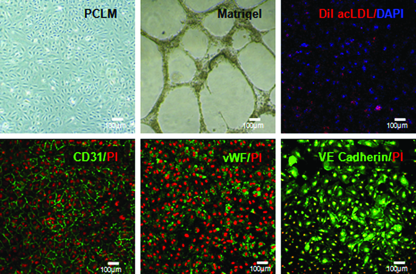
Characterization of umbilical-cord-blood endothelial progenitor cells (UCB-EPC) using phase-contrast light microscopy (pCLM) showed the emergence of cobblestony colonies, which differentiated and formed tube-like networks on Matrigel. Immunophenotype of EPC were demonstrated by its expression of CD31, vWF, VE Cadherin, and acLDL uptake, with cell nuclei counterstained with propidium iodide (PI) and 4′,6-diamidino-2-phenylindole (DAPI) accordingly. Color images available online at www.liebertpub.com/tea
Lentiviral transduction of EPC and hfMSC
The lentiviral vectors were produced as described previously. 20 Briefly, the transfer plasmids (pHIV-dTomato and pRRLSIN.cPPT.PGK-GFP.WPRE) were cotransfected with pHIV-Luciferase and pCMV.ΔR8.74 into HEK293T cells. The supernatant was collected at 48 and 72 h following transfection and concentrated by one round of ultracentrifugation at 50,000 g for 2 h, and the final pellet was dissolved in a small volume of 1% bovine serum albumin in phosphate-buffered saline (PBS) (1/100 of starting volume). The number of transducing units (TUs) of the vectors was determined by infecting 100,000 293T cells using serial dilution of the vector. The dilution resulting in<30% GFP- or RFP-positive cells was used to calculate the number of TUs per mL. For transduction, cells were seeded at 0.5×10⁴ cells/cm2 in T-25 flasks, and exposed to lentivirus with 4 mg/mL polybrene at multiplicity of infection of 5. GFP-labeled EPC and RFP-labeled hfMSC at>90% transduction efficiencies were used in the experiments.
Other media preparation
Bone media
Well-defined bone media (BM) was prepared using the D10 medium supplemented with bone inducing elements—10 mM β-glycerophosphate, 10−8 M Dexamethasone, and 0.2 mM Ascorbic acid (Sigma Aldrich).
Osteogenic assays
Calcium deposition was assayed as previously described. 8 Briefly, each sample well (n=3) was incubated with 0.5 N acetic acid overnight to dissolve calcium. A colorimetric calcium assay kit (BioAssay Systems) was used to quantitate the calcium content spectrophotometrically at 612 nm according to the manufacturer's instruction. Calcium from the PCL/TCP scaffolds was leached out from empty scaffolds and subtracted off from the measured calcium levels.
Alkaline phosphatase (ALP) was assayed as previously described. 8 Briefly, samples (n=3) were gently rinsed twice with PBS, and incubated in 1 mg/mL of Collagenase Type 1 (Sigma Aldrich) dissolved in a 0.1% Trypsin (Invitrogen) solution and incubated at 37°C for 2 h to digest the extracellular matrix completely. After three cycles of freeze–thaw, the cell lysate solution was assayed for ALP activity using the SensoLyte™ pNPP Alkaline Phosphatase Assay Kit (AnaSpec) following the manufacturer's instruction.
The Von Kossa staining was performed as previously described. 8 Briefly, samples were gently rinsed twice with PBS, then fixed with 10% formalin for 1 h, followed by two washes with distilled water, and stained with freshly made 2% Silver nitrate (Sigma Aldrich) in distilled water (w/v) for 10 min in the dark and exposed to sunlight for 30 min.
Preparation of cellular–scaffold constructs
Scaffold fabrication and treatment
Macroporous PCL-TCP (80/20) scaffolds (Osteopore International) with a lay-down pattern of 0/60/120°, porosity of 70% were fabricated using a fused deposition modeling technique under the cGMP environment (ISO 13485) (Fig. 2A, B). Scaffolds were cut into 4X4X4mm dimensions and treated with 5 M NaOH for 3 h to enhance hydrophilicity, washed thoroughly with PBS thrice before disinfection by immersion in 100% ethanol overnight.

Microarchitectural design of polycaprolactone tricalcium phosphate (PCL/TCP) scaffolds and the design of the biaxial bioreactor setup.
Cell loading and culture
Cells were suspended in Tisseel Fibrin Sealant (Baxter) before seeding into the porous scaffolds, keeping the EPC cell density constant at 3000 cells per mm3 of empty scaffold space in both study groups. EPC/hfMSC cocultures were used in a 1:1 ratio as optimized in our previous study. 22 The seeded cells were left to adhere to the scaffolds for 2 h under static conditions before transfer to sterile 24-well plates for static culture or a biaxial bioreactor culture for 14 days in BM culture.
Bioreactor setup
The bioreactor setup is as previously described in Zhang et al.16,17 Briefly, the biaxial bioreactor consists of a spherical culture chamber in which the cellular scaffolds are anchored to the cap of the bioreactor by pins, a medium reservoir and a perfusion system, which connects the culture chamber and the medium reservoir. The spherical culture chamber is designed to rotate in two perpendicular axes (Y and Z, pink block arrows) simultaneously and perfused with media flow circulating between the culture chamber and the medium reservoir (Fig. 2C, D).
In vivo transplantation and assays
Under general anesthesia, acellular, and cellular–scaffold constructs were implanted into subcutaneous pockets generated at the dorsal surface of each immunodeficient mice, and the skin closed with 5-O Vicryl sutures. Scaffolds were harvested at weeks 4 and 8 for analysis.
Imaging
Phase-contrast light microscopy
Cellular morphology, adhesion, and extracellular matrix production were examined daily and imaged at fixed time intervals using an attached camera unit.
Confocal microscope
Cryosections of harvested cellular–scaffold constructs and 3D cultures were viewed and scanned under a confocal laser microscope using laser wavelengths of 405 nm (green), 594 nm (red) (Olympus, FV300 Fluoview). Image Analysis software Imaris (Bitplane) were used for the quantification Lamins A/C staining of 10 different regions of the cryosections as well as for image analysis.
Scanning electron microscope
Glutaldehyde-fixed scaffolds were then dehydrated, air-dried, and gold sputtered with the SCD 005 gold sputter machine (Bal-Tec) for 70 s at 30 mA under high vacuum before imaging on the scanning electron microscope (SEM) (JSM 5660; JEOL).
Microcomputed tomography
The harvested cellular–scaffold constructs were fixed in 10% formalin for 1 week before analysis of bone or vasculature network using the microcomputed tomography (Micro-CT) scanner (SMX-100CT X-ray CT Sys; Shimadzu). The scan settings were X-ray voltage=45 kV, X-ray current=100 μA, detector size=9, scaling coefficient=50, and voxel resolution=0.008 mm. The scans were then reconstructed using VGStudioMax (Version 1.2; Volume Graphics) to create the 3D geometry and for quantitative histomorphometric analysis and calibrated against its background.
Vascularization assay
Micro-CT evaluation of vascularization was evaluated with the use of a blue vascular contrast, Microfil (MV-120; Flow Tech). Briefly, the mice were anesthetized, laid supine, and a midline incision extending across the thorax and abdomen was made. The left ventricle was cannulated and a perforation was made in the right atrial appendage before perfusion with heparinized saline. Upon complete exsangination, 10 mL of the microfil solution diluted in the MV-diluent (1:1) along with the MV-curing agent (10% of total volume) was then perfused via a syringe. The mice were then stored at 4°C for 2 h for the silicon compound to polymerize before the scaffolds were harvested and fixed in 4% paraformaldehyde for a week before Micro-CT imaging.8,23
Histology
Cellular–scaffold constructs were placed on crushed dry ice for slow freezing and sectioned at 10 μm thickness transversely with a Cryostat (CM 3050S; Leica). Sections were stained with Masson's Trichrome (MT) to visualize tissue morphology and evidence of new bone formation. Capillary density was quantitated by evaluating the entire area of histological sections. Luminal structures perfused with red blood cells were identified as capillaries. The capillary density was calculated by dividing the total number of red blood cell-filled capillaries by the area of each section (vessels/mm2).
Immunohistochemistry
Human mouse chimerism
Lamins A/C immunostaining was used to investigate chimerism of human cells in murine tissue. Cryosections from each sample were blocked with 5% normal goat serum for 1 h, and left to react with a monoclonal mouse anti-human Lamins A/C antibody (1:100; Vectorlabs) overnight; sections were then incubated with goat anti-mouse secondary antibodies (1:100 Alexaflour 488; Invitrogen) for 1 h, and counterstained with propidium iodide. Images were visualized through confocal microscopy as above.
Osteopontin staining
The contribution of human cells to in vivo bone regeneration was delineated with human-specific osteopontin staining (1:100; Abcam) as previously described. 8
Statistical analysis
Parametric data are presented as mean±standard deviation and were analyzed by the t-test or analysis of variance. *p<0.05, **p<0.01, ***p<0.001 indicate statistically significant differences between samples (n=3).
Results
Characterization of UCB-EPC
UCB-EPC emerged as colonies with a cobblestone morphology, which differentiated and formed tube-like networks on Matrigel. EPC expressed endothelial markers, such as CD31, vWF, VE Cadherin, and scavenged acLDL (Fig. 1). hfMSC had a spindle morphology, which were negative for hemopoietic and endothelial markers (CD14, CD34, and CD45) and were positive for CD73, CD105, and CD90. 22
Dynamic culture of EPC/hfMSC constructs led to greater mineralization in vitro, but did not exhibit vessel formation as compared to static culture
We explored the effects of introducing biomechanical forces with a biaxial bioreactor (Fig. 2C, D) on EPC/hfMSC cocultured scaffolds for 2 weeks before vascularization and osteogenic differentiation assays (Fig. 3A). Phase-contrast light microscopy showed enhanced mineralization within the pores of dynamically cultured scaffolds (Fig. 3B), with more prominent trabecular-like networks of extracellular matrix formation on day 14 (SEM; Fig. 3B). Quantitatively, 1.7-fold more calcium was deposited in dynamically cultured over statically cultured scaffolds on day 14 (p<0.05, Fig. 3C). Both treatment groups also maintained high degrees of cell viability over 14 days of culture as seen by the continuous expression of both fluorescent proteins (Fig. 3D). In terms of its vessel-forming ability, dynamic bioreactor culture of EPC/hfMSC was unable to induce any in vitro vessel formation over all 14 days. However, it was noted that EPC were able to form vessel-like networks upon coculture with hfMSC under static conditions in the 3D microenvironment on day 7
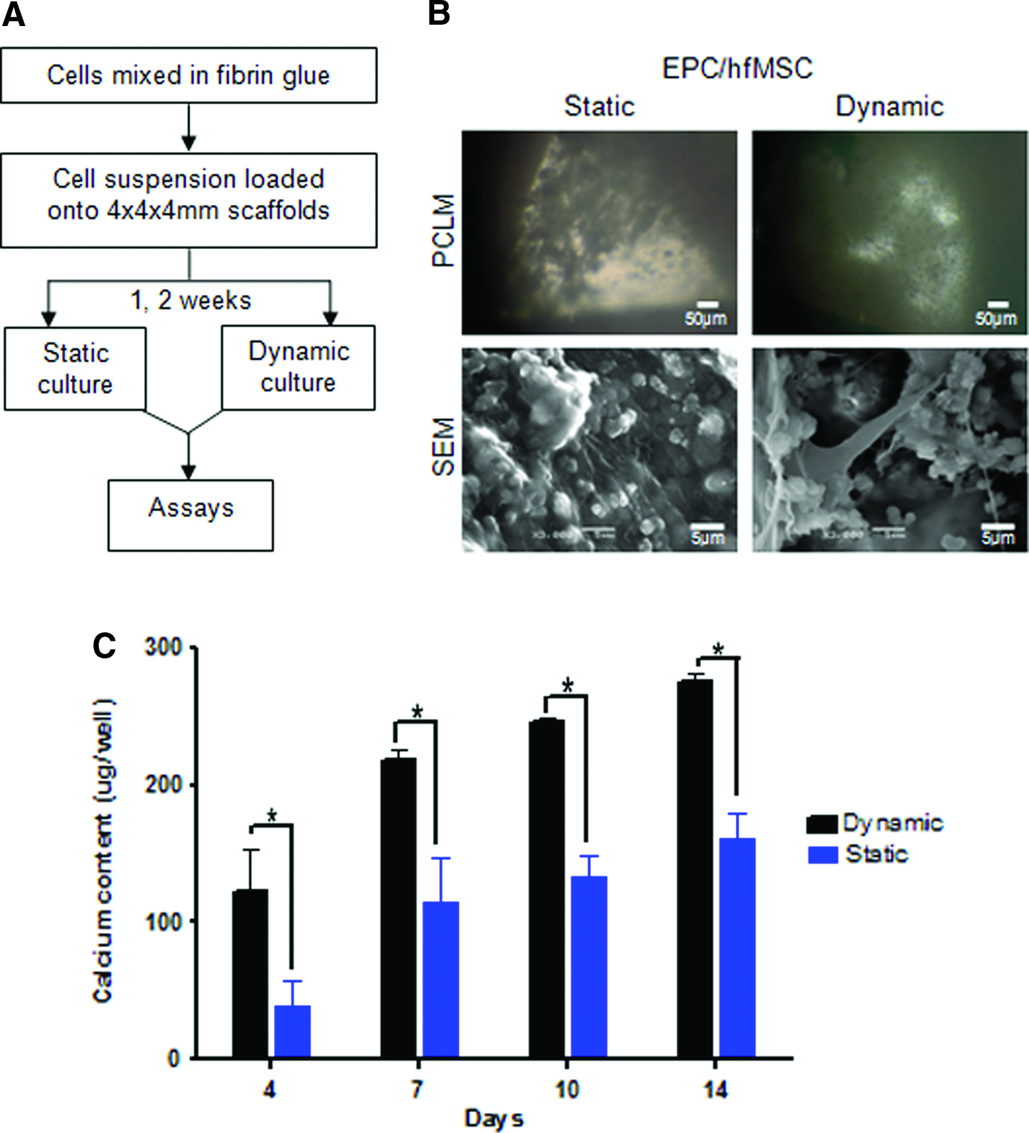
Dynamic bioreactor culture of EPC/hfMSC constructs induced greater mineralization, but did not exhibit vessel-forming ability in vitro as compared to static culture.
Dynamically cultured constructs exhibit earlier vasculogenesis and enhanced ectopic bone formation in vivo compared to statically cultured constructs
Next, we implanted all three scaffold groups (dynamic, static, and acellular) subcutaneously into immunodeficient mice to test their in vivo vascularization and osteogenic potential (Fig. 4A). Histological sections of MT staining exhibited larger regions of premineralizing collagen in dynamically cultured scaffolds on week 4. Capillaries, which can be seen as luminal structures filled with red blood cells, were most abundant in the dynamically cultured scaffolds, being 1.2- and 2.3-fold more numerous than the static and acellular controls, respectively (p>0.05; p<0.01 respectively; Fig. 4B). Cross-sectional analyses of contrast-perfused scaffolds on Micro-CT further revealed an extensive network of fine capillary structures in the central pores of dynamically cultured scaffolds as compared to fewer vessel occurrences in the static and acellular–scaffolds 8 weeks postimplantation (Fig. 4C).
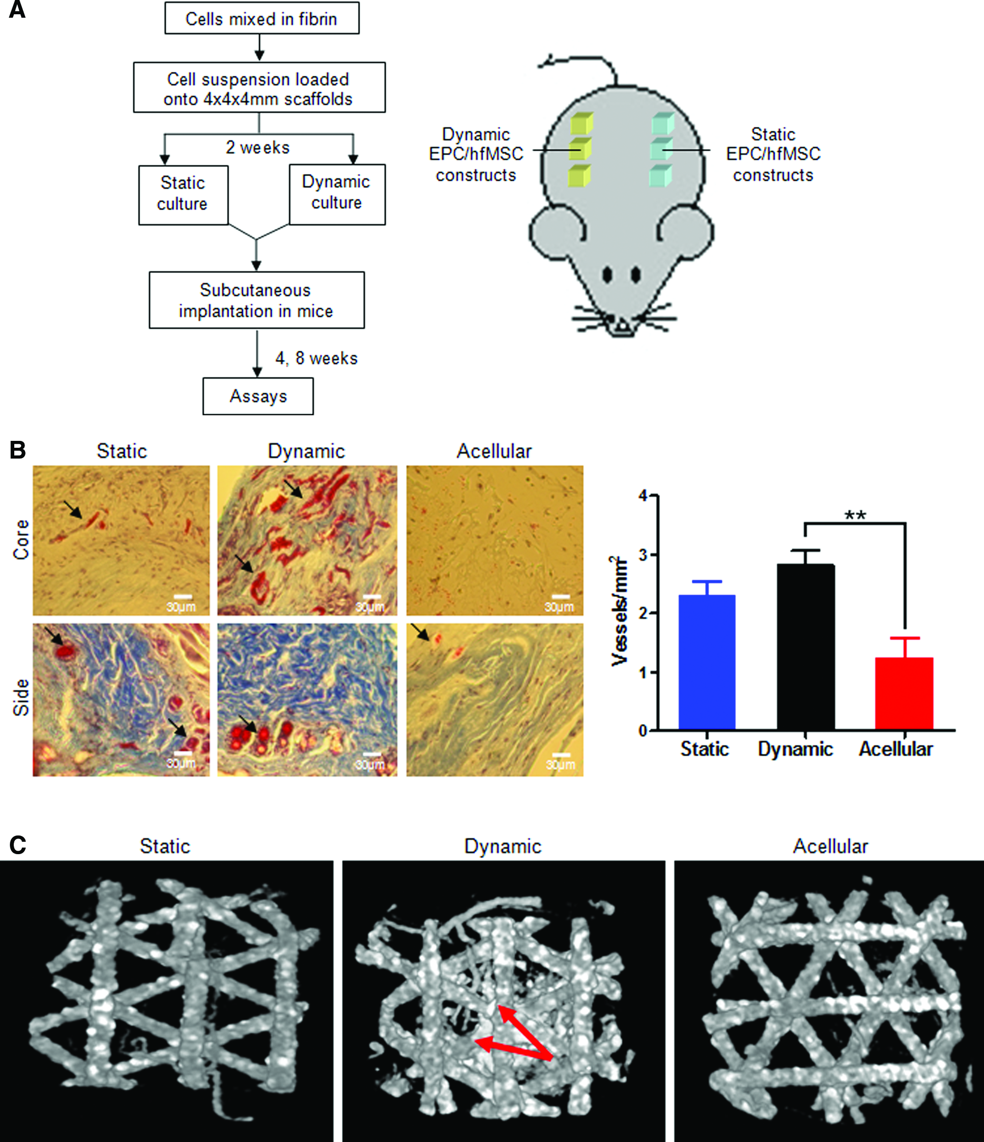
Dynamic bioreactor culture induced greater vascularization, new bone formation, and maintain higher cellular viability of EPC/hfMSC constructs in vivo compared to static culture.
Dynamically cultured scaffolds exhibited 1.4-fold higher human:mouse chimerism, which showed a more homogenous cellular distribution throughout the scaffold as compared to the statically cultured constructs, which had higher numbers of murine cells at the peripheral parts of the scaffold edges 8 weeks postimplantation (54.6%±15.7% vs. 77.7%±4.5%; p<0.001, Fig. 4D). Dynamically cultured EPC/hfMSC scaffolds also laid down more human osteopontin at the sides of the scaffolds, with larger areas of Von Kossa staining compared with statically cultured scaffolds (week 8, Fig. 4E).
Discussion
The lack of angiogenesis in large bone grafts remains a key challenge for clinical translation for BTE, with several strategies being undertaken to prevascularize osteogenic constructs before site implantation 24 for accelerating fracture repair. Despite the growing interest in the use of coculture systems for in vitro prevascularization in BTE in the recent years,10–15,24,25 and the markedly improved performance of dynamically cultured MSC-based tissue-engineered bone grafts,17,26,27 a dynamic culture system has not been explored for optimization of coculture systems for vascularized BTE. In this study, we leveraged the osteogenic and vasculogenic potency of primitive cell sources, namely, hfMSC and EPC, respectively, cultured within a 3D dynamic system to mechanically stimulate and prime the cellular constructs. We observed improved mineralization of scaffolds in vitro, the laying down of more human osteopontin and formation of more compact bone in vivo in dynamically cultured scaffolds compared to statically cultured scaffolds. In addition, dynamically cultured scaffolds resulted in improved vascularization and a significantly higher donor cell survival postimplantation in vivo.
Currently, various groups have utilized the approach of in vitro prevascularization through a coculture of endothelial lineages and osteoblast-like cell types. Some have demonstrated enhanced neovascularization with the formation of highly organized prevascular structures upon coculture in vitro, which anastomosized and integrated with host blood vessels in vivo as compared to the use of singular cell sources.12,22,25 Only a few groups have reported an accompanied improvement in bone formation.13,14,22 Usami et al. showed a 1.6-fold increase in capillary density and a slight increase by 1.3-fold in bone formation in canine-EPC/MSC compared to MSC alone 14 ; Orthotopic implantation of the EPC/MSC construct led to earlier vascularization and significantly more bone formation in a critical-sized femoral defect rat model, with higher ultimate loading than the MSC-alone group. 13 In contrast, Koob et al. showed no improvement in bone formation upon coculture of human umbilical vein endothelial cells (HUVEC) with MSC, despite a higher neovessel formation in HUVEC/MSC spheroids in a critical-sized mouse calvarial defect, 28 while Geuze et al. showed that goat-EPC/MSC did not have a higher bone content compared to MSC alone upon ectopic implantation in a goat model. 29 This poor correlation of vessel formation and inconsistent functional contributions toward aiding bone formation could possibly be influenced by the use of functionally different species-specific cell types/sources. In addition, the lack of biomechanical stimulation is likely necessary for osteogenic maturation and functional vessel network formation, 30 which require further experiments for verification.
To date, there had been no investigations into the efficacy of a dynamic culture in coculture systems for the purpose of improving vascularization and bone formation as compared to a static environment for vascularized BTE. Grellier and coworkers are thus far, the only team that utilized a dynamic culture for the culture of cell-encapsulated alginate microspheres in a spinner flask before assaying or implantation, 15 investigating cellular metabolic activity and osteogenic gene expression of its cocultures of human osteoprogenitors and HUVEC to its monocultures. However, there was no comparison made with a 3D static environment to demonstrate its contributions toward both vasculogenic and osteogenic efficacy. Dynamic culture conditions may expose endothelial cells to the physiological shear forces, which control many aspects governing the fate of endothelial cells, such as the mediation of transcriptional changes, endothelial sprouting, 31 proliferation, 32 and vessel homeostasis.33,34 Indeed, dynamic culture conditions have already been demonstrated to be advantageous in BTE, with our group and many others demonstrating profound improvements in cellular viability and bone formation in dynamically cultured scaffolds.17,35
In our biaxial bioreactor culture, we observed consistently a higher calcium deposition across all 14 days of in vitro culture, with a more prominent extracellular matrix formation. While both dynamic and static culture demonstrated a high level of cellular viability in vitro, dynamically cultured constructs exhibited higher human:mouse chimerism over statically cultured EPC/hfMSC-scaffolds 8-weeks postimplantation. The higher degree of human cell chimerism distributed homogenously throughout the core of dynamically cultured scaffolds suggests the rapid establishment of a vascular blood supply upon implantation. Mechanical stimulation of the bioreactor could have induced greater EPC viability, hence, promoting vessel formation in the dyamically culture scaffolds in vivo. This is particularly important in larger grafts where poor mass transport is poor toward the central core of the scaffold. In agreement with our previous work with hfMSC-scaffolds, Zhang et al. demonstrated the ability of dynamic culture in sustaining cellular viability after 28 days of dynamic culture, avoiding massive necrosis seen in statically cultured scaffolds loaded with hfMSC. 17
The inability to form a vessel network within the dynamically cultured constructs as compared to the extensive vessel network formation under static conditions was unexpected, although we speculate that the increased mass transport in the bioreactor may have reduced the potent hypoxic gradient in the core of the scaffold. As theoretically modeled by Muschler et al., 36 an oxygen gradient from the scaffold centre to the surface is created based on a balance between cellular density, their oxygen consumption, as well as diffusion distance within the scaffold. Experimentally, Volkmer et al. demonstrated a central hypoxic gradient in 3D scaffolds, which had a corresponding effect on cellular viability. 37 Furthermore, it has been well documented that angiogenesis is driven by hypoxia, 38 which directs endothelial tip cells to migrate toward the direction of low oxygen tensions, and initiates capillary outgrowths. 39 In addition, the transient generation of a complex vascular network under static conditions in vitro could have been influenced by suboptimal culture conditions, leading on to poor EPC survival. In agreement with our previous study, we showed a high level of EPC confluence at day 7 of both dynamic and static cultures, which subsequently declined in cell numbers by day 14 of BM culture. 22 Due to the complexity of coculture systems and the different requirements of each cell type governing cellular behavior and differentiation, the choice of optimal culture conditions remains a challenge.40,41
Although dynamic bioreactor culture could not induce in vitro vessel formation, subsequent implantation in vivo demonstrated earlier initiation of neovasculogenesis upon dynamic culture, where a higher capillary density was noted as well as a fine capillary network existing within the central pores of the dynamically cultured scaffolds over 8 weeks postimplantation. This result suggests the rapid formation of vessels, possibly mediated by the secretion of vasoactive cytokines upon biomechanical stimulation as well as a higher cellular viability observed 8 weeks postimplantation. This, in turn, could have assisted in the formation of a greater ectopic bone formation. Alternatively, direct mechanostimulatory contributions exerted by biomechanical stresses onto the cells could have triggered the induction of vasculogenesis42,43 and enhanced osteogenic differentiation of MSC. 44 Here, we verified our previous in vivo data on the efficacy of biaxial bioreactor priming for the osteogenic maturation of BTE constructs,16,17 but also highlight its ability to induce earlier vasculogenesis in the EPC/hfMSC constructs.
Conclusion
Angiogenesis plays a critical role in aiding bone formation and accelerating fracture repair. Current studies using coculture systems have failed to show maturity of vessel and bone formation possibly due to the lack of biomechanical stimulation onto coculture constructs. Using an integrated approach of bioresorbable polymeric scaffold and bioreactor technologies, we demonstrated the efficacy of a EPC/hfMSC coculture system on prevascularizing a bone graft under dynamic conditions with early initiation of neovasculogenesis of EPC/hfMSC construct upon mechanical stimuli from the biaxial motion of the bioreactor. Future work will investigate the influence of oxygen tension in establishing the vascular network in vitro as well as optimize the time of exposure to dynamic bioreactor culture before implantation to derive the maximal response in vessel formation and osteogenic regeneration. Successful demonstration of vascularization and accelerated bony repair in a large orthotopic animal model in future studies will present viable options for the treatment of large bony defects in the clinical realm.
Footnotes
Acknowledgments
We acknowledge Mr. Chong Woon Shin, Mr Foo Toon Tien, Miss Chng Yhee Cheng, and Miss Soh Hui Lun from Singapore Polytechnic and Quinxell for their support in the bioreactor setup; Ms. Tan Lay Geok and Mr. Dedy Sandikin for the assistance with the animal and cell culture work, respectively. This work was supported by National Medical Research Council of Singapore (NMRC/1179/2008 and NMRC/1268/2010). MC and JKYC received salary support from NMRC (CSA/007/2009 and CSA/012/2009), respectively.
Disclosure Statement
No competing financial interests exist.
References
Supplementary Material
Please find the following supplemental material available below.
For Open Access articles published under a Creative Commons License, all supplemental material carries the same license as the article it is associated with.
For non-Open Access articles published, all supplemental material carries a non-exclusive license, and permission requests for re-use of supplemental material or any part of supplemental material shall be sent directly to the copyright owner as specified in the copyright notice associated with the article.