
Abstract
Control of retinal progenitor cell (RPC) survival, delivery, and differentiation following transplantation into the retina remains a challenge. This is largely due to the use of culture systems that involve poorly defined animal products and do not mimic the natural developmental milieu. We describe the use of hyaluronic acid (HA) based hydrogels to encapsulate mouse RPCs and a delivery system for injectable tissue engineering. We selected HA because of its role in early development and as a feeder layer in stem cell cultures, and the relative ease with which various parameters can be controlled (e.g., hydrogel architecture, mechanics, and degradation). When encapsulated in three-dimensional HA hydrogels, RPCs maintained their undifferentiated state and readily formed neurospheres. These hydrogels were viscous solutions, exhibiting properties ideal for delivery to a subretinal space. The transplants caused very little disruption to the host retinal architecture. Hydrogels were completely degraded and RPCs distributed evenly in the subretinal space by week 3 and expressed the mature photoreceptor marker recoverin. HA hydrogels, with their developmentally relevant composition and malleable physical properties, provide a unique microenvironment for self renewal and differentiation of RPCs for retinal repair.
Introduction
Several studies, including our own, have shown that transplantation with a scaffold can improve RPC survival and differentiation.6,7 Scaffolds can also direct the organization of the RPC and RPE cell populations. Although important advances have been made, these solid scaffolds do not match the modulus of the retina and lack the flexibility required for subretinal delivery across the damaged retina.
In an effort to improve cell delivery to the subretinal space, some researchers have begun to explore the use of injectable materials to avoid retinectomy and to normalize the regional distribution of the transplanted cells.8–10 Such an approach offers the potential for a minimally invasive, catheter-based treatment that can be administered during surgery. We examined a retinal repair approach that used an injectable hydrogel to deliver cells directly into the subretinal space. 11 We hypothesized that hyaluronic acid (HA), the simplest glycosaminoglycan (a class of negatively charged polysaccharides) and a major constituent of the extracellular matrix (ECM), would support mouse RPC growth in vitro and in vivo. HA regulates cell adhesion and motility and mediates cell proliferation and differentiation, making it not only a structural component of tissues, but also an active signaling molecule. 12
The purpose of this study was to develop an in situ cross-linkable hydrogel/RPC system for injectable tissue engineering. We explored the use of a commercially available HA-based hydrogel that can transform from a liquid to a gel in less than 30 min, thus easily allowing the addition of cells and its injection through a small-bore cannula.13,14 Our goal was to mimic the natural in vivo environment and to improve retinal repair in a rhodopsin−/− (rho−/−) retinal degenerative mouse model. This information will be extremely important in developing the optimal material for treating human retinal degeneration.
Methods
Materials
Glycosan HyStem HA-based polymers used for casting hydrogels were obtained from BioTime, Inc. green fluorescent protein (GFP) positive (GFP+) C57Bl6 mice (postnatal age 0 days, P0) were used as RPC donors (Jackson Laboratory). Rho−/− mice (Peter Humphries) were used as recipient animals with retinal degeneration. All experiments were conducted with the approval of the Schepens Eye Research Institute Animal Care and Use Committee and adhered to the ARVO Statement for the Use of Animals in Ophthalmic and Vision Research.
Preparation of hydrogels
The quantities of HyStem, Extralink, and cell medium were varied to get an ideal cast composition for cell survival (Table 1). The lyophilized solids, HyStem and Extralink, were dissolved in deionized water to produce 1% (w/v) solutions and neurobasal medium (Invitrogen) added to achieve working solutions with different stiffness. Cells were encapsulated by resuspending a mouse RPC pellet in a HyStem working solution at a 1:10 ratio and adding Extralink to it at a 1:4 volume ratio to form the hydrogel. The solutions were mixed by pipette to ensure an even cell distribution. After solidification (gelation time was estimated using an inverted tube test 13 ), culture medium was added to the top of the hydrogel. The shear elastic modulus in gigapascals (G′) for each hydrogel formula was obtained at room temperature (RT) using a parallel-plate rheometer.
Alexa Fluor 350 C5-maleimide (a blue fluorophore; Invitrogen) at a concentration of 2.5 μg/μL in deionized water was used to label the hydrogels (generally 10 μL/mL of hydrogel).
RPC isolation and culture
Eyes from postnatal day 0 (P0) GFP+ mice were enucleated, placed in Hank's Balanced Salt Solution, and the neural retinas carefully dissected away from the optic disc and ciliary marginal zone. Retinal tissue was minced and digested with 0.1% type 1 collagenase (Sigma) for 20 min at RT. Liberated cells were collected after passing the solution through a 100 μm mesh strainer, centrifuged at 1000 rpm for 5 min and resuspended in neurobasal medium containing 20 ng/mL epidermal growth factor (Promega), 2% B-27 (Invitrogen), 1% N2 (Invitrogen), 100 mg/mL penicillin/streptomycin (Sigma), 2 mM
Proliferation assay
Cell proliferation was identified with an MTT kit (Sigma) according to the manufacturer's instructions. Briefly, undifferentiated RPCs were seeded in three-dimensional (3D) hydrogel at a final density of 1×10⁵ cells/mL, and 100 μL quickly pipetted into each well of a 96-well plate. For 2D cultures, 100 μL of the cell suspension (1×10⁵ cells/mL) was seeded into each well. Samples of 3D- and 2D cultures were analyzed after 0, 5, 10, and 15 days in culture. Because MTT can be adsorbed by the hydrogels (making it difficult to accurately quantify cell proliferation), a 1×Collagenase/Hyaluronidase enzyme solution was used to dissociate the RPCs from the 3D hydrogels. After dissociation, 20 μL MTT (5 mg/mL) solution was added to each 3D and 2D cultural well, and the cells incubated for 4 h, followed by centrifugation, and removal of the medium. The cells were resuspended in a 96-well plate with 150 μL dimethyl sulfoxide. The wavelength of the microplate reader was set at 560 nm.
Immunocytochemistry
3D cultures were fixed with 4% paraformaldehyde in 0.1 M cacodylate buffer for 1 h and washed in 1×phosphate buffered saline (PBS). The fixed gels were infiltrated with Tissue-Tek (Miles Diagnostic Division) and sections of the embeded cells cut on cryostat (10 μm). Sections were rinsed thrice for 5 min in PBS and blocked in PBS containing 10% goat serum, 3% bovine serum albumin, and 0.3% Triton-X for 1 h. After rinsing for 5 min in PBS, sections were incubated with the primary antibodies overnight at 4°C (CD44 [1:500; BD Bioscience], Pax6 [1:200; Santa Cruz], and Nestin [1:500; BD Bioscience]). Normal serum was used as negative control. Sections were subsequently incubated in Cy3-conjugated secondary antibodies (1:300; Jackson Immunochem). Cell nuclei were counterstained with 4′,6-diamidino-2-phenylindole (DAPI) and sections analyzed using a confocal microscopy.
Reverse transcription-polymerase chain reaction
RNA was extracted from 2D and 3D cultured cells using two-step reverse-transcription-polymerase chain reaction kit (Qiagen), and analyzed according to the manufacturer's instructions. Total RNA was quantified by UV spectrophotometry, and 1 μg was used for each reverse transcription. The reaction conditions were 5 min at 94°C and 35 cycles at 94°C for 30 s, and annealing temperature 60°C for 30 s and 72°C for 30 s. A final 7 min extension at 72°C was applied. The primers used included Nestin: sense, 5′-AACTGGCACACCTCAAGATGT-3′ and antisense, 5′-TCAAGGGTATTAGGCAAGGGG-3′; glial fibrillary acidic protein (GFAP): sense, 5′-AGAAAACCGCATCACCATTC-3′ and antisense, 5′-TCACATCACCACGTCCTTGT-3′; rhodopsin: sense, 5′-CTCAGAGCCTGTGAAGTACCC-3′ and antisense, 5′-CACGTCCTGAATGTTCTTGG-3′; GAPDH: sense, 5′-CTTAGTCTCACAGAGGCCAC-3′ and antisense, 5′-TACACACTCCTTGCTCCTGG-3′.
The amplified products were separated on 2% agarose gels containing ethidium bromide.
Subretinal transplantation
Rho−/− mice (n=15) were placed under general anesthesia with an intraperitoneal injection of ketamine (5 mg/kg; Ketaset; Fort Dodge Animal Health) and xylazine (10 mg/kg; TranquiVed; Vedco, Inc.), followed by a local anesthetic (0.5% proparacaine; Accutome) and then the pupil dilated with 1% tropicamide (Akorn). Body temperature was maintained at 37°C using a heating pad and heat lamp during surgery. Transplantation was performed under an operating microscope. Hydrogels were prepared using the group 4 ratio (HyStem:medium:Extralink=40:20:20). The tip of a 30-gauge needle (BD) was inserted through the sclera into the intravitreal space to reduce intraocular pressure and then withdrawn. Hydrogel containing 2×10⁴ RPCs (2 μL) was loaded into a glass micropipette attached to a 50 mL syringe (Hamilton) and inserted tangentially through the sclera into the photoreceptor–RPE interface. Pressure was applied to the pipette via polyethylene tubing and the gel slowly injected to produce a retinal detachment in the superior hemisphere around the injection site. Following transplantation, mice were sacrificed on day 7 (n=2) and day 21 (n=10) post-transplantation.
Spectral domain-optical coherence tomography imaging
Spectral domain-optical coherence tomography (SD-OCT; Bioptigen, Inc.) was designed for in vivo retinal structure in small animals and provides resolution of 2 μm. At 1 and 3 weeks after transplantation, mice were anesthetized with an intraperitoneal injection of a mixture of ketamine (100 mg/kg) and xylazine (9 mg/kg). Both pupils were dilated with topically applied drop of 1% tropicamide. Radial volumetric images, centered on the injection area of the transplanted eye, were acquired with SD-OCT. All SD-OCT images consisted of 1000 A-scans×100 B-scans.
Immunohistochemistry after transplantation
Following the acquisition of SD-OCT images, the eyes were enucleated and fixed in 4% paraformaldehyde in PBS, then cryoprotected in sequential 10% and 25% (w/v) sucrose solutions and processed as described above. Tissue was immunostained with recoverin (Chemicon; 1:1000), rhodopsin (Chemicon; 1:250), and Tuj-1 (β-tublin III) as primary antibodies and subsequently labeled with Cy3-conjugated secondary antibodies before confocal microscopy.
Statistical analysis
The data were expressed as mean±standard deviation. Data between groups were compared using a Student's t test and p≤0.05 was considered statistically significant.
Results
Sphere cultures
To achieve an ideal ECM environment for mouse RPC growth, we compared six different formulations of the hydrogel. The concentration of the chemically modified HA and the cross-linking density were the main determinants of gel stiffness; diluting the effective concentration of the HA or Extralink component causes a reduced gel stiffness. 15 Gel stiffness and viscosity was gradually reduced with formulas 1–6 (Table 1). Mouse RPCs did not survive in formulas 1–3 (Fig. 1), whereas, formula 4 allowed cells to form nonadherent spheres that increased in size over time. In addition, gelation time was about 30 min and this provided enough time for the subsequent transplantation. Formula 5 resulted in neurospheres that became looser (grape clusters), and formula 6 was too thin to allow proper gelation to occur. RPCs encapsulated in the HA hydrogels were uniformly distributed throughout the gel, forming cell colonies and extending processes that closely resembled cells in 2D culture (Fig. 1). Cell proliferation assays in 2D and 3D culture systems showed that the cells contained in formulas 4–6 retained their metabolic activity and maintained a consistent growth rate. These results favorably compared to those in 2D culture systems measured by MTT assay except on day 10 (p>0.05, Fig. 2). These results indicate that mouse RPCs can survive in HA-based hydrogels of optimal stiffness. Formula 4 (40% HyStem, 20% Extralink, and 40% medium) was used in subsequent studies to maintain neurosphere self-renewal.
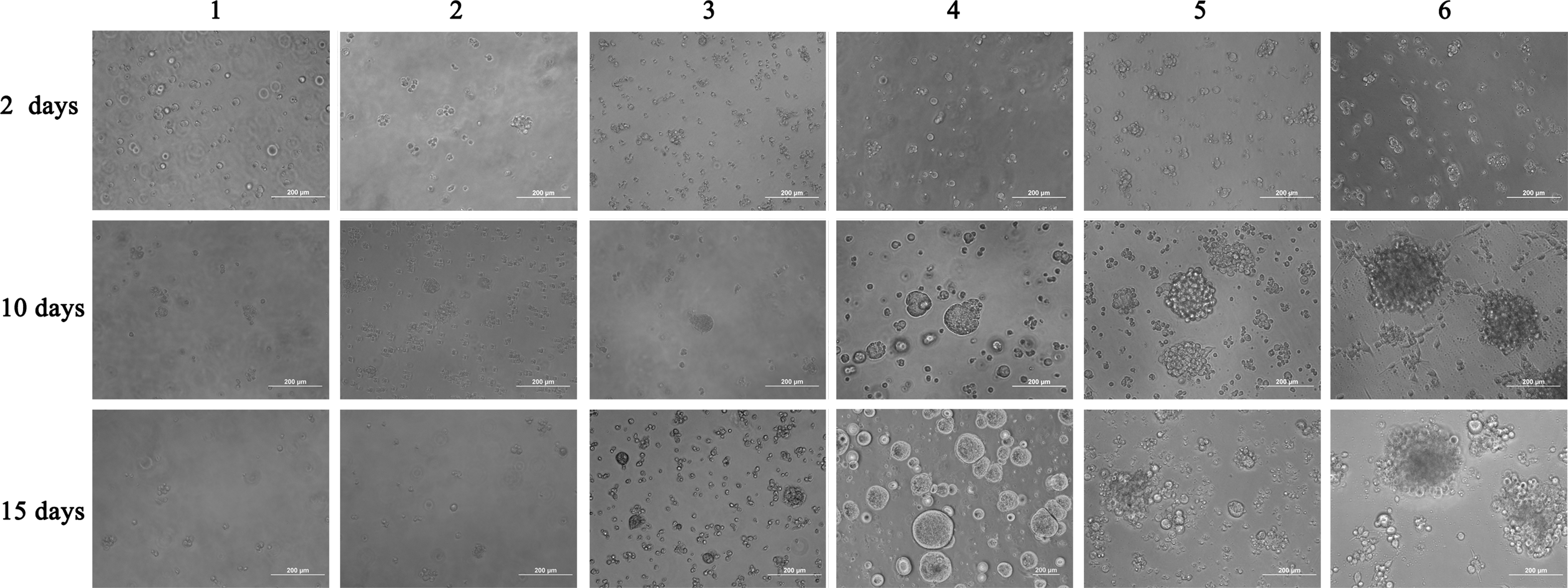
Mouse RPCs survival, proliferation, and characterization in hydrogels with different composition ratios. Most cells died or did not propagate in groups 1–3. In groups 4–6, cell proliferation was robust. Typical neruoshpere formation was observed in group 4, which was to be used in subsequent studies. RPCs, retinal progenitor cells.

MTT assay showed comparable growth rates of mouse RPCs encapsulated in different HA hydrogels with that of cells grown in 2D conditioned dishes. At the beginning of culture, there were no appreciable differences in the number of cells between all the groups, but most of the cells had died in groups 1–3 after 5 days in culture. Cells in other 3D groups showed consistent proliferation rates (except on day 10) compared with that of 2D culture system (n=5 for each group). *p<0.05, compared with that of 2D cultures, NS, nonsignificant; HA, hyaluronic acid; 2D, two-dimensional; 3D, three-dimensional.
Morphology and gene expression in 3D cultured cells
Morphologic results suggest that HA hydrogels provide a biocompatible niche for mouse RPCs in culture (Fig. 3). During development, cellular interactions with HA are mediated by some adhesion factors, including CD44. CD44 is a mediator for HA-induced cell proliferation and survival pathways and is present in cumulus cells, oocytes, early embryos, and prehatched blastocysts [12]. CD44 is also involved in the initial binding of HA to the cell surface before its internalization and degradation by acid hydrolysis. Mouse RPC colonies cultured in hydrogels showed clear evidence for CD44 double labeling with RPC and HA (Fig. 3).
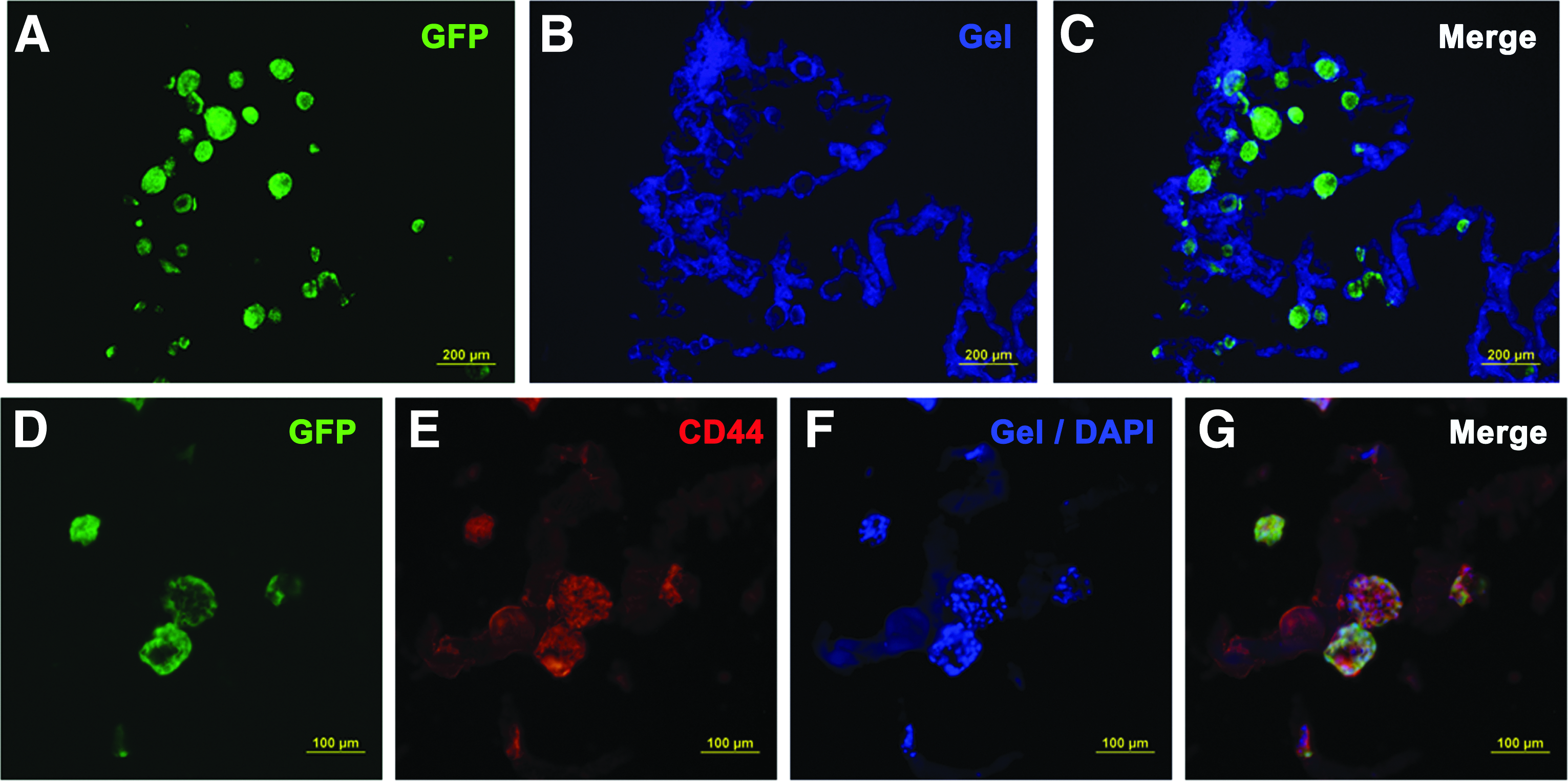
HA hydrogels supported the maintenance of viable mouse RPC neurospheres.
The RPCs maintained a typical undifferentiated morphology in colonies within the HA matrix, and after 15 days in culture most cells in the spheres expressed Pax6 and nestin (Fig. 4). Pax6 is essential for proliferation and pluripotency of retinal progenitors, and nestin is a protein marker for neural stem cell. 5 Cells in spheres also expressed many of the genes characteristically co-expressed by 2D cells, which included nestin, rhodopsin, and GFAP (Fig. 4), indicating they maintained their stemness (nestin) and pluripotency for differentiation into photoreceptors (rhodopsin) and glial cells (GFAP). Thus, we have demonstrated that these HA hydrogels provide a suitable microenvironment for self-renewal and pluripotency of mouse RPCs.
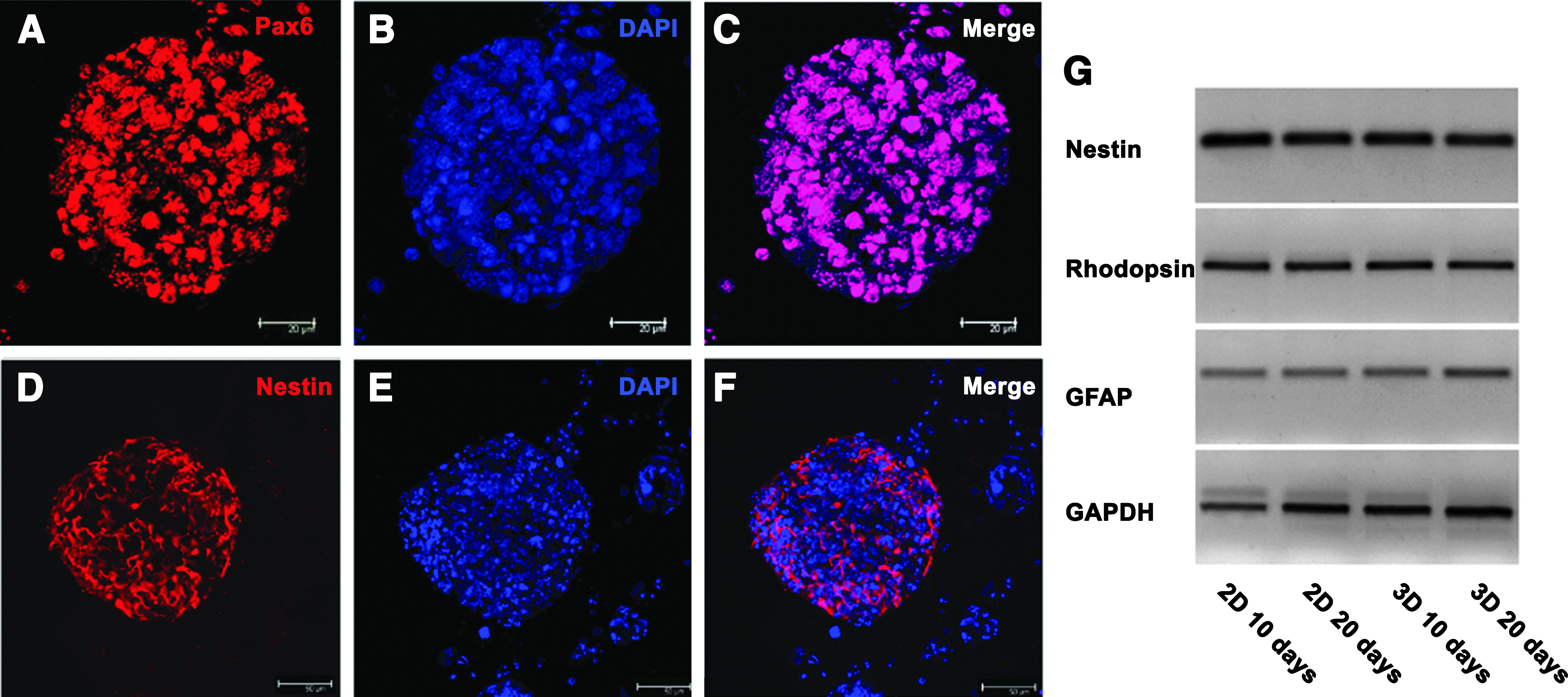
Characteristics of different gene expression levels in the neurospheres from mouse RPCs.
Characterization of RPCs encapsulated in hydrogels in the subretinal space
Twelve of the 15 transplants were successful based on the OCT scanning following injection. The grafts showed some vacuoles, resembling the holes in a sponge at 1 week (Fig. 5A). Based on their blue fluorescence staining pattern, these vacuoles were found to consist of the hydrogel (Fig. 5D) containing patches of viable transplanted cells. In contrast, the hydrogel was entirely degraded after 3 weeks (Fig. 5B, E), and the transplanted cells had become a denser and more continuous layer sitting under the outer nuclear layer (ONL). This change was qualitatively seen across all 12 mice. OCT also showed that there had been minimal damage at the injection site (Fig. 5C).
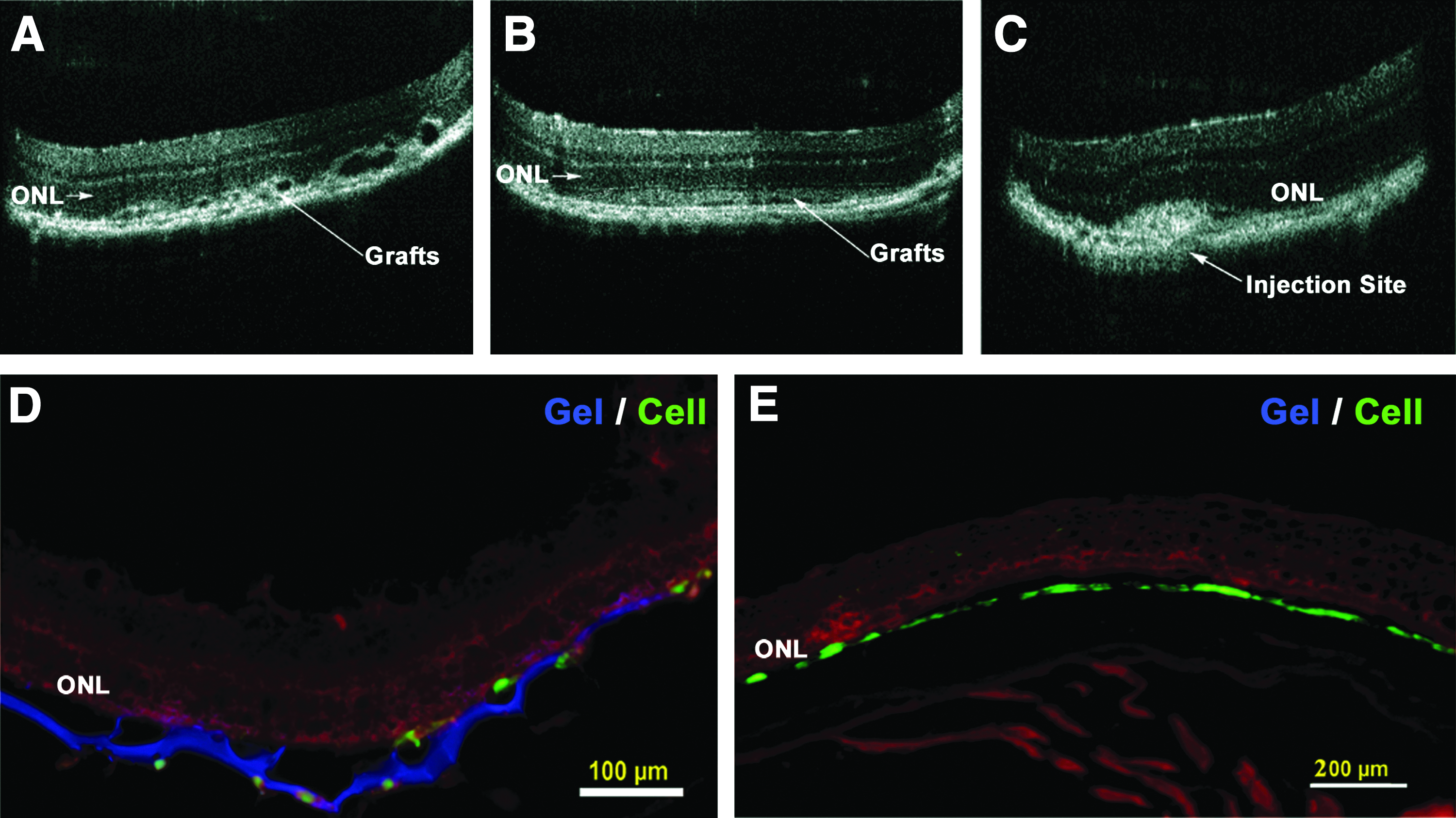
Delivery of retinal progenitor cells to dystrophic retinas via HA-based hydrogels leads to even distribution of cells in the newly created subretinal space.
A series of immunohistochemical studies were performed to determine whether cells had differentiated in vivo and expressed specific phenotypic makers characteristic of retinal cells. As shown in Figure 6, a large population of cells in the subretinal space was labeled by recoverin (a retinal calcium-binding protein found in photoreceptors). However, rhodopsin-positive cells were not found within the transplant cohort (figure not shown). Cells that leaked into the vitreous cavity expressed Tuj-1, a characteristic marker of retinal ganglion cells, and some of them migrated into the inner plexiform layer. We did not quantify the proportion of cells that develop the above characteristics.
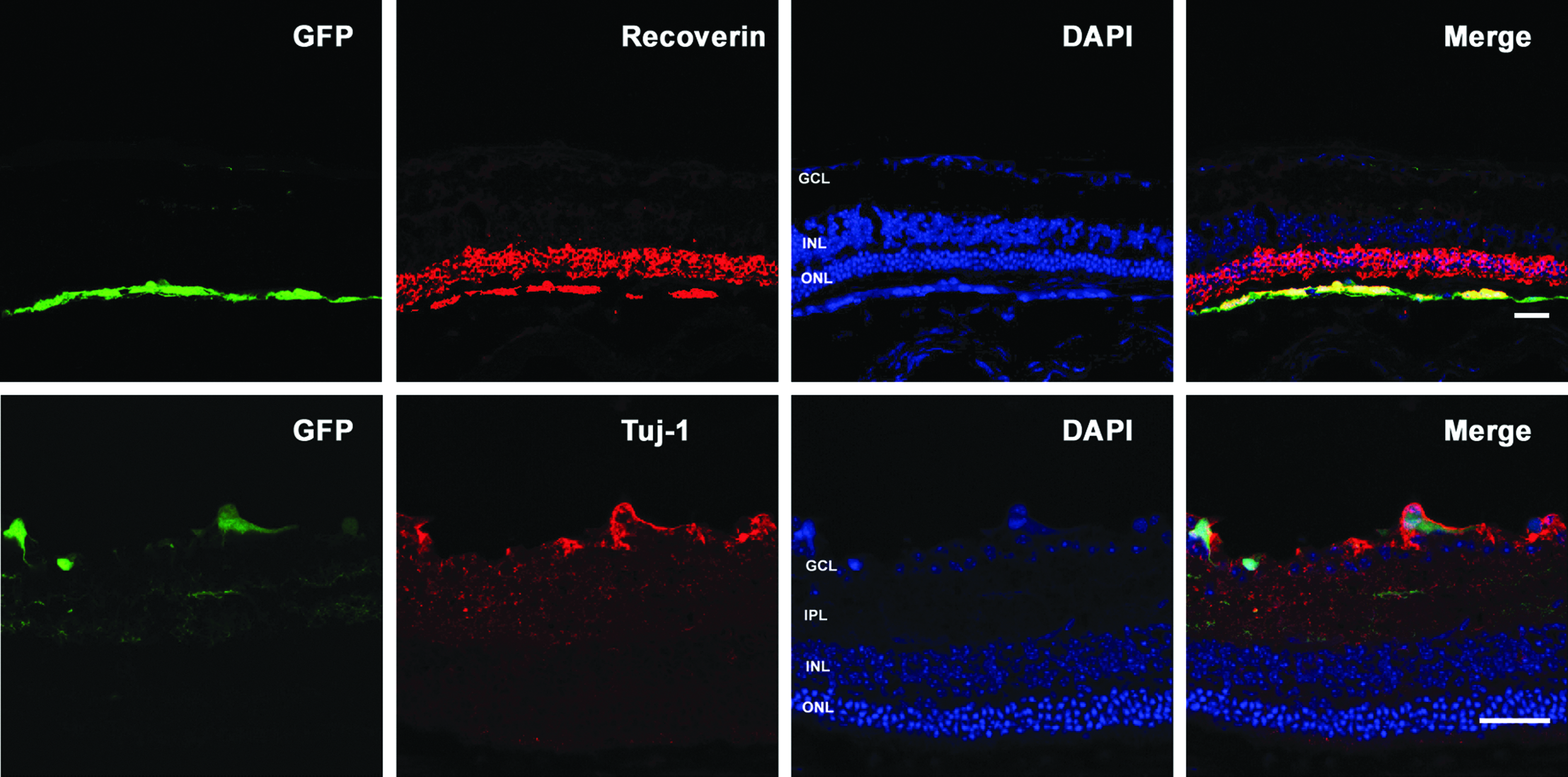
Differentiation of RPCs 3 weeks after transplantation of hydrogel grafts into the subretinal space of rho−/− mouse retinas. Transplanted cells contained GFP+. Retinas were immunostained for the presence of recoverin (red) or Tuj-1 (red; marker for retinal ganglion cells) and cell nuclei labeled with DAPI (blue). Recoverin coexpressing RPCs were found in the subretinal space of rho−/− mice. GFP+ cells with Tuj-1 expression were found in the ganglion cell layer (GCL); some cells migrated into the outer plexiform layer (OPL). No GFP+ cells were found in the inner nuclear layer (INL) and ONL. Scale bar: 20 μm. Color images available online at www.liebertpub.com/tea
Discussion
To achieve successful cellular transplantation for AMD or RP in a clinical setting, a more efficient means of cellular delivery and enhanced cellular integration and survival is needed. We show that biodegradable HA-based hydrogels can maintain sphere growth of RPCs in vitro. Injection of hydrogels containing RPCs promoted an even cellular distribution in the created subretinal space without disruption of the retinal architecture. Moreover, cells in the subretinal space expressed the mature photoreceptor marker recoverin. The use of this cell delivery vehicle is a step toward the development of safe biomaterials for cell-based treatments of retinal diseases.
In this study, ECM density, predominantly determined by the concentration of HyStem, may play a crucial role in cell growth. Three of the formulas used (1–3) did not support the growth and survival of mouse RPCs probably due to the higher concentration of HyStem (80%), which would substantially dilute or block nutrient and oxygen supply. Hydrogels with 40%, 20% HyStem (groups 4–6) effectively supported self-renewal of RPCs, without obvious differences in cellular proliferative abilities between the polymer blends. Additionally, the mechanical properties of HA hydrogels were optimized through a simple alteration in the amounts of the two reactive components (HyStem and Extralink); a change that is easily made during synthesis. In groups 4–6, the extent of cross-linking led to variations in the stiffness of the hydrogels, and these variations correlated with changes in the morphologic properties of the RPCs. Sphere formation within the pores, a characteristic 3D culture system, was observed using formula 4. Importantly, since progenitor cells take cues from the stiffness of its microenvironment, 16 the stiffness of the implanted material should roughly match that of the tissue it replaces and be appropriate for cell growth. Previous work has shown that the stiffness of retina is about 300–800 Pa in 37°C saline. 17 HA-based hydrogels that are more flexible (low modulus with 65 Pa) can localize the cells to a desired anatomic site and provide instruction to the cells.
3D cell culture has become an increasingly important tool for culturing and differentiating adult, embryonic, and induced pluripotent stem cells.18–20 Using identical media and growth factor stimulation on parallel cultures, mouse RPC maintained their undifferentiated state when encapsulated in 3D HA hydrogels. This is consistent with the results of Gerecht et al. 14 Importantly, this system can ensure an even distribution of cells in hydrogel due to its high viscosity before and after gelation. 10 Delivery of a cell suspension resulted in the formation of rosettes within the retina, 21 which contained abnormal orientations of photoreceptors that aggregated together and thus did not integrate with the host tissue. 22 Delivery of HA hydrogels overcomes the problems of cellular aggregation and provides a greater opportunity for cellular integration with the host tissue. Further, OCT demonstrated that hydrogels slowly degraded in 3 weeks without leaving evidence of its presence.
Efficient cell differentiation and migration are major barriers to successful cellular transplantation in retinal degenerative diseases. RPCs have demonstrated an ability to integrate into the injured and adult rodent retina. 23 When isolated from mouse retinas at a time that corresponds to photoreceptor development, these cells can differentiate into photoreceptors in vivo.4,24 The data presented here show that RPCs in the subretinal space can indeed develop into recoverin-positive cells. However, the photopigment rhodopsin was not detected in the grafted cells. A similar result was also obtained in co-transplantation of biodegradable matrix metalloprotenase-2 and poly(D,L-lactic-co-glycolic acid) (MMP2-PLGA) microspheres with RPCs into the subretinal space of rho−/− mice. 25 This may relate to the complete absence of rhodopsin expression in rho−/− hosts or the delivery system may inhibit this line neuronal differentiation. Since HA hydrogels can maintain cells in an undifferentiated state, attempts are being made to speed up the biodegradation processes after transplantation.
We did not observe RPC integration into the ONL of the host retina with the delivery of HA hydrogels after transplantation. However, this cell delivery strategy may be useful for the treatment of widespread or advanced maculopathy, where large areas of the RPE are destroyed. 26 The lack of cellular integration, even in the face of highly efficient cell delivery, is likely to be due, in part, to the deposition of inhibitory ECM proteins. 27 Some adhesion molecules, such as CD44 and neurocan are present at the outer surface of the degenerative retina, and separate subretinal grafts from the host retina.28–30 Previous studies in our lab have shown that biodegradable MMP2-PLGA microspheres with RPCs could enhance cellular integration via degradation of CD44 and neurocan at the outer surface of the degenerative retina. 25 HA, combined with CD44, may make the culture medium more viscous and thus inhibit cell migration into host retina. Given that the hydrogel is absorbed by 3 weeks, maybe the cells must migrate within a defined window and this is shorter than the time needed to absorb the gels. Therefore, the balance between initial stress exposure upon implantation, proliferation, differentiation, and HA degradation is critical for integration and longevity of RPCs. Ballios et al. reported that HA exhibited a rapid degradation over time, falling to a minimum concentration of approximately 3% after 1 week in the subretinal space. 10 Our cross-linkable HA is more stable than a blend of HA and methylcellulose as used by Ballios et al., 10 so it does not degrade in 1 week. Interestingly, some injected cells leaking into vitreous can migrate into host retina; the likely reason is that the leaking hydrogel was diluted by vitreous fluid. Collectively, it is critically important to determine more details about the interaction between the gel and the cells, such gel degradation time and the role of MMP2 or hyaluronidase in the degradation of HA hydrogels and how this affects cell integration after transplantation.
Conclusions
We have developed an injectable delivery system that combines a commercially available HA-based hydrogel and mouse RPCs. These hydrogels are naturally degraded in the subretinal space, enhance neurosphere growth, and maintain the pluripotency of RPCs in vitro. Delivery of hydrogels containing RPCs promoted an even distribution of cell without a major disruption to the host retina, and cells differentiated toward photoreceptors. Further research will focus on extending these studies to human RPCs with the intent of pursuing its clinical use.
Footnotes
Acknowledgment
The authors thank Dr. T. FitzGibbon for comments on revised drafts of the article.
Disclosure Statement
No competing financial interests exist.