
Abstract
Injectable calcium phosphate cements (CPC) are frequently used for filling of bone defects due to their excellent osteocompatibility. Their poor degradability, however, limits complete regeneration of bone defects. Organic additives that produce acid by-products are particularly attractive to create macroporosity in situ since CPC degrade by acid dissolution. The aim of the current study was to investigate whether glucono-delta-lactone (GDL) can be used as acid-producing microparticles for incorporation into CPC without compromising its osteocompatibility. Characterization studies confirmed that CPCs containing either low or high amounts of GDL were injectable and self-setting, while a considerable amount of porosity was formed already within 1 day of incubation in phosphate buffered saline due to dissolution of GDL. Histomorphometrical evaluation after 2 weeks of implantation revealed that CPC containing 10% of GDL degraded faster and was replaced by more bone tissue than CPCs containing either Poly (lactic-co-glycolic acid) (PLGA) or gelatin microspheres. Summarizing, the current study showed that CPCs containing appropriate amounts of GDL display accelerated degradation and new bone formation compared with CPCs containing microparticles made of conventional polymers such as PLGA or gelatin.
Introduction
B
Still, apatitic CPC is poorly resorbable and cannot be substituted by new bone tissue in time. To enhance the speed of CPC replacement by newly formed bone, various polymeric additives have been included in CPC. Provided that these microparticles degrade at a faster rate than the ceramic CPC, a porous scaffold can be obtained that is accessible for fluid flow and ingrowth of new bone tissue, thereby enhancing its degradation rate. An ideal organic additive is biocompatible, fast-degradable, cost-effective, available off-the-shelf and easy to handle by surgeons in the operation room.
Organic additives can be classified depending on their degradation pathways. Additives such as poly-trimethylene-carbonate 7 or gelatin 8 undergo enzymatic digestion that leads to slow surface erosion and degradation. Gelatin is well known for its excellent biocompatibility and therefore it is being used in various medical areas. Upon use as additive in CPC in vivo, however, very slow degradation rates were observed. 9 On the other hand, organic additives such as poly (lactic-co-glycolic) acid (PLGA) degrade by hydrolysis. 10 PLGA has been used frequently as pore generator because, among several other advantages, it degrades into lactic and glycolic acid that are eliminated from the body by the Krebs cycle as carbon dioxide and water once the pores have been created in the CPC material. These acidic lactic and glycolic acid monomers were recently shown to enhance CPC dissolution both in vivo and in vitro.11,12
Despite the fact that CPC formulations containing PLGA microspheres revealed enhanced degradation and abundant new bone formation throughout the scaffold, degradation rates of PLGA containing CPCs are still in the order of several months depending on the chemical nature of the PLGA microparticles.13,14
In contrast to the relatively slow degradation rates of PLGA, inclusion of glucono-delta-lactone (GDL) into CPC would allow for fast creation of macroporosity within apatitic CPC owing to its fast dissolution in aqueous media. GDL is a neutral compound that gradually hydrolyzes in aqueous systems to gluconic acid, thereby leading to a local acidification, which facilitates the dissolution of surrounding CPC matrix and formation of macroporosity. 15 No major adverse biological effects were expected since GDL is a naturally occurring food-additive that is metabolized to glucose. GDL is classified as GRAS (Generally Recognized As Safe) for use as a curing and pickling agent, pH control agent, or as sequestrant. Finally, GDL is commercially available as fine powder in contrast to gelatin or PLGA, which adds to the attractiveness of GDL since it can be directly mixed with the powder phase of CPCs without the need for further processing into microspheres.
Therefore, the aim of the current study was to investigate the effect of incorporation of GDL microparticles into CPC in vitro and evaluate its in vivo performance. First, characterization studies have been performed using MicroComputed Tomography, Reverse Phase High-Performance Liquid Chromatography (RP-HPLC), X-Ray Diffraction (XRD), and Scanning Electron Microscopy (SEM) to optimize the cement formulations containing GDL. Second, two CPC-GDL formulations containing either low (10%) or high (30 wt%) amounts of GDL (abbreviated as CPC-10%GDL and CPC-30%GDL) were selected for further in vivo evaluation in an established orthotopic implantation model to test the bone response and degradation behavior of CPCs containing GDL. These GDL-containing formulations were compared to CPCs containing microparticles made of either gelatin or PLGA. CPC degradation and new bone formation were evaluated histologically and histomorphometrically after 2 and 12 weeks of implantation.
Materials and Methods
Materials
CPC powder consisted of a mixture of 85% alpha-tricalcium phosphate (α-TCP; CAM Bioceramics BV), 10% dicalcium phosphate anhydrous (DCPA; Sigma Aldrich), and 5% precipitated HA (pHA; Merck). Na2HPO4 was purchased from Merck.
PLGA Purasorb® materials were obtained from Purac Biomaterials. Purasorb PDLG 5002A (Mw=17 kDa, acid terminated, L:G=50:50) was used to prepare microspheres. Polyvinyl alcohol (PVA; 88% hydrolyzed, MW=22 kDa) was obtained from Acros and isopropanol (IPN; analytical grade), dichloromethane (DCM; analytical grade), and D-(+)-GDL were obtained from Merck.
Anionic gelatin (from bovine skin, Type B Bloom 225, IEP∼5) in addition to acetone and glycine was purchased from Sigma-Aldrich. Olive oil and glutaraldehyde (GA, 25 wt% solution in water) were commercially available from Acros Organics.
Preparation and characterization of PLGA and gelatin microspheres
Dense PLGA microspheres were prepared by a single emulsion technique. Briefly, 0.2 g of PLGA was dissolved in 2 mL of DCM in a 20 mL glass tube. Then, this solution was transferred into a stirred beaker containing 100 mL of 0.3% PVA solution. Subsequently, 50 mL of 2% IPN solution was added. The solution was stirred for 1 h. The microspheres were allowed to settle for 1 h and the clear solution was decanted. The remaining suspension was centrifuged and the clear solution on top was aspirated. Finally, the microspheres were frozen, freeze-dried for 24 h and stored at −20°C.
The morphology and size distribution of the PLGA microspheres was determined by light microscopy. Microspheres were suspended in H2O and pictures were taken with an optical microscope equipped with a digital camera (Leica/Leitz DM RBE Microscope system; Leica Microsystems AG). The size distribution of the microspheres was determined by digital image software (Leica Qwin®; Leica Microsystems AG) using a sample size of at least 300 microspheres. In addition, morphology of the microspheres was observed by SEM (JEOL 6310) at an accelerating voltage of 10 kV.
Gelatin microspheres were prepared using a surfactant-free water-in-oil emulsion method. A 10 w/v% gelatin solution was obtained by dissolving gelatin in deionized water at 45°C. Thereafter, 10 mL of this gelatin solution was added dropwise into a 500 mL round bottom flask containing 300 mL olive oil while stirring at 500 rpm (upper stirrer) at 45°C. Spontaneous gelation of the gelatin droplets was then obtained by moving the emulsion into an ice bath under continuous stirring. After 30 min, 150 mL of chilled acetone (4°C) was then added to the emulsion followed by another 15 min of stirring. Gelatin microspheres without crosslinking were collected by filtration (Whatman 90 mm filterpaper) and washed with chilled acetone to remove residual olive oil. Subsequently, gelatin microspheres were resuspended in water/acetone (1/3 in volume ratio) solution, and further cross-linked using GA with a molar ratio of GA relative to the amount of free amine groups present in gelatin of 0.5. After cross-linking for 16 h, glycine solution (100 mM) was added into the microsphere suspension to block unreacted aldehyde groups from GA. The suspension was then subjected to three cycles of centrifugation (5000 rpm for 5 min) and re-suspension in deionized water by vortexing. After freeze-drying, microspheres were stored at 4°C until further use.
Preparation of cement formulations containing PLGA, gelatin or GDL
First, the amount of CPC additives was optimized to yield comparable volumetric percentages for PLGA, gelatin, and GDL. Several CPC-microparticles combinations were scanned using μCT (Skyscan −1072 X-ray microtomograph, TomoNT version 3N.5, Skyscan®, Belgium; X-ray source was set to100 kV, current to 98 μA and the resolution was 7 μm pixel). Cone beam reconstruction was performed and the data were analyzed by CT analyzer software (version 1.4; Skyscan). The resulting weight percentages of the additives to achieve comparable CPC porosity were equal to 5 wt% for gelatin (CPC-gelatin), 43 wt% for PLGA (CPC-PLGA), and 30 wt% GDL (CPC-30%GDL) yielding volumetric percentages of additives in the CPC preset discs of 53%±3% for CPC-gelatin, 50%±4% for CPC-PLGA, and 48%±2% for CPC-30%GDL. In addition, a fourth CPC-GDL formulation was fabricated by reducing the amount of GDL to 10 wt% corresponding to a volumetric percentage of 19%±2% (CPC-10%GDL) to test whether low amounts of GDL can produce sufficient acidity to degrade CPCs without compromising the biocompatibility of CPC-GDL formulations.
Subsequently, the amount of liquid phase was optimized to obtain cement formulations that exhibited comparable injectability and setting properties. It was observed that considerably lower amounts of liquid phase (2 w/v% aqueous solution of Na2HPO4) were needed for GDL-containing formulations to yield handling properties similar to composites containing PLGA or gelatin. Therefore, 350 μL of aqueous Na2HPO4 solution were added to composites containing gelatin or PLGA, whereas 290 μL or 210 μL of aqueous Na2HPO4 solution were added to the powder precursor phase for CPCs containing 10% or 30% of GDL, respectively.
After combining all liquid and powder precursor compounds and additives, the mixtures were vigorously mixed for 30 s in 2 mL syringes (Silamat® mixing apparatus, Vivadent, Schaan, Liechtenstein) and the cement was injected into Teflon molds (6×3 mm). Initial and final setting times of the different CPC pastes were assessed using Gillmore needles (ASTM C266). Briefly, a bronze block was used as mold containing holes of 6 mm diameter and 12 mm height. The mold was placed in a water bath at 37°C. Samples of each formulation were mixed and injected into the mold and initial and final setting time was determined (see Table 1), confirming that initial setting times were equal for CPC-PLGA, CPC-gelatin, and CPC-10%-GDL, whereas slightly longer initial setting times were observed for formulations containing the highest amount of GDL (CPC-30%GDL).
CPC, calcium phosphate cements; PLGA, poly (lactic-co-glycolic acid); GDL, glucono-delta-lactone.
To investigate the rate of GDL hydrolysis, preset composites containing either 10% or 30% of GDL were incubated at 37°C in 1.5 mL of phosphate buffered saline (PBS) for 30 min, 1, 3, and 5 days. The amount of gluconic acid in the incubation media of the different CPC-GDL composites was analyzed by RP-HPLC as a measurement of GDL degradation. The system consisted of a Hitachi L2130 HPLC pump, Hitachi L-2400UV detector, and Hitachi L-2200 auto sampler, and an Atlantis dC18 column (Waters) 250×4.6 mm, 5 μm. Two mobile phases were used; 1% acetonitrile in 20 mM NaH2PO4 pH=2.2 (mobile phase A) and 100% acetonitrile (mobile phase B). The flow was 0.5 mL per minute, the injection volume 40 μL, and the UV detection wavelength 210 nm.
The crystal structure of the CPC containing 10% and 30% GDL was evaluated after crushing preset samples (24 h at room temperature followed by 24 h of freeze drying) before and after soaking in PBS for 2 weeks at 37°C with a mortar until the discs were reduced to a fine powder and subsequently analyzed using XRD (PW3710 Philips, The Netherlands) using a Cu Kα radiation source at a wavelength of 1.5405 Å, an accelerating voltage of 40 kV, and a current of 30 mA. Diffractograms were collected for 2θ values between 3° and 45°.
Morphology of the CPC materials was observed by SEM (JEOL 6310) at an accelerating voltage of 10 kV after being lyophilized for 24 h.
The compressive strength (σc) and modulus of elasticity (E) of the samples were measured with a mechanical testing bench (858 MiniBionixII®; MTS) at a cross-head speed of 0.5 mm min−1. Cylindrical samples were tested after 0, 1, 2, and 12 weeks of immersion in 1.5 mL of phosphate buffer saline at 37°C.
Fourier transform infrared spectroscopy (FTIR; Perkin-Elmer) was used to characterize the molecular structure of the set cements. For this purpose, the injected samples were soaked in PBS for 72 h at 37°C. After curing, samples were removed from the bath, dried in air, and crushed in mortar.
Preparation and characterization of injectable CPC composites for in vivo implantation
To prepare CPC-PLGA composites, 0.6 g of dense PLGA microspheres was added to 0.8 g of CPC powder yielding a mass percentage of 43 wt% PLGA.
Two different CPC-GDL composites were prepared containing either 10 wt% GDL or 30 wt% GDL. Briefly, 0.1 g of GDL was mixed with 0.9 g of CPC (abbreviated as CPC-10%GDL) or 0.3 g of GDL were mixed with 0.7 g of CPC (abbreviated as CPC-30%GDL). All formulations containing PLGA or GDL were sterilized in the 2 mL syringes (sealed with a closed tip) using gamma radiation (25–50 kGy; Isotron BV). Gelatin microspheres, on the contrary, were sterilized prior to mixing with the CPC precursor powder by exposure to ethylene oxide (Isotron BV) to avoid excessive additional cross-linking of gelatin microspheres due to gamma irradiation. Ethylene oxide sterilization was carried out as a mixed cycle consisting of 10 h preconditioning, 3 h dwell time, 10 h of sterilization, and 48 h of postconditioning. About 0.05 g of sterile gelatin microspheres were mixed with 200 μL of filter-sterilized dH2O and subsequently mixed after 10 min of microsphere swelling with 0.95 g of CPC powder (sterilized previously using gamma irradiation under the conditions as described above) in a 2 mL plastic syringe, yielding a gelatin mass percentage of 5 wt%.
CPC was formed by adding 350 μL of aqueous Na2HPO4 solution to composites containing gelatin or PLGA, whereas 290 μL or 210 μL of aqueous Na2HPO4 solution were added to the powder phase for CPCs containing 10% or 30% of GDL, respectively. Subsequently, the mixtures were mixed vigorously for 30 s and the cement was injected into the bone defects.
Surgical procedure
Twenty-four skeletally mature female 8-week-old New Zealand White rabbits with a weight between 3.2 and 3.6 kg were used as experimental animals. The protocol was approved by the Animal Ethical Committee of the Radboud University Nijmegen Medical Centre (Approval no: RU-DEC 2010-174) and national guidelines for the care and use of laboratory animals were applied.
Surgery was performed under general inhalation anesthesia. The anesthesia was induced by an intravenous injection of Hypnorm (0.315 mg/mL fentanyl citrate and 10 mg/mL fluanisone) and atropine, and maintained by a mixture of nitrous oxide, isoflurane, and oxygen through a constant volume ventilator. To reduce the peri-operative infection risk, the rabbits received antibiotic prophylaxis (Baytril®, 2.5% (Enrofloxacin), 10 mg/kg). The animals were immobilized on their back and the hind limbs were shaved, washed, and disinfected with povidone-iodine. After exposure of the distal femoral condyle a 1.0 mm pilot hole was drilled. The hole was gradually widened with drills of increasing size until a final defect size of 4 mm in width and 6 mm in depth was reached. Low rotational drill speeds (max. 450 rpm) and constant physiologic saline irrigation were used. After preparation, the defects were thoroughly irrigated and packed with sterile cotton gauze to stop bleeding. Surgery was performed in both legs of the rabbits and one defect was created in each condyle.
The four different CPC formulations were injected into the created condylar defects in a randomized manner (N=6). After injection, the superfluous cement was removed with a scalpel blade and soft tissues were closed layer by layer using resorbable Vicryl sutures after approximately 10 min of setting.
After 2 and 12 weeks of implantation, rabbits were euthanized using an overdose of Nembutal® and the femoral condyles were harvested for evaluation.
Histological procedures
After harvesting of the femoral condyles and removal of surrounding soft tissues, each condyle was divided into medial and lateral halves along its longitudinal axis. Subsequently, the medial half was fixed in 4% formaldehyde for 2 days, dehydrated in a graded series of ethanol and embedded in methylmethacrylate. After polymerization, at least three 10 μm sagittal cross sections of the condylar part containing the composite implants were prepared using a sawing microtome technique. Sections were stained with methylene blue/basic fuchsin and examined with a light microscope (Leica Microsystems AG).
Histological and histomorphometrical analysis
In addition to a subjective histological description, quantitative histomorphometrical analysis was performed. Therefore, a 4 mm diameter circular area was superimposed onto the bone defect area as region of interest (ROI) for further quantifications of new bone formation (Fig. 1). Digital images (magnitude 5×) were recorded, and bone and cement content were determined by color recognition using image analysis techniques (Leica Qwin Pro-image analysis system).

Four millimeter circular region of interest (ROI) used for the histomorphometrical measurements. Color images available online at www.liebertpub.com/tea
Next to conventional bone measurements by color recognition, measurements of new bone formation were also expressed relative to a ROI consisting of 100% trabecular bone (TB) to avoid underestimation of new bone formation due to the presence of bone marrow and fat tissue in the TB as present in femoral condyles. An ROI consisting of 100% TB corresponded to a value 70 vol% of TB tissue in the rabbit femoral condyles, the remaining 30 vol% consisting of bone marrow or fat tissue.
Statistical analysis
All statistical analyses were performed with SPSS® software (IBM Corporation). Significant differences between the groups were determined using analysis of variance. A Tukey–Kramer (Multiple Comparisons) Test was applied for comparison of the groups at different implantation periods. Results were considered significant if p<0.05.
Results
Physicochemical characterization of additives and composites
The average microsphere size was 42±5 μm for PLGA microspheres and 37±10 μm for swollen gelatin microspheres. In contrast, the size of commercially available GDL microparticles was 595±40 μm (Fig. 2). SEM micrographs of the preset CPC composites revealed the presence of micron-sized additives within a nanoporous CPC matrix for composites containing PLGA or gelatin, whereas single microparticles were not recognizable in formulations containing 10% GDL. A remarkably compact morphology was observed for CPC-30%GDL formulations in which the amount of nanoporosity was considerably reduced; likewise, the initial GDL particle size (Fig. 3).
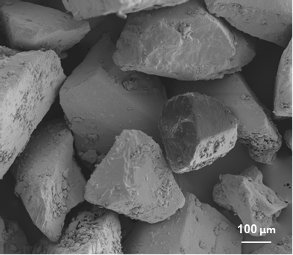
Scanning Electron Microscopy (SEM) picture of glucono-delta-lactone (GDL) microparticles.

SEM pictures of calcium phosphate cements (CPC)-Poly (lactic-co-glycolic acid) (PLGA)
The rapid dissolution rate of GDL upon incorporation into CPC can be observed in the RP-HPLC results (Fig. 4). After 1 day of incubation in PBS the majority of the GDL was released and detected by HPLC (96.0%±2.0% for CPC-30%GDL and 96.5%±1.3% for CPC-10%GDL).

Detection of gluconic acid release from preset CPC-10%GDL and CPC-30%GDL composites by means of Reverse Phase High Performance Liquid Chromatography.
Regarding the XRD results (Fig. 5), it was observed that the diffraction peaks related to the CPC phase (i.e., α-TCP, dicalcium phosphate anhydrous [DCPA] and HA) were unaltered by inclusion of 10 or 30 wt% GDL after injection (Fig. 5A). Except for a slight delay in initial and final setting of the CPC containing 30 wt% GDL, the incorporation of GDL did not hamper the setting of the CPC (see Table 1), resulting into a phase transformation of α-TCP to HA for both pure CPC samples and GDL-containing CPCs after 2 weeks of soaking in PBS (Fig. 5B).
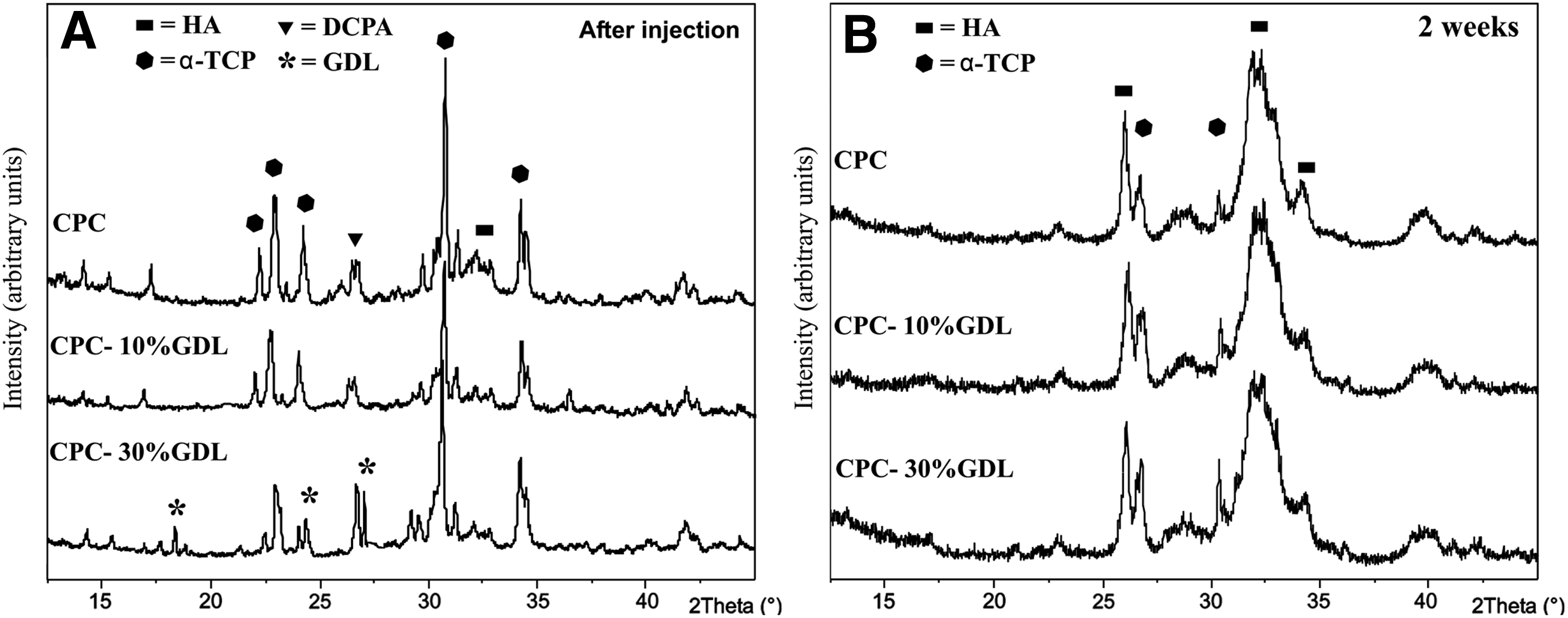
X-ray diffraction (XRD) analyses of CPC and CPC containing 10% and 30% GDL evaluated after injection (24 h at room temperature followed by 24 h of freeze drying)
The different GDL, PLGA, and gelatin combinations with CPC indicated that after injection, all four composites exhibited similar mechanical properties (Fig. 6). After 1 week of incubation, the samples containing 30% GDL disintegrated. As a consequence, the mechanical properties of these materials could not be determined after 1 week of incubation. Samples containing 10% GDL microparticles exhibited higher compressive strength values that were constant in time due to the higher percentage of the cement phase compared to cements containing PLGA and gelatin microspheres. In addition, it was observed that the mechanical properties of CPC-PLGA decreased after 1 week of incubation due to the onset of PLGA degradation. In contrast, mechanical properties of CPC-gelatin samples only started to decrease after 12 weeks of incubation due to the absence of the enzyme collagenase.
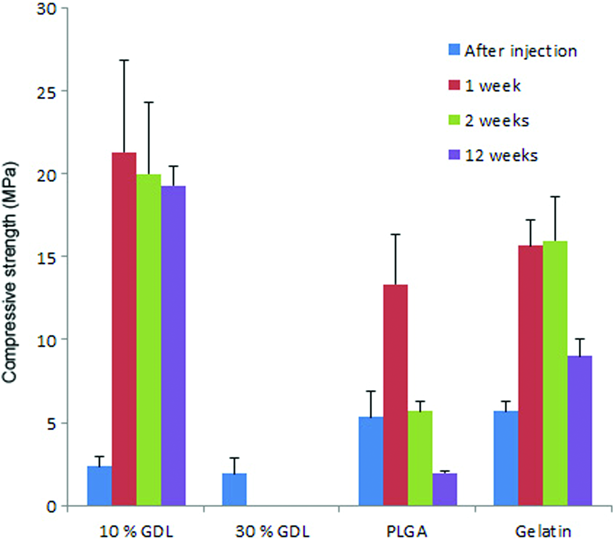
Mechanical evaluation of the different composites. Color images available online at www.liebertpub.com/tea
FTIR analysis (Fig. 7) confirmed that the setting of CPC was not affected by the various additives. The main absorption characteristic for HA were observed at 559 cm−1 (υ4b), 600 cm−1 (υ4a), 1021 cm−1 (υ3c) and 1087 cm−1 (υ3a), while an absorption peak at 860 cm−1 could be attributed to protonated phosphate HPO42− (v5). 16 The C=O stretching vibration of the PLGA polymer was observed at 1750 cm−1 while the peak at 1645 cm−1 was related to the stretching of the C=C bonding.17,18
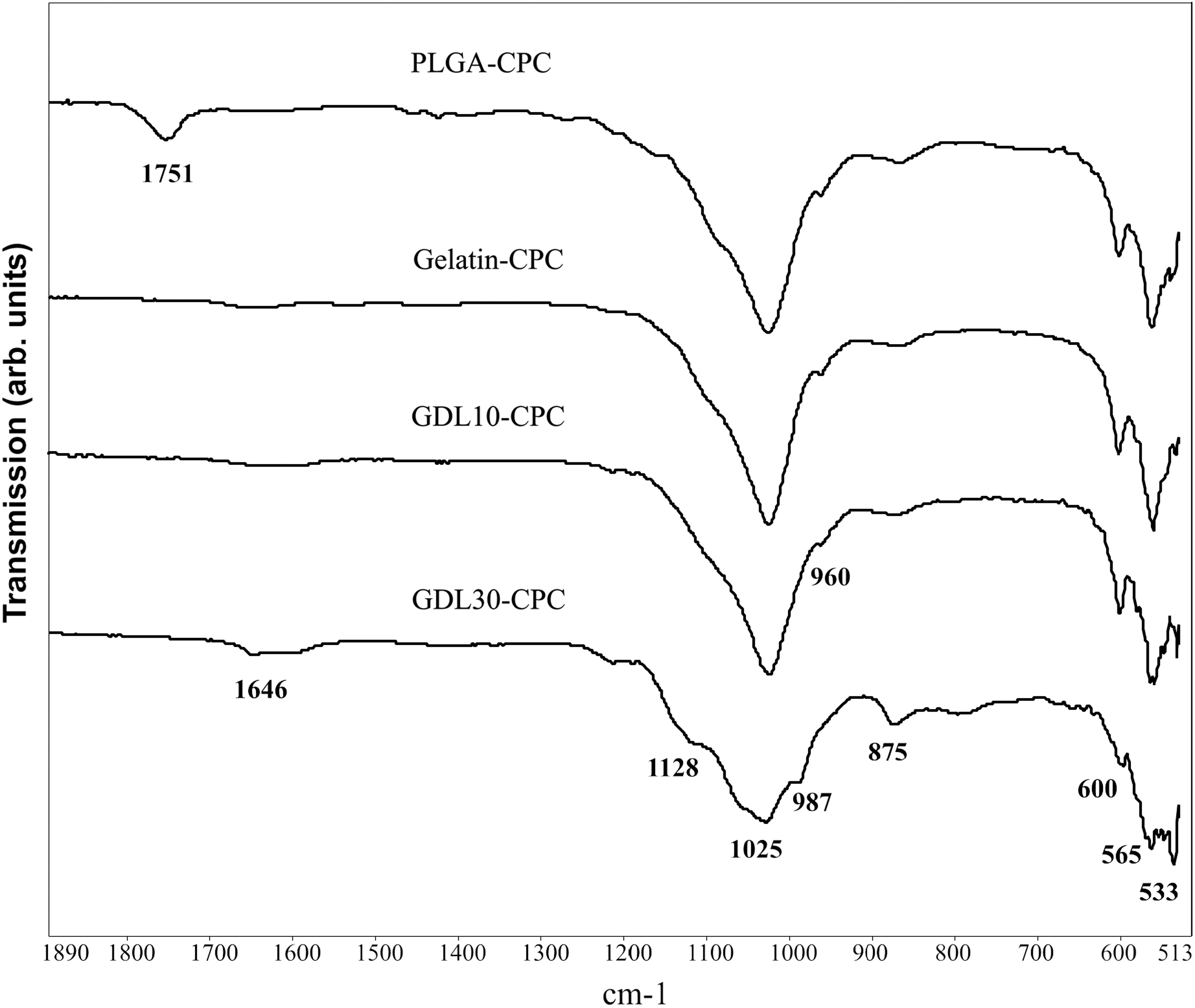
FTIR measurements of CPC containing PLGA, gelatin, 10% GDL, and 30% GDL after 72 h incubation in PBS at 37°C.
Preclinical observations
All 24 rabbits exhibited good health and did not show any wound complications. At the end of the two implantation periods, a total of 48 implants were retrieved (12 rabbits×24 implants after 2 weeks of implantation and 12 rabbits×24 implants after 12 weeks of implantation). At retrieval, no visual signs of inflammatory or adverse tissue reactions were observed.
Descriptive light microscopy
A uniform tissue reaction was observed for specimens of the same experimental group and implantation time. Representative light microscope sections corresponding to 2 and 12 weeks implantation time are presented in Figure 8 and Figure 9, respectively.
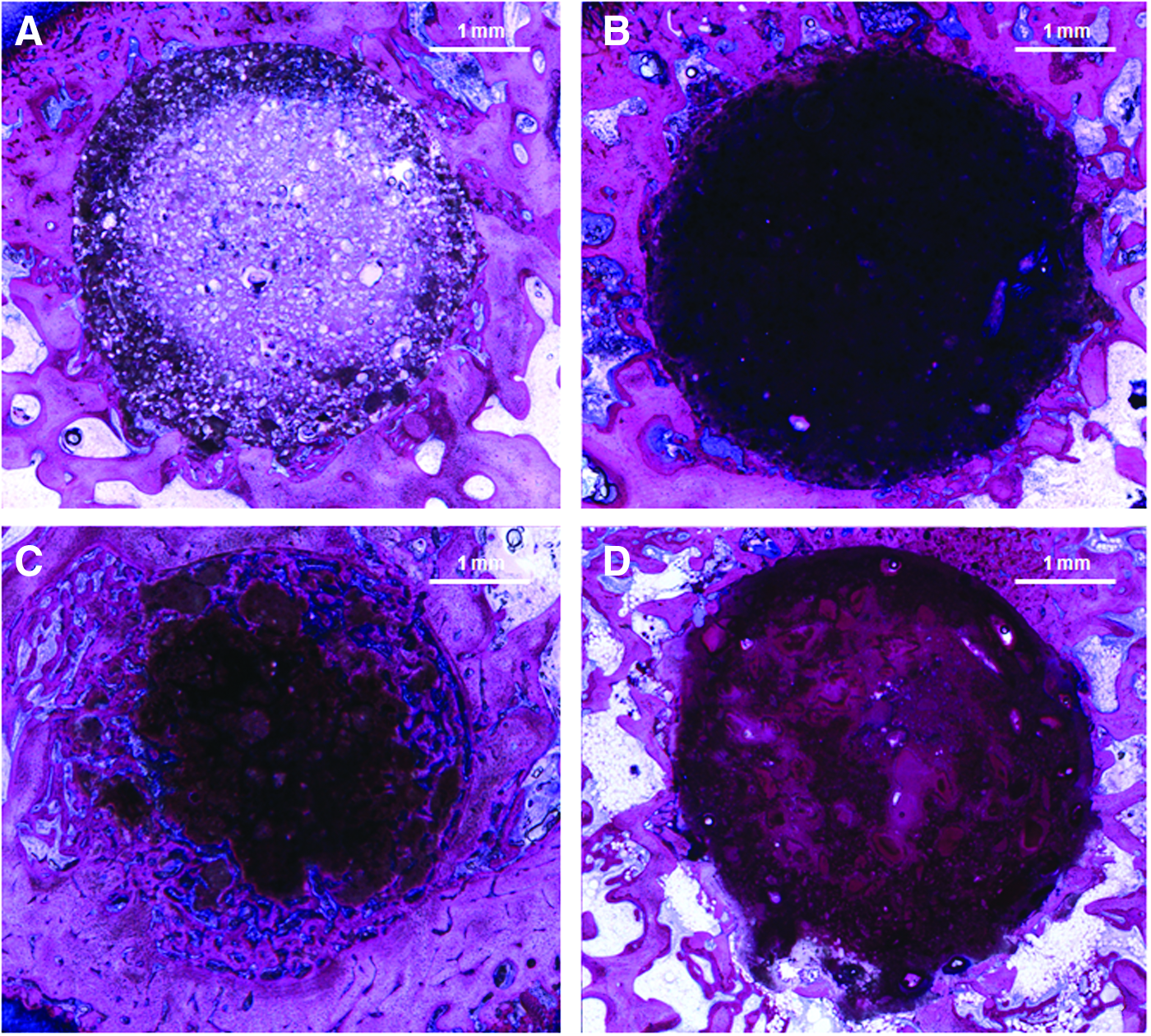
Histological sections at 2 weeks implantation of CPC-PLGA
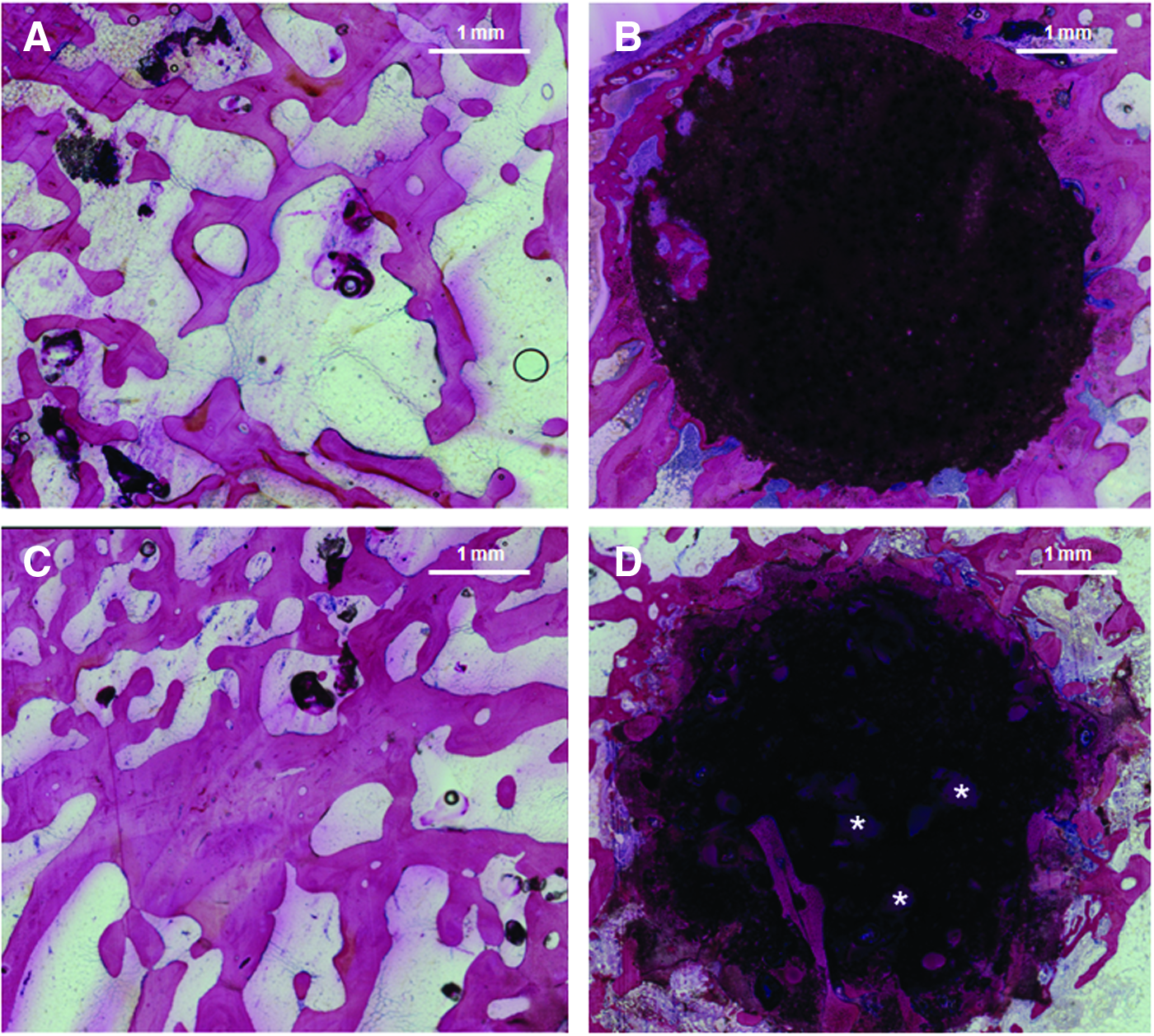
Histological sections at 12 weeks implantation of CPC-PLGA
Examination of the sections after 2 weeks implantation revealed variable amounts of cement degradation and new bone formation in the ROI. The newly formed bone displayed a cancellous structure similar to the pre-existing bone in the vicinity of the defect location. CPC-PLGA and CPC-gelatin composites presented an intact circular shape, which confirmed that these CPC-based formulations did neither degrade nor were substituted by new bone tissue. In contrast, CPC degradation was clearly observed in the peripheral areas of the circular defects that were filled with CPC-10%GDL. These degraded areas were substituted with newly formed bone tissue. For CPC-30%GDL, on the contrary, only minor degradation was observed in the peripheral areas of the defects.
After 12 weeks of implantation, the majority of the CPC-PLGA composites were completely degraded and only small remnants of CPC were observed in between the newly formed interconnected bone network. CPC-gelatin composites revealed minor degradation of the implant peripheral area and limited new bone formation occurred throughout these degraded implant margins. Regarding CPC-10%GDL composites, major degradation of the material was detected and excessive new bone formation was observed throughout the entire ROI. Similar to the CPC-PLGA, a cancellous bone network was formed in the defect location after degradation of CPC-10%GDL implants. In CPC-30%GDL composites, cement degradation and concomitant new bone formation was only observed in the outer regions of the circular implant, whereas big undissolved GDL particles with and approximated size of 500 μm were observed within the CPC matrix (Fig. 9D).
For both CPC-10%GDL and CPC-30%GDL, no signs of inflammatory response were observed.
Histomorphometry
Bone formation
Quantitative evaluation of new bone formation within the ROI revealed that the amount of newly formed bone varied considerably among the experimental groups (Fig. 10).
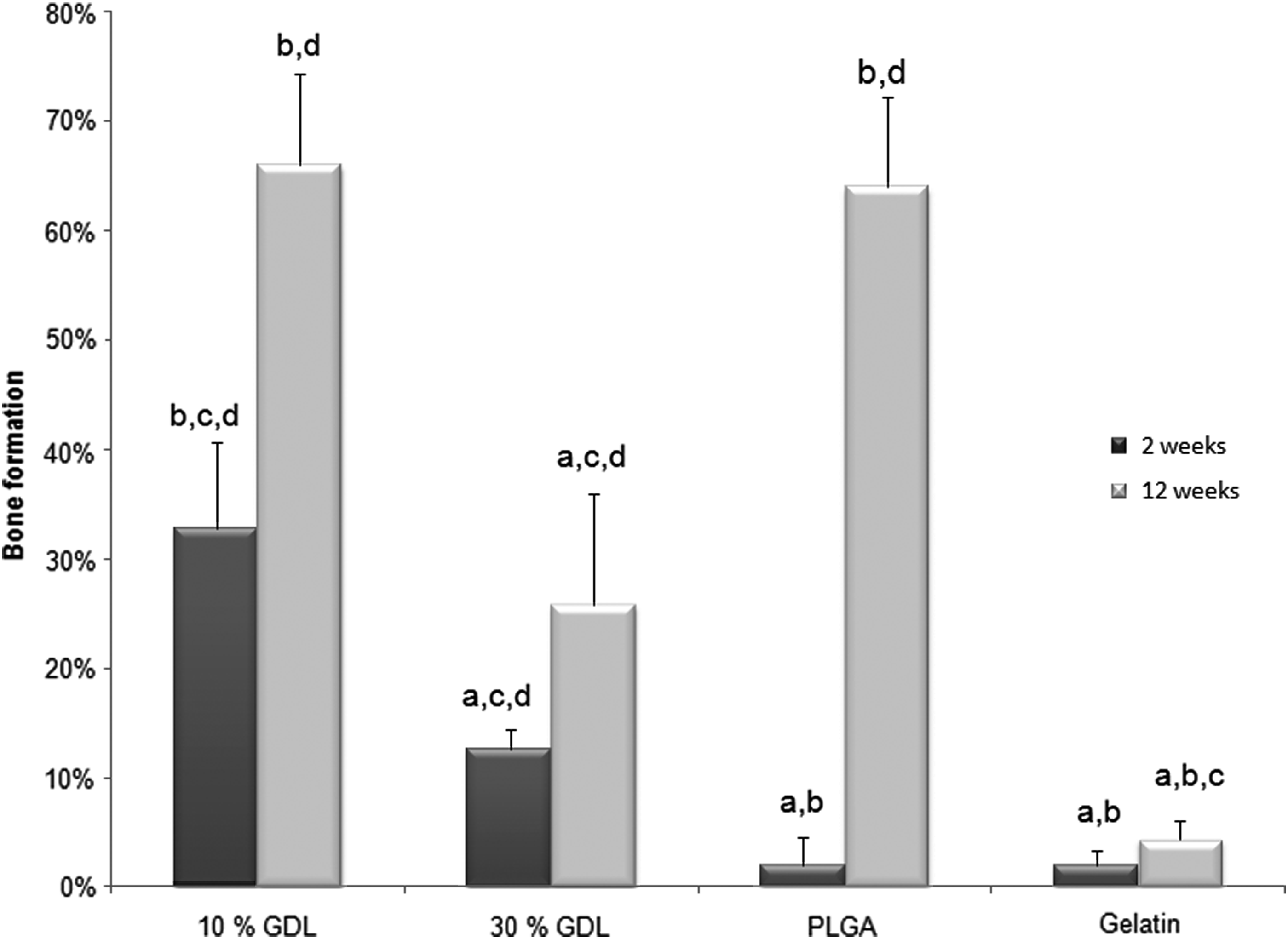
Quantitative evaluation of bone formation: Bone formation in the defect area (ROI) in the four composite types after 2 and 12 weeks of implantation. (a) Significantly different compared with CPC-10%GDL. (b) Significantly different compared with CPC-30%GDL. (c) Significantly different compared with CPC-PLGA. (d) Significantly different compared with CPC-gelatin.
After 2 weeks implantation CPC-10%GDL showed significantly higher amounts of new bone formation (32.8%) compared with all other experimental groups (p<0.001). CPC-30%GDL presented significantly more newly formed bone (12.5%) than composites with PLGA (2.0%) or gelatin (1.9%) microspheres (p<0.05).
After 12 weeks implantation CPC-PLGA (64.0%) and CPC-10%GDL (66.0%) revealed significantly (p<0.001) enhanced bone formation in comparison to CPC-30%GDL (25.8%) and CPC-gelatin (4.4%). CPC-30%GDL composites displayed significant more bone formation than CPC-gelatin (p<0.01).
Bone formation relative to TB
After 2 weeks implantation, values for new bone formation relative to a ROI solely composed of TB equaled 46.8%, 17.9%, 2.9%, and 2.8% for defects that were filled with CPC-10% GDL, CPC-30% GDL, CPC-PLGA, and CPC-gelatin, respectively.
After 12 weeks implantation both CPC-PLGA (91.4%) and CPC-10%GDL (94.3%) specimens revealed similar new bone formation values as when the ROI is filled for 100% with bone, confirming that TB was regenerated almost completely using these implants. On the contrary, significantly less bone formation occurred for CPC-30%GDL (36.8%) and CPC-gelatin composites (6.2%) (p<0.01) (Fig. 11).
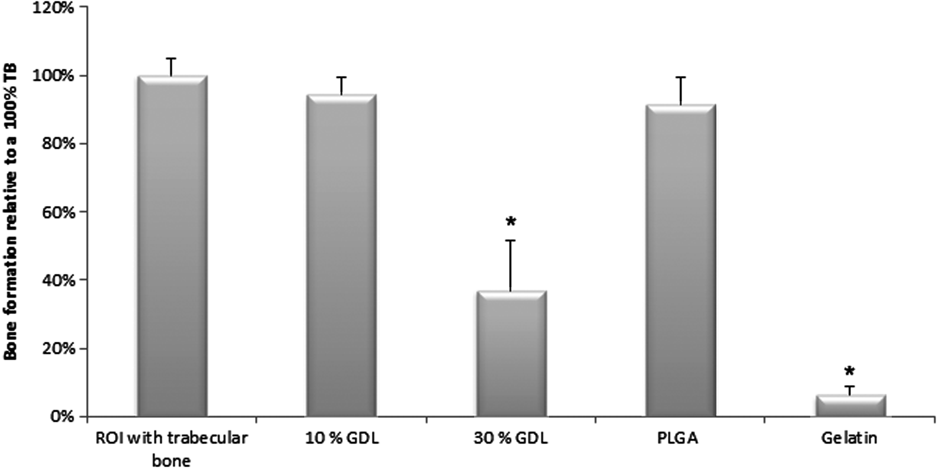
Bone formation relative to a ROI filled with trabecular bone (TB) after 12 weeks implantation. Significantly (*) less bone formation occurred within CPC-30%GDL and CPC-gelatin samples when compared with a ROI filled with TB.
Discussion
The aim of the current study was to evaluate the application of GDL within CPCs and to render these injectable bone fillers resorbable within weeks rather than months. It was hypothesized that the incorporation of GDL would result into a fast generation of porosity within apatitic CPCs, thus enhancing CPC degradation rates and subsequent replacement by newly formed bone compared to formulations containing conventional additives such as PLGA. The results indeed confirmed that incorporation of GDL resulted into enhanced CPC degradation at early time points (2 weeks) and almost complete resorption of the CPC and substitution by newly formed bone tissue at later time points (12 weeks).
GDL is a commonly used additive in the food industry and it is applied in a variety of products such as a.o. milk, 19 cheese, 20 or cookies. 21 Recently, the use of GDL in biomedical applications has also been investigated for applications as acidifier in alginate hydrogels for tissue engineering applications 22 or as a part of cell-biomaterials constructs for regenerative medicine. 23 In the current study, it was observed that GDL positively influences the injectability of CPCs since GDL-containing CPCs were injectable at considerably lower liquid-to-powder ratios without compromising the final setting times. Inclusion of GDL resulted in local acidification, thereby increasing the dissolution rate of α-TCP precursors. In other studies the addition of acidic components such as citric acid was also shown to improve CPC injectability and setting properties.24,25 Local acidification of the microenvironment of the setting cement will have resulted into increased (positive) charges of the precursor powders and precipitating apatitic nanoparticles, thereby increasing the interparticle repulsion forces that are responsible for the observed improved injectability.
Upon inclusion of 30% of GDL microparticles, the morphology of the resulting composites was less granular and more compact than the nanoporous morphologies of other experimental groups. This compaction was also suggested to relate to the acidification caused by the incorporation of GDL. GDL might have acted as dispersant by increasing the positive surface charge of precipitating apatitic nanoparticles. Compaction of ceramic slurries and cements depends on achieving the dispersion of fine ceramic particles in an aqueous medium by electrostatic/steric stabilization. As a result, the formation of a heterogeneous structure containing large powder agglomerates can be avoided while a uniform particle packing will result into a better microstructure. Acidic additives such as hyaluronic acid were earlier shown to improve the mechanical strength and cohesion of CPCs.26–28
The PLGA microspheres used in the current study were fabricated from 17 kDa acid-terminated polymers. Microspheres of these chemical and morphological characteristics were the material of choice due to their recently observed beneficial properties with respect to CPC degradation and new bone formation. 12 As earlier, the results from the current study confirmed the efficacy of dense PLGA microspheres as an additive for CPCs.
Based on the results of the characterization studies, two time points where selected for the evaluation of the in vivo implantation, that is, 2 and 12 weeks. An implantation time of 2 weeks was selected as early time point since GDL had induced the formation of porosity after 1 day of incubation in PBS whereas degradation of PLGA takes several weeks. Therefore, an implantation time of 2 weeks was considered a suitable early time point to discriminate between the fast degradation rate of GDL versus the slow degradation rate of PLGA. Degradation of the CPC-PLGA composites indeed did not occur after 2 weeks of implantation (2% of new bone formation), whereas a large part of the CPC-10%GDL materials was degraded and replaced by newly formed bone (32.8% of new bone formation). These differences in the rate of new bone formation for CPCs containing GDL versus PLGA can be attributed to differences in the time necessary to generate CPC porosity owing to the different physicochemical characteristics of PLGA and GDL. The relatively slow degradation of PLGA microspheres is related to the lag time between initial exposure of the hydrophobic PLGA to water and the actual hydrolytical degradation of the water-insoluble polymer into lactic and glycolic acid monomers. GDL, on the other hand, is a highly hydrophilic monomer with a solubility of 47% in water. 29 Upon contact with an aqueous solution, GDL dissolves gradually followed by hydrolytical cleavage of the cyclic ester into gluconic acid. The current results revealed that GDL undergoes almost complete hydrolytical degradation in vitro within 24 h in PBS upon incorporation into CPC. In contrast, hydrolytical cleavage occurs at much later time points for 17 kDa PLGA microspheres within CPC 11 since it was observed that comparable degrees of hydrolysis (90%) were observed after 9 weeks only for PLGA. Moreover, GDL microparticles were about 10 times bigger than PLGA microspheres.
Interestingly, inclusion of high amounts (30%) GDL did lead to less bone formation (12.5%) than 10% GDL after 2 weeks of implantation. This observation can be related to the reduced fluid flow throughout the CPC-30%GDL composites due to the low liquid-to-powder ratio and the compact microstructure for CPC-30%GDL (see Fig. 3). Using histology, it was observed that dissolution of GDL microparticles was incomplete (see Fig. 9D), confirming that a poor access of water to the interior of compact CPC-30%GDL composites might have caused the residual presence of undissolved GDL microparticles. Regarding the short presence of GDL, we would like to point to the fact that large differences were observed between in vitro and in vivo results. In vitro, GDL dissolved almost completely within less than a day for cements containing either 10% or 30% of GDL, whereas dissolution of GDL proceeded slower in vivo.
After 12 weeks of implantation, the amount of CPC degradation and new bone formation were comparable for CPC-10% GDL and the CPC-PLGA. This confirms that although initial degradation rates (and thus acid production rates) of PLGA were slower than GDL, this initial lag time in degradation was compensated for at later time points. Both CPC-10%GDL and CPC-PLGA composites exhibited a very good biocompatibility and replacement by new bone tissue, whereas only CPC-30%GDL hardly degraded after 12 weeks of implantation. Even though these formulations of high GDL content did not degrade in vivo, no toxic or inflammatory events were observed, confirming the biocompatibility of GDL-CPC in bone tissue. In detail, less cement degradation and bone formation was observed after 2 and 12 weeks for CPC-30%GDL versus CPC-10%GDL, indicating that the denser CPC matrix of CPC-30%GDL limited the perfusion of body fluids into the implant materials, thereby limiting GDL degradation (which was still partly present after 12 weeks of implantation) and new bone formation.
CPCs containing gelatin microspheres did not degrade after 2 or 12 weeks of implantation. In a previous study from our group adverse inflammatory responses were reported upon injection of CPCs containing gelatin microspheres into bone defects. 9 In the current study, inflammation of gelatin-containing CPCs was not observed. Several reasons can be held responsible for these differences such as the absence of proper sterilization methods or preparation steps to block residual uncross-linked glutaraldehyde groups in this previous study. The procedure to sterilize gelatin microspheres using ethylene oxide gas and the glycine washes performed to block unreacted GA prior to implantation as employed in the current study resulted into a considerably improved response of the bone tissue in the vicinity of the injected material. Nevertheless, gelatin-containing CPCs did not degrade, which can be attributed to the poor accessibility of proteolytic enzymes such as collagenases to CPC-gelatin constructs. Without these enzymes, degradation of cross-linked gelatin microspheres will not occur. These results confirm that acid production by additives is a prerequisite to obtain significant degradation of apatitic CPCs in vivo.
Quantification of the histological slides revealed abundant bone formation of up to 66 vol% of the ROI when 10% GDL or PLGA were used as organic additives. However, it was observed that the majority of the ROI was filled with TB and no implant remnants were found. Due to the porous structure of TB, the amount of new bone formation was underestimated since histomorphometrical control experiments on rabbit femoral condyles without any defect revealed that TB consists of about 70 vol% of mineralized bone tissue, whereas the remaining 30% consists of interstices containing bone marrow or fat. As a consequence, the above-mentioned values for new bone formation of about 66% correspond to almost complete bone regeneration after 12 weeks of implantation of CPC-10%GDL and CPC-PLGA implants.
The current study reveals the potential of organic additives to promote degradation of CPCs. However, degradation of CPC materials is known to be a combination between chemical dissolution and cell-mediated processes. 30 Additional studies would be necessary to elucidate whether the cell-mediated degradation of CPC is influenced by the physicochemical nature of the used organic additives, for example, by decalcifying the explants followed by paraffin embedding and specific staining for osteoclasts using tartrate resistant acid phosphatase.
Summarizing, GDL was successfully introduced in the current study as acid-producing organic additive for CPCs without compromising its osteocompatibility. Based on the data reported herein, GDL emerges as a promising candidate organic additive to promote the degradability of CPCs and allow subsequent rapid bone formation. Moreover, CPC-GDL composites can be used as off-the-shelf injectable materials without the need for additional and often expensive preparation steps to produce particles of appropriate size and shape that are necessary for inclusion of gelatin and PLGA (microspheres). In addition, CPC-GDL is cheap and can be stored at room temperature, which warrant further studies on the mechanism by which GDL microparticles create in situ porosity into CPCs and the biological response in animal models that are more relevant for clinical application.
Conclusion
The current study has shown that GDL can be used as organic additive in injectable apatitic CPCs to render these bone fillers resorbable within weeks rather than months. Self-setting, GDL-containing CPCs were developed by simple mixing of GDL with calcium phosphate powder precursors and lowering the liquid-to-powder ratios, thereby producing cements with setting times comparable to conventional CPCs. Histomorphometrical evaluation after 2 weeks of implantation in femoral condyles of rabbits revealed that CPCs containing 10% of GDL degraded faster and were replaced by more bone tissue than CPCs containing PLGA, gelatin, or high amounts (30 wt%) of GDL. After 12 weeks, CPC degradation and concomitant new bone formation was equal for CPC-10%GDL and CPC-PLGA. Summarizing, the current showed that CPCs containing appropriate amounts of GDL display accelerated degradation and new bone formation compared with CPCs containing conventional polymers such as PLGA or gelatin.
Footnotes
Acknowledgments
SEM was performed at the Microscope Imaging Centre (MIC) of the Nijmegen Centre for Molecular Life Sciences (NCMLS).
The authors gratefully acknowledge the support of the TeRM Program of the Netherlands Ministry of Economic Affairs and the Netherlands Ministry of Education, Culture and Science.
Disclosure Statement
No competing financial interests exist.