
Abstract
Calcium phosphates (CaP) are considered as biomaterials of choice for the treatment of critical-sized bone defects. Novel injectable CaP materials integrating poly(epsilon-lysine) generation 3 dendrons tethered with phosphoserine were obtained by sol-gel synthesis. This type of dendron was integrated to mimic the biochemical structure of noncollagenous proteins present in the forming osteoids during bone repair. Sol-gel synthesis was coupled with a dialysis process able to equilibrate the materials at a physiological pH value. Fourier transform infrared spectroscopy (FTIR) showed the successful retention of the dendrons after gel dialysis, whereas X-ray diffraction analysis demonstrated both the pH-tuned formation of a hydroxyapatite crystalline phase within the gel and the complete removal of ammonium nitrate deriving from the sol-gel reaction solvent. Scanning electron microscopy images confirmed the presence of crystalline domains in gels synthesized at pH 9.0. Injectability tests showed that the optimized formulations fulfilled the rheological properties required to minimally invasive surgical procedures. Cytotoxicity tests on osteoblast-like MG-63 cells as well as morphology and viability studies showed that the dendrons induced a significantly higher level of cell proliferation at early incubation time. Differentiation of the cell was also clearly enhanced at longer incubation time as demonstrated by both alkaline phosphatase activity and expression of typical markers. Altogether, the data from this work indicate the clinical potential of the osteoid-mimicking CaP cements in minimally invasive bone surgery.
Introduction
D
Materials and Methods
Synthesis of CaP gels
CaP gels were synthesized at room temperature using calcium nitrate tetrahydrated [Ca(NO3)2·4H2O, 99.0%; Sigma-Aldrich] and diphosphorus pentoxide (P2O5 99.99%; Sigma-Aldrich) as precursors and using ethanol as the solvent. In particular, the procedure (Fig. 1A) consists of adding phosphorus pentoxide (P2O5) to ethanol, and after 30 min, to obtain different degrees of crystallinity, the medium alkalinity was adjusted by drop-wise addition of NH4OH up to pH of 9 and 11 (P solution). Calcium nitrate tetrahydrated was dissolved in ethanol at room temperature (Ca solution). After mixing for 20 min, P solution was added drop wise to Ca solution in their required amounts to achieve a Ca/P ratio of 1.67. CaP solutions were placed in a shaking incubator at 100 rpm, at 37°C until gelling occurred. After gelling, the gels were dialyzed in 0.01 M phosphate-buffered saline (PBS), pH 7.4, until equilibrated to the buffer pH.

Illustration of
Synthesis of dendrons G3-K PS
G3-K PS were synthesized by the conventional solid-phase peptide synthesis method using a microwave synthesizer (Biotage Initiator)35,36 (Fig. 1B). The resin support and all amino acids (aa) were obtained from Iris Biotech GmBH. Rink amide linker (0.4 mmoles; Iris Biotech GmBH) was attached to a Tentagel-S-NH2 resin (Iris Biotech GmBH) in a glass vial; 1 mL of 0.45 M HBTU in N,N-dimethylformamide (DMF) and 0.5 mL of 33% DIPEA in DMF were added to the reaction vessel to activate the exposed amino groups of the resin and after each coupling step to activate the deprotected group of the coupled aa. The aa coupling was performed after stirring the reaction suspension for 7 min in a Microwave synthesizer (Power 13W; Temperature 60°C; stirring rate 900 rpm) 37 ; the solvent was removed and the mixture was washed with DMF (Aldrich). Each aa coupling step was performed using 0.4 mmoles of Fmoc-Lys-OH in DMF until the third generation (G3) was obtained. Fmoc-protecting group was removed from the coupled aa by incubating the resin with 20% v/v piperidine in DMF for 3 and 7 min. The obtained G3-K was finally tethered with Fmoc-phosphoserine following the same deprotection and activation procedure. Following coupling of the final aa, the resin-bound G3-K PS were washed several times with dichloromethane, methanol, and diethyl ether. Finally, G3-K PS was cleaved from the resin support by exposure to TFA/EDT/TIS/H2O mixture for 3 h and precipitated in a cold diethyl ether. The cleaved products were characterized by analytical HPLC and mass spectrometry (micro-TOF; Burker) before and after purification by preparative HPLC. 36
Preparation of hybrid CaP/G3-K PS
The preparation of hybrid CaP/G3-K PS gels was obtained by resuspending freeze-dried G3-K PS in ethanol to reach a 1% w/w concentration in the gelling CaP solutions. CaP hybrids at pH of 9 and 11 were placed in a shaking incubator at 100 rpm, 37°C until gelling occurred. After gelling, the gels were dialyzed in PBS until equilibrated to pH 7.4.
Morphology investigations
The surfaces of gel materials prepared at different pHs with and without G3-K PS were analyzed by scanning electron microscopy (SEM; JEOL 6310) before and after dialysis.
For SEM analysis, the materials were mounted by a double adhesive tape to aluminum stubs. The stubs were sputter-coated with gold to a thickness of around 20 nm. SEM analysis was performed at different magnification at 20 keV.
X-ray diffraction and Fourier transform infrared spectroscopy analyses
Phase analysis was conducted using X-ray diffraction (XRD) to detect phase composition and crystallinity of CaP gels (at pH 9 and 11). An X-ray diffractometer (XRD-PANalytical X'Pert Pro) was used; on powders taken from specimens were scanned from 2Θ=0° to 70° using CuKα radiation. Fourier transform infrared spectroscopy (FTIR) spectroscopy was performed on a Nicolet Nexus spectrophotometer with KBr discs in the 500–4000 cm−1 region (4 cm−1 resolution, average 64 scans). The XRD and FTIR investigations were performed on dry materials before and after dialysis process.
Injectability test
Material viscosity and cohesion are the most important properties for injectable CPC. As there are no standard procedures that exist to assess these properties, a specific testing method was developed. A 10-mL syringe was filled with the gels prepared at pH 9 and 11 before and after their dialysis. The syringe was fitted vertically in a fixture and put under the plate of the Universal Testing machine (INSTRON 4204) set as compressive mode. The compressive force was recorded during the extrusion of the gel at a cross-head speed of 15 mm/min with a load cell of 1kN. Tests were repeated at n=3.
Biological studies
Cytotoxicity test in vitro
Indirect cytotoxicity assay was performed using the MG-63 human osteosarcoma cell line that is widely used to study the biocompatibility of orthopaedic and dental materials (LONZA). MG-63 osteoblast-like cells were previously seeded at 1.2×10³ cells onto 24-well cell culture plates and incubated in Dulbecco's modified Eagle's medium (DMEM), supplemented with 10% (v/v) fetal calf serum, 1% (w/v) nonessential amino acids,
Cell adhesion and morphology
Cell adhesion and morphology assays were performed using human mesenchymal stem cells (hMSCs) obtained from LONZA. hMSCs were cultured in 75-cm2 cell culture flask in Eagle's alpha minimum essential medium (α-MEM) supplemented with 10% fetal bovine serum, antibiotic solution (streptomycin 100 μg/mL and penicillin 100 U/mL; Sigma Chem. Co.), and 2 mM
For cell adhesion, hMSCs were plated at concentration of 1.6×10⁴ in triplicate onto gel materials with and without G3-K PS sterilized by gamma-irradiation at a dose of 2.5 Mrad for 15 min at room temperature. The cell adhesion was checked by the Alamar Blue assay for 1 and 3 days of culture, and the fluorescence was measured at 540 and 600 nm.
For representative cell morphological analysis, hMSCs were grown on culture flask until 80% of confluence; thereafter, hMSCs were washed with PBS to remove all media and avoid the presence of phenol red. Then, hMSCs were incubated with cell tracker Red CMPTX in phenol red-free medium at 37°C for 30 min. Subsequently, the cell culture was washed with PBS and incubated for 1 h in complete medium. After recovery, hMSCs were trypsinized and counted to the desired cell concentration, seeded at 1×10³ onto gel materials with and without G3-K PS, and incubated for 24 h. The cell morphology of the hMSCs was then evaluated using a confocal laser scanning microscopy (LSM 510; CarlZeiss).
Cell proliferation
Cell proliferation assays were performed on hMSCs obtained from LONZA. hMSCs were cultured in 75-cm2 cell culture flask in Eagle's α-MEM supplemented with 10% fetal bovine serum, antibiotic solution (streptomycin 100 μg/mL and penicillin 100 U/mL; Sigma Chem. Co.), and 2 mM
hMSC differentiation studies
The differentiation of hMSC was tested by measuring their alkaline phosphatase (ALP) activity upon culture onto gel materials with and without G3-K PS composite materials after 7, 14, and 21 days (SensoLyte pNPP ALP assay kit; ANASPEC). Briefly, at the end of each time point, cultures were washed gently with PBS followed by washing twice with cold 1×assay buffer (BDBiosciences). For extract cell layers, the cultures were lysing with 1×lysis buffer with 0.2% of Triton X-100. The ALP activity was analyzed onto the cell lysates (50 μl). Sample absorbance was measured in a 96-well plate at 405 nm. The ALP activity was calculated from standard curve after normalization to the total protein concentration, which was determined using a kit Pierce MicroBCA Protein Assay Kit (Thermo Fisher). The results of ALP activity were reported as nanograms of ALP normalized to the milligrams of total protein content. ALP experiments were repeated twice and three gel materials were used in each experiment.
Assay for gene expression
hMSC cultures onto CaP gel materials grown in the medium for 21 days were characterized by reverse transcription—polymerase chain reaction (RT-PCR) for gene expression of bone-related markers.
At the end of the culture period, total RNA was extracted from hMSC cultured onto gels with and without G3-K PS composite materials using the Trizol reagent, according to the manufacturer's protocol (Invitrogen). The RNA was precipitated with isopropyl alcohol and the final pellet resuspended in DEPC-water and DNAse I digested (Invitrogen) to remove contaminating of genomic DNA. The absorbency at 260/280 nm was measured to determine the RNA concentration. One microgram of total RNA was used to perform one-step RT-PCR reaction (Invitrogen) according to the manufacturer's protocol. Briefly, cDNA synthesis program was one cycle at 60°C for 30 min followed of denaturation cycle of 94°C for 2 min. cDNA was amplified at 94°C for 15 s, 55°C for 30 s, and 68°C for 1 min for 35 cycles in a thermal cycler (Applied Biosystem). Primers used for the amplification of bone-related molecules were upstream and downstream as follows for ALP: 5′-GGAGGGACCCTCGCCAGTGCT-3′, 5′-AGAGGGCCACGAAGGGGAACT-3′; osteocalcin (OCN): 5′-CAGCAGAGCGACACCCTAGACC-3′, 5′-CATGAGAGCCCCTCACACTCC-3′; osteopontin: 5′-TTCGGATGAGTCTGATGAGACC-3′, 5′-GGAAGAACAGAAGCAAAGTGC-3′. Glyceraldheyde-3-phosphate dehydrogenase (GAPDH): 5′-CCACCCATGGCAAATTCCATGGCA-3′, 5′-TCTAGACTGGCAGGTCAGGTCCACC-3′ was utilized as housekeeping gene. Reaction products were separated using gel electrophoresis on 1.2% agarose gel stained with ethidium bromide. Bands were visualized using ultraviolet illumination and captured with BioRad Imaging System (BIORAD). Moreover, the image density of amplified bone-related products was semiquantified and represented as a ratio of the respective PCR product/GAPDH PCR product.
Statistical analysis
One-way analyses of variance were performed to detect significant effects among treatment with post-Bonferroni t-test. Results were considered to be significant at p<0.05.
Results
Synthesis of gel materials
A preliminary investigation showed a difference in the gelification time of the biomaterials prepared at different pHs. In particular, gel prepared at pH 9 reached a final setting after 24 h incubation. The gels prepared at pH 11 showed a significant delay; their gelation time was achieved only after 3 days. It was not possible to ascertain whether the longer gelation time of CaP gels was affected by the pH changes or by the dilution factor caused by the addition of different volumes of NH4OH during titration. However, the integration of G3-K PS into the setting gels induces a slight delay at all pH (from 30 min to 2 h) with the pH 11 being the most affected. Both the gels synthesized at pH 9 and 11 could be successfully equilibrated at pH 7.4 without any significant dissolution, thus ensuring their compatibility with biological systems.
Morphological and chemicophysical investigations
SEM imaging performed on the gel material after dialysis treatment showed crystal plates clearly emerging from the amorphous material bulk (Fig. 2-ii, iv). These crystals were more evident in the cross-section biomaterials synthesized at pH 11 as showed in the square (Fig. 2-ii). The presence of microporosity due to solvent evaporation and ammonium nitrate removal was also observed (Fig. 2-iv). However, the presence of G3-K PS, either before or after dialysis, induced no significant alteration of the morphological properties of the materials (Fig. 2-i–iv). As expected, the XRD analysis (Fig. 3) of the freeze-dried CaP gels showed that the increased pH during the synthesis favored the transformation of the ceramic phase into HA. This is confirmed with SEM images (Fig. 2), which showed a more pronounced crystal plate formation in the gels formed at pH 11. This was also evident after equilibration of the materials in PBS. In addition, XRD of the freeze-dried gels synthesized at alkaline pH values showed the successful removal of ammonium nitrate residues by dialysis in PBS. The ammonium nitrate residues observed in the XRD spectra before dialysis (Fig. 3A–C) disappeared when the analysis was performed on samples that had been equilibrated with PBS (Fig. 3B–D). The XRD analysis of CaP synthesized at pH 11 also showed that the relatively higher pH favored the formation of HA more than DCP and that the gel included calcium carbonate in its composition. The presence of calcium carbonate was confirmed using FTIR spectroscopy through the presence of the peak at 1456 cm−1. FTIR also confirmed the XRD data when CaP gels prepared at pH 11 were examined before the dialysis treatment; the typical peak of NO3− at 1385 cm−1 was indeed observed in these samples (Fig. 4A) and disappeared after dialysis in PBS (Fig. 4B). Furthermore, the spectra of the gel after the dialysis (Fig. 4B) showed peaks between 500 and 1100 cm−1 that was assigned to PO43−. The stretching and bending modes of PO43− appeared approximately at 562 cm−1, HPO42− peak appeared at 920 cm−1 suggesting the presence of DCP. In the freeze-dried gels, the hydroxyl bending band was present at 3660 cm−1, thus supporting the presence of HA.27,35
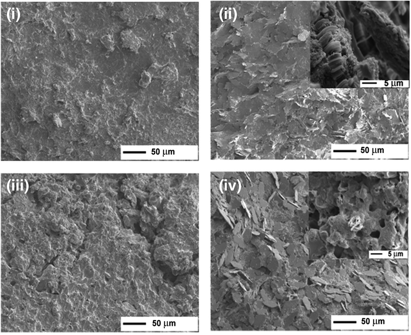
Scanning electron microscopy micrographs of: CaP G3KPS at pH 9 before
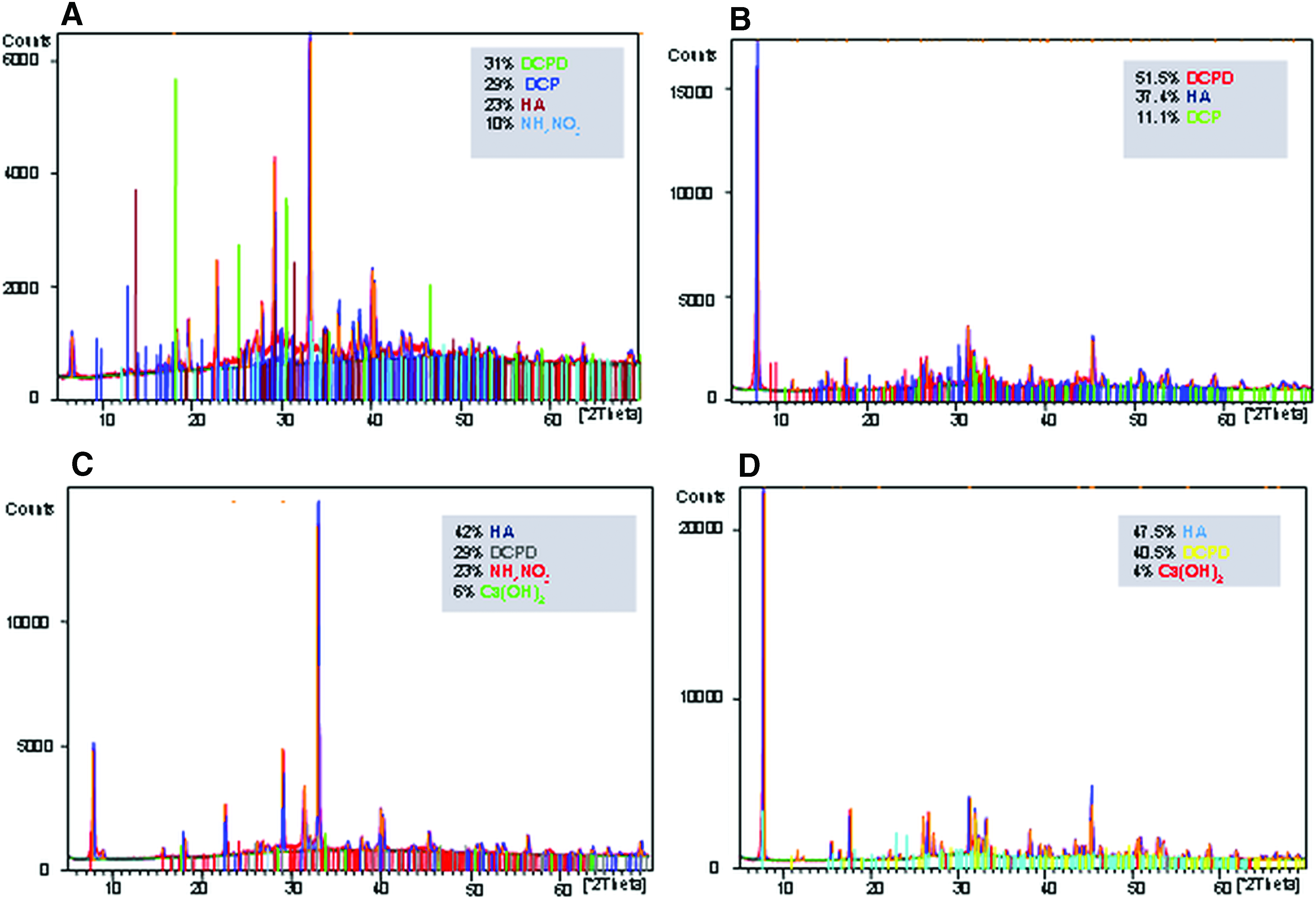
X-ray diffraction patterns of CaP9 gel materials before and after dialysis
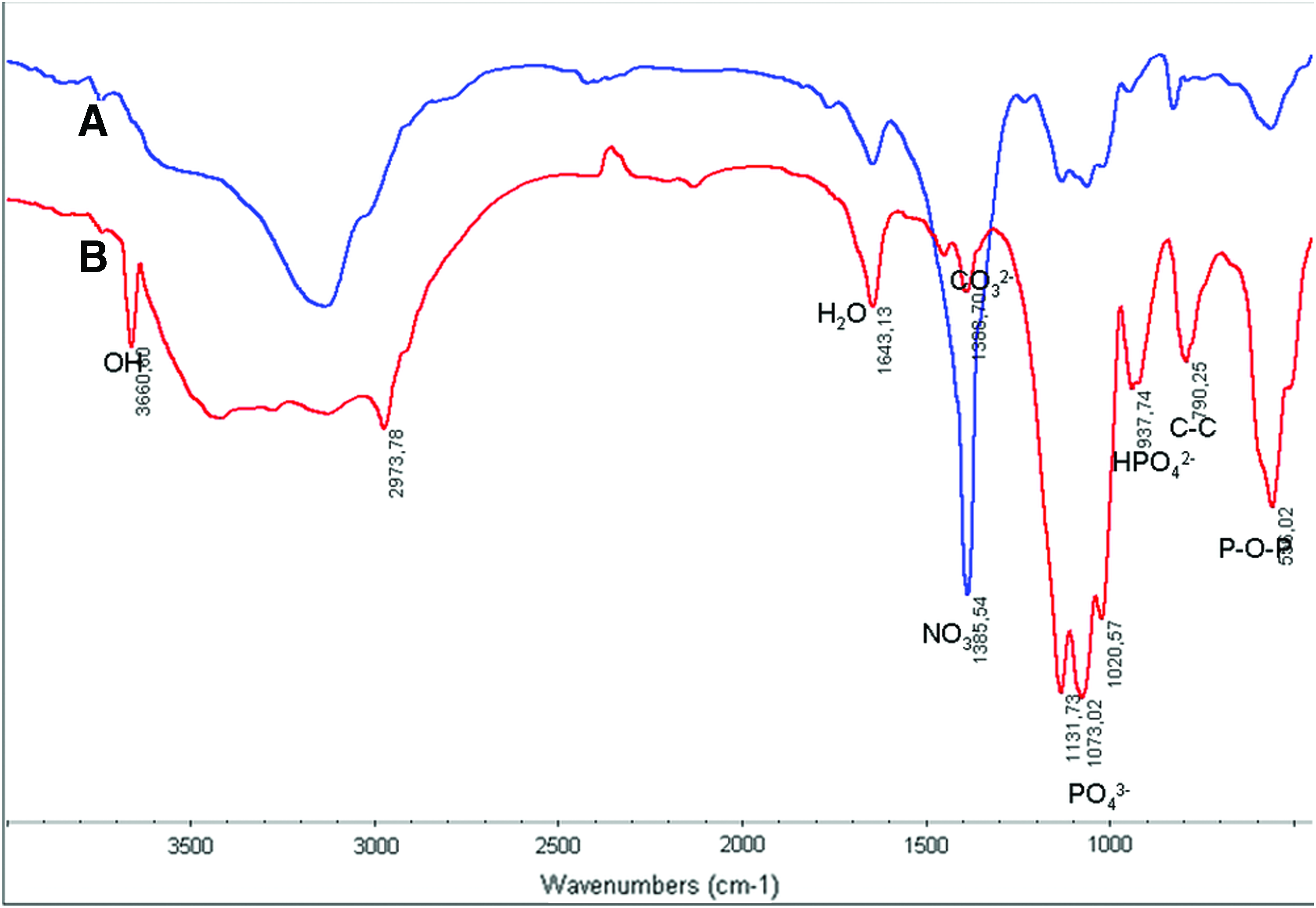
Fourier transform infrared spectroscopy (FTIR) spectra of CaP11 gel before
FTIR also showed typical signals of CO32− due to chemiosorptions of atmospheric CO2 on the surface of HA. 25 Van der Houwen et al. have reported that the dissolution of atmospheric CO2 in CaP gels yields CO32− appearing as peaks at 1456 cm−1 after crystallization at the alkaline value of pH 10.5. 38 Since the present experiment was also done in alkaline conditions, a similar process might also have occurred; while there is no narrow FTIR peak at 1456 and 873 cm−1 in the FTIR spectrum, close observation shows a shoulder at about 1456 cm−1. As evidenced in Figure 5B, the peaks at 556 and 3570 cm−1 are the characteristic peaks for stoichiometric HA. 39 It is possible to observe the HPO42− peak demonstrating the presence of DCP. In addition, the spectrum of the G3-K PS hybrid gels prepared at pH 9.0 clearly revealed the integration of the dendron in the gel with strong amide I, II, and III bands appearing in the range of 1016 to 1044 cm−1, which do not appear in the spectrum of gel without G3-K PS (Fig. 5A). The shift of peaks in the range of 550 to 940 cm−1 suggested the occurrence of interactions between the mineral phases with the integrated dendrons. An NH band was also visible at 3436 cm−1 alongside an NH3+ band at 3134 cm−1 (Fig. 5B). 40 Similar results were obtained for CaP gel synthesized at pH 11 suggesting no main effect of the pH on the level of interactions between the CaP gel and the dendrons (data not shown).
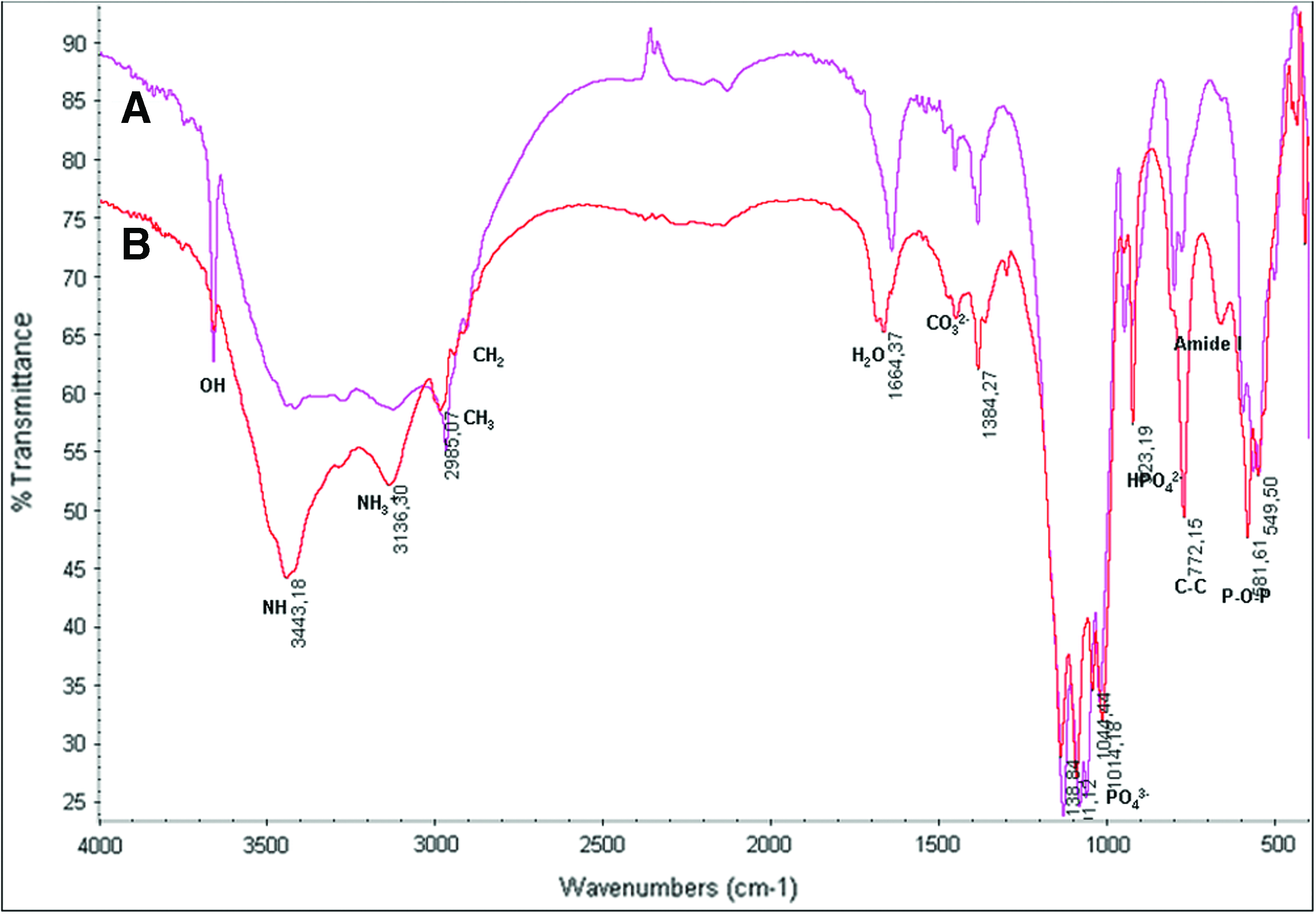
FTIR spectra of CaP9
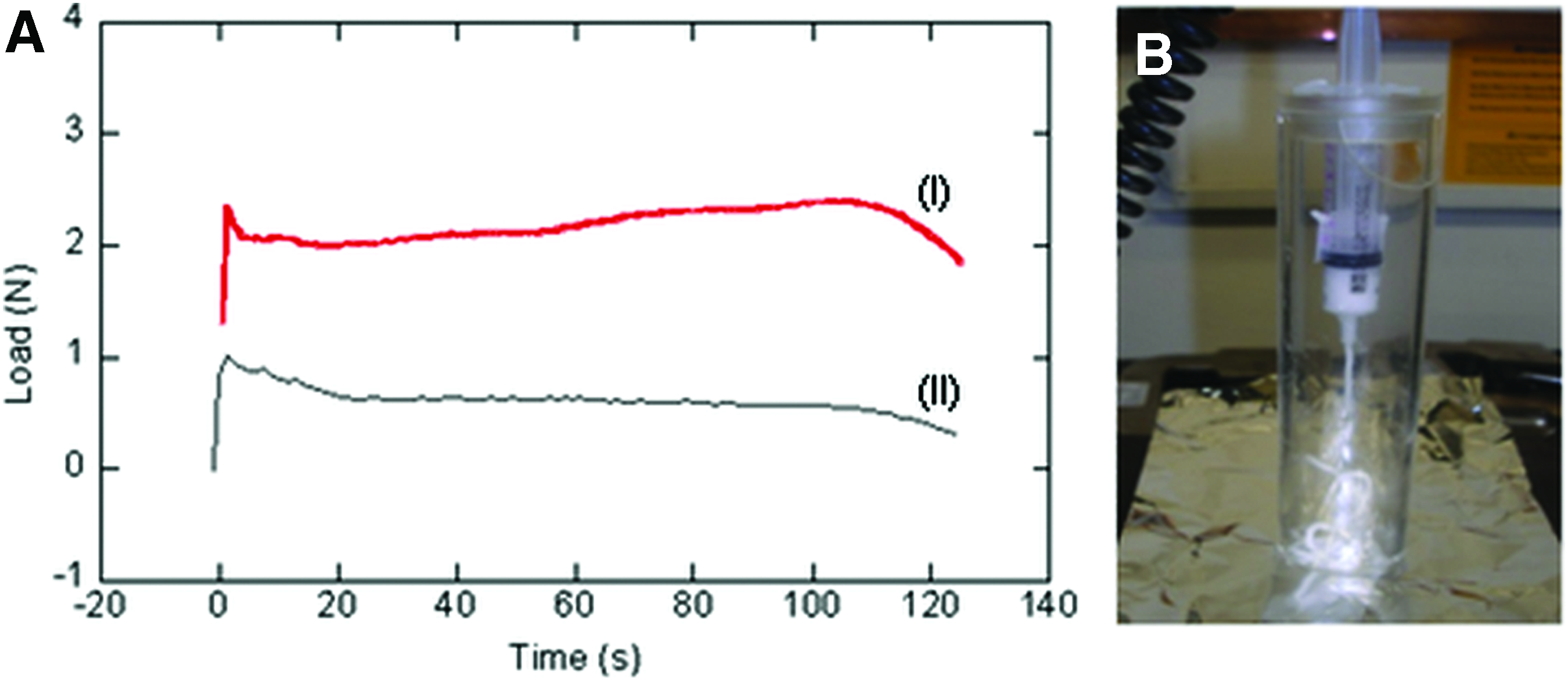
The injectability tests demonstrated that the gels both with and without dendrons maintained injectable properties suitable for clinical applications. Figure 6A has reported a result of CaP9 with G3-K PS dendrimers (I) and of empty syringe (II) was observed with a similar mechanical property (data not shown). The applied force versus piston displacement curves (Fig. 6A) show that the applied force increases at the beginning of the injection. After the vanishing of transient state, the force soon reaches an almost constant level close to 2 N necessary to inject all the material. Moreover, the flow in the syringe took place without a phase separation phenomenon (Fig. 6B). The dialysis treatment used to equilibrate the pH and to remove the ammonium nitrate did not seem to compromise the injectability; all samples require the application of load, which is within the range of force applied during hand applied pressure, thus supporting the feasibility of the system for surgical purposes.
Biological studies
Tests of cell metabolic activity on CaP materials synthesized either at pH 9 or 11, with and without the integration of G3-K PS, were performed using MG-63 cell line. The results of the indirect contact assay (Alamar Blue) after 48 h exposure of the cells to the supernatants of leaching studies are shown in Figure 7. The results indicated that the gel materials with and without G3-K PS did not have cytotoxic effect; indeed, it is possible to evaluate the effect of materials on the cells by an increasing of the metabolic activity.

Alamar Blue indirect test on MG63 cells after 48 h exposure time. Results were calculated from at least three independent experiments. Asterisk (p<0.05) indicates statistically significant differences between the material groups and control (CTR).
Figure 8A and B show the effect of the materials on the adhesion and metabolic activity of hMSC at different times. Cell adhesion values after 1 and 3 days (Fig. 8A) are presented as the cellular percentage of attached cells in relation to control tissue plates. The cellular attachment is the first step in evaluating the biocompatibility of hMSC onto the CaP gel material with and without G3-K PS. It shows that a short incubation in the presence of the dendrons improved the cell adhesion. In particular, the representative morphology of adhering cells after only 1 day of culturing on CaP gels showed an elongated morphology of the cells (Fig. 9). Our results demonstrated that cell adhesion and spreading of cells showed by representative morphology were optimal in all cases. However, the cell attachment and spreading were consistently higher on CaP9G3-K PS (Fig. 9B) and CaP11G3-K PS (Fig. 9D) when comparing to control and bulk gel materials. This effect on G3-KPS materials could be for the presence of poly(epsilon-lysine) that is a homopolymer that improves cell adherence, and this is important for cell–material interaction. 41
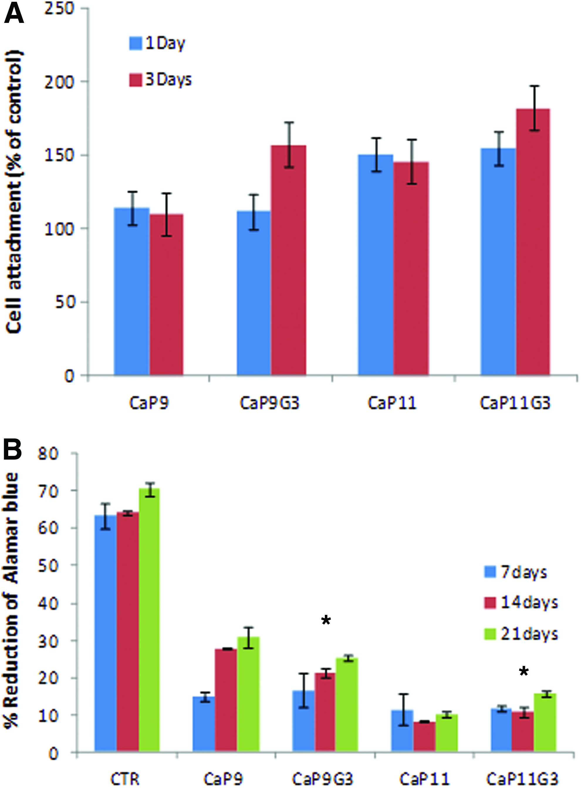
Cell adhesion after 1 and 3 days

Staining of morphology and spreading of hMSC culture for 1 day at lower and higher magnifications onto gel materials
At long times (7 to 21 days), a reduced proliferation was observed. This behavior might due to the increase of differentiation rate of hMSC adhering on the surface of samples integrating the G3-K PS (Figs. 9 and 10).
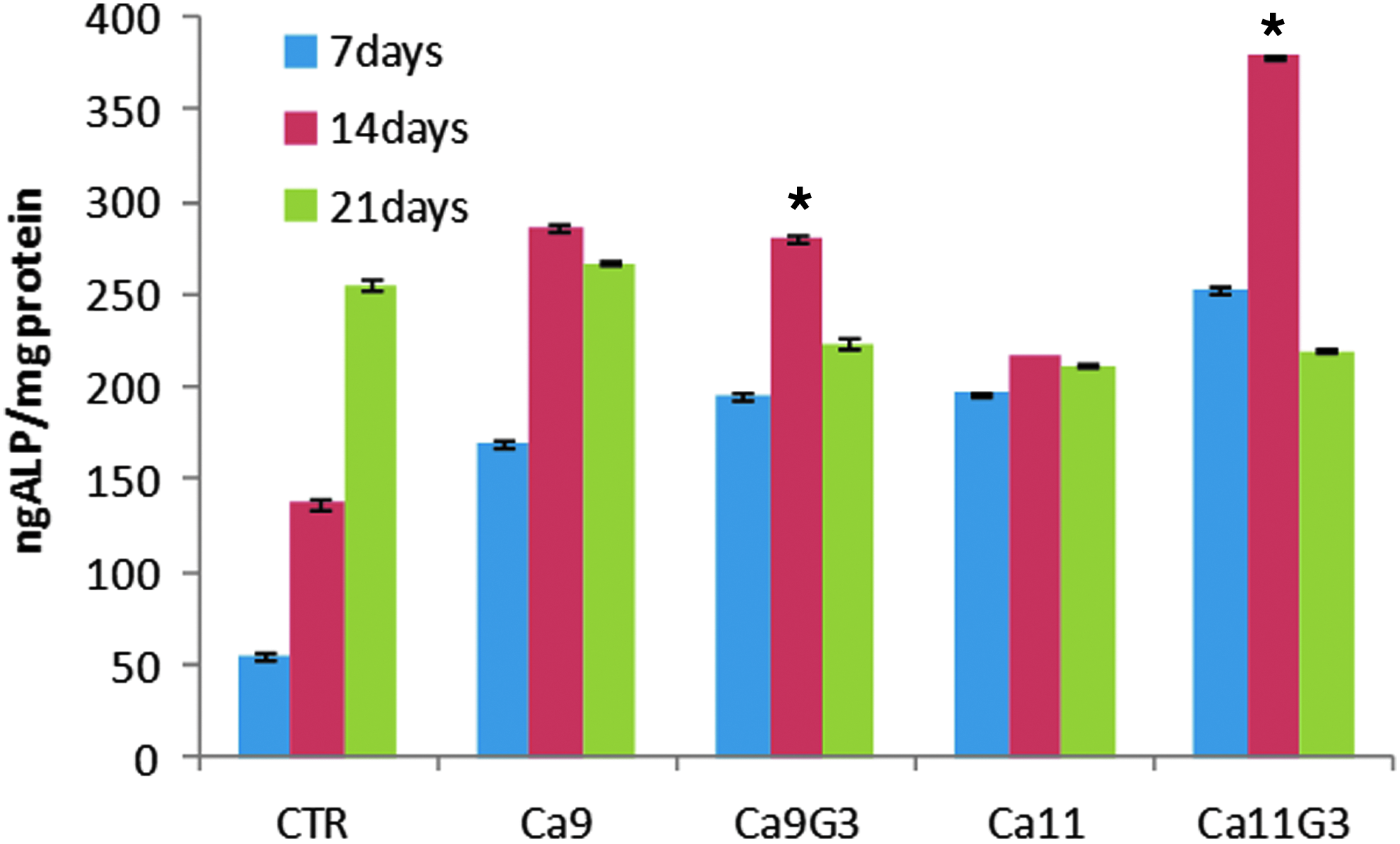
ALP activity per mg protein of hMSCs cultured on gel materials at 7, 14, and 21 days. Asterisk (p<0.05) indicates statistically significant differences between the material groups with and without G3-K PS at different times. ALP, alkaline phosphatase. Color images available online at www.liebertpub.com/tea
The ALP activity was determined on cell cultures, and it showed that the cells adhering on CaP11G3-KPS had the highest differentiation level with the enzyme activity reaching its peak at 14 days (Fig. 10). Indeed, ALP activity on cultures of hMSC onto the gel materials with the presence of G3-K PS was significantly higher at days 7 and 14 (p<0.05), but not after 21 days of cultures on all experiments. Furthermore, the qualitative and semiquantitative RT-PCR analyses performed on cells grown on gels with and without G3-K PS composite materials revealed a more intense bands on agarose gel for the materials with the presences of G3-K PS in all the investigated genes and particularly for the transcripts specific for ALP and OCN (Fig. 11A, B). Noticeably, the presence of dendrons completely changed the cell substrate properties of the cements synthesized at pH 11.0 that otherwise resulted less suitable than those synthesized at pH 9.0.
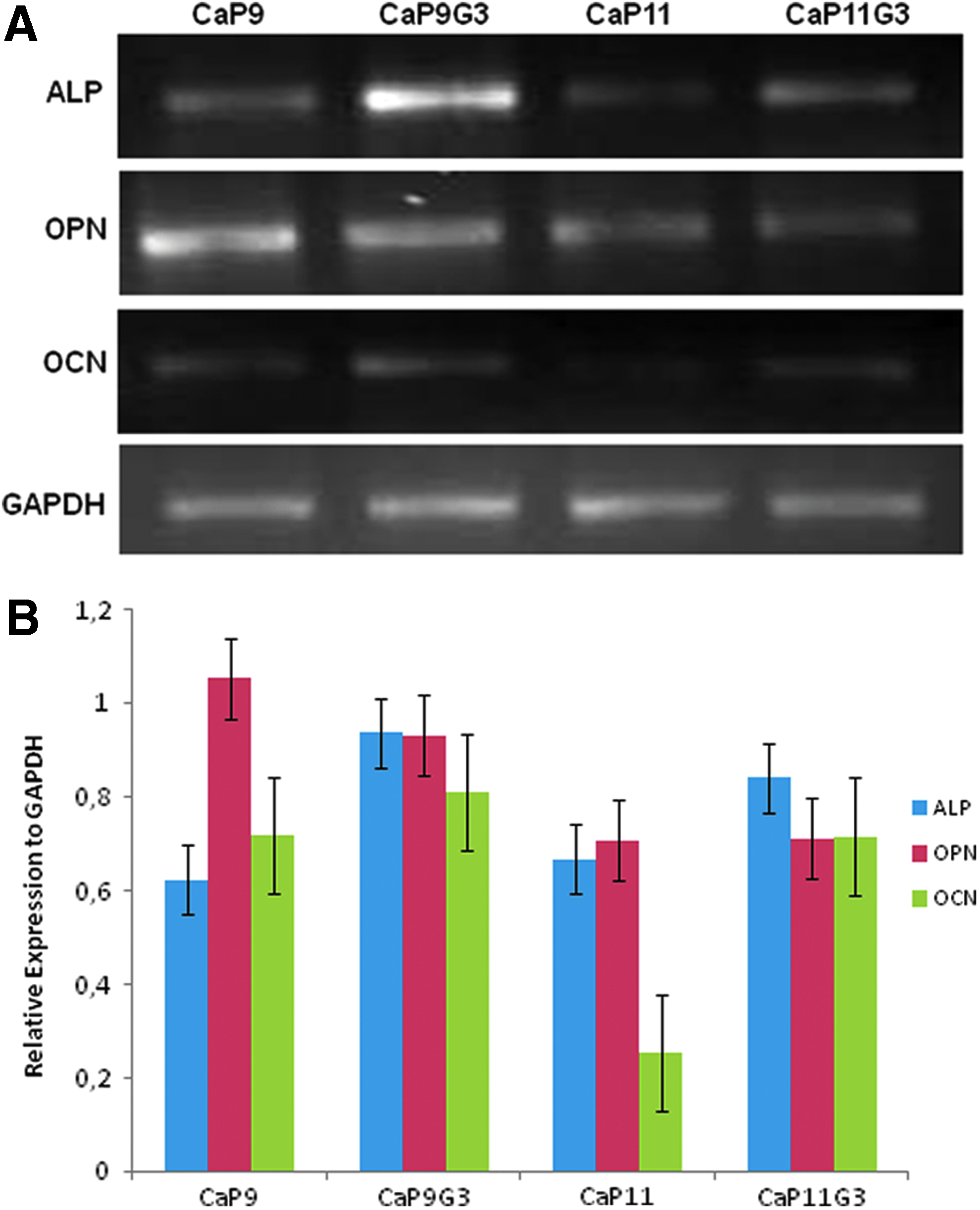
Gene expression of CaP and CaPG3-K PS gels after 21 days of culture in alpha minimum essential medium
Discussion
Synthetic bone fillers with osteoconductive and injectable properties are widely considered as materials of choice for the repair of large bone defects.42,43 Currently, the most widely used injectable bone cement is poly(methyl methacrylate) (PMMA), but this type of biomaterial acts as a filler with no tissue regeneration properties. Rather, the hardening PMMA paste leads to tissue necrosis of the surrounding tissue and its lack of biodegradation does not allow the in-growth of newly formed tissue. 44 CPC offer good osteoconductivity and bone tissue repair properties, but their injectability is poor unless additives including surfactants and biopolymers are included in their formulations.45,46 Furthermore, it is suggested that for injectable bone cements the setting time should not be too short as a rapid setting paste will increase the paste thickness and rigidity upon injection. 47
The present study investigated the synthesis and in vitro osteoconductivity properties of sol-gel CaP composite gels obtained at room temperature where the HA percentage could be controlled to modulate bioresorption. Indeed, although not focus of this study, the resorption kinetics of CPC have been shown to be regulated by changing the ratio of the more and the less soluble ceramics phases (HA and DCP, respectively) in the gel material. 48 In this article, to achieve tunable resorption, the gels has been re-equilibrated at various pH values, thus allowing the control of the biphasic CaP phases into different DCPD/HA ratios. DCPD is one of the precursors of HA that is the main component of hard tissues in vertebrates. It was proved that a more soluble CaP might be better for fixation during the early period following implantation. 49 Brushite can gradually dissolve at physiological conditions to provide increased levels of calcium and phosphate ions near the tissue–implant interface. 50 In this case, it is possible to identify the gel material like a biphasic implant where DCPD component stimulates the early bone formation while HA domains with slower resorption rate act as anchorage points for the newly formed bone tissue. Such an approach eliminates the need for macroporous interconnecting materials that have been deemed necessary to encourage tissue in-growth in monophasic materials with slower resorption rate. 22 In this study, the synthesis of the CaP gels at different pH values (i.e., pH 9 and 11), followed by their equilibration at neutral pH was shown to produce pastes of comparable injectability properties, but with different percentages of HA. In particular, the synthesis at pH 11 was shown to enhance the formation of HA domains in forms of crystal plates that could act as anchorage points for the growing bone. The extension of the setting time, together with clear changes in the FTIR profiles of the cements, showed that the phosphoserine-tethered hyperbranched dendrons were interacting with the ceramic phase.41,51 Indeed, phosphoserine is known to be the most potent biocatalyst of biomineralization in nature. 52 This potent catalyst role derives from the zwitterionic character of this modified amino acid that induces the formation of calcium ions coordination spheres and their availability to form clusters with phosphate ions, thus nucleating the CaP crystals. Furthermore, poly(epsilon-lysine) is a well-known cell adhesion promoter and their presence in the hybrid gel is expected to further enhance cellular adhesion and bioactivity to the implant.53,54 In addition, recent articles have shown the ability of the phosphoserine-tethered poly(epsilon-lysine) dendrons to encourage both osteoblasts and stem cell adhesion, early proliferation, and accelerated differentiation.55,56 It can be speculated that this substrate properties could emerge from the combination of the ability of the dendrons to rapidly induce the formation of a CaP clusters at the material surfaces with the nanostructured topography generated by the grafted dendrons. When integrated in the CaP gels, these dendrons could therefore play both a role into the gel setting and biphasic properties modulation and be at some extent exposed to the surface to enhance their tissue regeneration potential. Indeed, these combined chemical features would therefore result into an enhanced osteoblastic colonization of bone fillers. 57 In particular, the integration of G3-K PS dendrons in the gels obtained at pH 11.0 increased the substrate properties of this type of CaP that was otherwise showing limited cell substrate properties. Therefore, it can be speculated that the increase of HA domains were not able to improve cell adhesion and that only the presence of dendrons mimicking noncollageneous calcium-binding protein could enhance the material cell substrate properties. This hypothesis appears to be supported by the ability of the dendrons to improve the hMSC adhesion in both CaP9 and CaP11 (Fig. 8A). Moreover, the presence of phosphoserine-tethered poly(epsilon-lysine) dendron in the CaP gels enhances the differentiation of hMSC in the osteoblastic phenotype increasing the expression of ALP and OCN, two osteoblastic markers of the osteogenic differentiation as reported in Figures 10 and 1. The presence of the biomimetic dendrons appeared to couple with the CaP11 physicochemical properties to provide a very suitable substrate for hMSC. These properties, combined with the suitable injectability properties, make these composite biomaterials a very attractive alternative for the manufacturing of injectable osteoconductive synthetic bone fillers 16 suitable for surgical procedures aiming at treating in situ fractures, filling root canals, and sealing furcation perforation in endodontic and in spinal surgery.
Conclusions
The present work demonstrates that injectable CaP gels with different percentage of HA can be synthesized at room temperature by the sol-gel method. The optimization of a dialysis process allows the equilibration of the gels at physiological pH value that removes the presence of toxic ammonium nitrate without altering the physicochemical properties of the materials. The integration of phosphoserine-tethered hyperbranched molecules increases the proliferation of mesenchymal stem cells at an early time point during cell culture and enhanced their long-term ALP and OCN activity, thus conferring a bone cell stimulatory potential to the injectable pastes. The high reproducibility of the manufacturing process of these composite materials together with their enhanced bioactivity on relevant cell types suggests their high clinical potential in the treatment of bone defects.
Footnotes
Acknowledgments
This study was supported through funds provided by the PNR-CNR Aging Program 2012–2014. The authors also thank Ms. S. Zeppetelli for facilitating biological analysis.
Disclosure Statement
No competing financial interests exist.